Learn more about diseases starting with the letter “B”: Basilar impression, Basilar migraine, Beriberi, Bettolepsy, Amyotrophic lateral sclerosis, Alzheimer's disease, Wilson's disease, Hallerwarden-Spatz disease, Hamstorp disease, Hippel-Lindau disease, Canavan disease, Creutzfeldt disease -Jacoba, Lafora's disease, Machado-Joseph disease, Moya-moya disease, Morton's disease, Parkinson's disease, Pick's disease, Refsum's disease, Fahr's disease.
Description and causes of the disease
The pathology is characterized by symmetrical deposition of calcium elements in the structures of the brain - in the subcortical region, meninges and cerebellar zone. The disease includes only cases of primary calcification; mineral accumulations against the background of other pathologies (secondary cases) are not included in the clinical picture of the disease. For the first time, the most complete description of the anomalous process was created in the 30s of the last century by the German doctor K.T. Farom.
The disease is classified as rare, as only one case is recorded per million people on the planet. The male half of humanity is susceptible to these disorders twice as often as the female half. The phenomenon can appear at any age, but the most common age category is the period from 30 to 60 years. Since the disease is practically asymptomatic, and obvious signs are very similar to other neurological diseases, only about 2% of pathological cases are detected during the patient’s lifetime.
External and internal factors in the development of the disease have not yet been established. Most medical scientists tend to believe that there is a congenital, genetic etiology, since a hereditary factor can often be traced in the same family. There are studies that prove the theory of mutations in the genetic code affecting chromosomes 14, 2 and 8; this issue is still being studied. This theory is counterbalanced by cases where the disease manifested itself in people who did not have relatives in the older generation with this phenomenon. The first manifestation of symptoms occurs already at the stage of critical filling of the brain fibers with calcium inclusions, which does not allow studying the course of the disease during a person’s lifetime.
Pathological studies reveal the symmetrical presence of calcifications in the tissues of the frontal lobes and the cerebellar zone. Most of the accumulations are present in the subcortical region, less often deposits are seen on the vascular canals (mostly on the arterial walls). In appearance, the deposits resemble microscopic thread-like channels interspersed with metal elements of various types.
In the medical literature, there are two forms of pathology, divided by age and inherent symptoms. The first form is represented by the so-called juvenile form, since primary manifestations occur in childhood or early adolescence. There is a slight decline in intellectual and cognitive abilities, and a slowdown in the functionality of the muscular system. A frequent “accompanying” of this form of anomaly is parallel mental retardation. Over time, symptoms develop into signs of Parkinson's disease.
The second form is characterized as cyanide and manifests itself in middle or old age. The main symptoms are signs of parkinsonism with obvious impairments in the decline of intellectual abilities. Over time, profound dementia progresses.
Archives
Short Wiklad
The brain is stolen from infusions of various toxic agents by the blood-brain barrier. The basal ganglia, which are sensitive to mineral factors that cause a lot of disorders, are deprived of the blame. Examples include accumulation of copper for Wilson's disease, saliva for Gallenvorden-Spatz disease, organic mercury for Minamata disease and manganese for toxic parkinsonism. Calcification of the basal ganglia and other basal ganglia underlies a significant clinical problem associated with a variety of neurological disorders. This article is a short overview of current knowledge from this “neuroradiological” illness.
1. HISTORY
In 1850, Delacour first described vascular calcifications in the basal ganglia in a 56-year-old man, which was clinically associated with muscle rigidity of the lower ends and tremor.
He died from severe illness, including diarrhea, hypotension and coma. Pathomorphological examination revealed the presence of binary calcification of the brain and sclerosis. Bamberger in 1855 predicted a histopathological single type of calcification of the small cerebral vessels in a woman with inhibition of mental development and vessels. In 1930, Fahr describes an 81-year-old patient with advanced dementia, who was hospitalized with fever, cough, bedsores, “dementia without paralysis,” and who died 3 days later. The section revealed a thickened granular cortex, scutulae filled with serous tissue, and calcification of the periphery and striatum. This has always been associated with all forms of binary calcification of the basal ganglia and other parts of the brain, regardless of the fact that it was not the first to describe such a disorder. Thus, Fritzsche insisted on noting the radiological manifestations of the remainder of the 1935 birth. There are a lot of terms (about 35) to describe the named illness, which creates significant difficulties (Table 1). Table 1. Common terms found in medical literature to describe binary calcification of the striatum, pallidum and dentate nucleus
| Rick | Descriptive term |
| 1939 | Symmetrical cerebral calcification |
| 1951 | Calcification of the dark body and dental nuclei |
| 1954 | “Idiopathic” calcification of cerebral capillaries |
| 1957 | Symmetrical (or familial) calcification of the basal ganglia |
| 1960 | Familial idiopathic calcification of the brain |
| 1961 | Idiopathic non-atherosclerotic calcification of the medullary vessels |
| 1964 | Familial vascular calcification of the central nervous system |
| 1968 | Calcification of the striopalidosdental system |
| 1969 | Idiopathic familial cerebrovascular ferocalcinosis |
| 1976 | Symmetrical calcification of Stovburian ganglia |
| 1982 | Fahr syndrome |
| 1983 | Progressive idiopathic striopalidosdental calcification |
| 1985 | Symmetrical intracranial progressive pseudocalcinosis |
| 1997 | Idiopathic calcification of the basal ganglia |
“Hvoroba Fara” is an incorrect name, unimportant in its wide stagnation. In this case, fragments of calcification tend to form in the dentate nuclei and basal ganglia, so the term “binary striopalidosdental calcification” (DSDC) is no longer appropriate.
2. PATHOMORPHOLOGY
Pathomorphological studies indicate that calcium is the main element responsible for radiological manifestations of illness. It also reveals traces of mucopolysaccharides, aluminum, amygium, cobalt, copper, molybdenum, saliva, lead, manganese, magnesium, phosphorus, sawdust and zinc. Calcium and other mineral deposits are evident in the walls of capillaries, arterioles, veins and perivascular spaces. Information about neuronal degeneration and gliosis in these areas. Electron microscopy confirms the presence of mineral elements in pericytes.
Although the details of the pathological process in DSPCD are not unknown, it is assumed that hyperintense MRI images in T2 mode depict a progressively progressive metabolic and inflammatory process in the brain, which eventually leads to calcification and consistent with clinical manifestations. It is also important to note that the accumulation of calcium and calcium is a reaction to the extravasation of the complex of acidic mucopolysaccharides - protein. MRI follow-up suggests that damage to the side of the vessel membrane is consistent with the leakage of plasma with further effects on the neuropil and mineral accumulation.
And while the exact reasons why the basal ganglia are so sensitive to calcium accumulation have not been elucidated, they act as targets for the accumulation of a large number of other non-mineral substances, for example, bilerubin in nuclear neonatal supplementation with MPTP and carbon monoxide for toxic parkinsonism.
3. CLINICAL MANIFESTATIONS
DSPZK is a rare disorder. This clinical verification is clouded by many factors. Symptoms described in the literature are based on single clinical reports. Their analysis of the data from the registries is conclusive that out of 99 individuals with obvious calcium accumulations, 67 were symptomatic in the middle age 47 ± 15, and 32 were asymptomatic in the middle age 32 ± 20 years. The relationship between men and women becomes 2: 1. The most widespread manifestations of DSPCD were lexical disorders (55%), of which half were parkinsonism, and half were hyperkinesis (chorea, tremor, dystonia, athetosis, orofacia alna dyskinesia). Cognitive impairment is another part of the syndrome, followed by cerebellar symptoms and brain disorders. Often there are overlaps in various clinical units, such as hypokinesia, cognitive dysfunction and cerebellar signs. Other neurological microsymptoms include pathological pyramidal signs, psychiatric phenomena, impaired walking, sensitive disorders and pain.
4. NEUROPHISIOLOGICAL DEVELOPMENTS
EEG, examination of neural conductivity and visual response potential in DSPPC are normal. Stovbur's auditory evocation potentials demonstrate minor changes, for example, the interpinal latencies between the I and V lines and the increased latency of the V. The somato-sensory potentials remain unchanged.
5. NEURORADIOLOGICAL DEVELOPMENTS
Before the CT scan, instrumental precautions for this disease were based on the data from radiography of the skull and autopsy. With the development of tomographic techniques, the number of such precautions has increased dramatically. It is important that in case of DSPCD, CT is a more sensitive diagnostic approach than MRI. When analyzing a large number of scans (19,080 in 3 studies), it was found that the frequency of calcification of the basal ganglia ranged from 6–7.49 per 1000 individuals. Most of these changes, no matter how insignificant, are separated by a pale sack; in other plots, the smell is rare. The lesions most often affect the basal ganglia, dentate nucleus, thalamus and centrum centrum. No specific patterns typical for a particular subgroup were identified in the patient population. In one study, calcium levels were measured using CT in 31 patients, in which cross-sectional differences were not observed. The average volume of calcification in the basal ganglia became 1.39 ± 0.28 cm3; thalamus - 0.26 ± 0.05 cm3; dentate core - 1.02 ± 0.35 cm3; for the circumferential center - 0.64 ± 0.22 cm3 (for the whole - 3.16 ± 0.64 cm3). Also, no consistent differences in calcification levels were found between groups of symptomatic and asymptomatic and young and older patients.
Single-photon computed tomography with a radiomarker showed a double decrease in perfusion in the area of the basal ganglia and cerebellar cortex in DSPCC. Positron emission tomography with fluorodopa did not show a significant difference between these patients and controls.
6. DIAGNOSTICS
The clinical manifestations of the disease may vary, and the diagnosis can be made with the help of neuroimaging and exclusion of calcium metabolism disorders and developmental defects.
Regardless of the widespread availability of CT and MRI and the frequent detection of calcium deposits in the basal ganglia in asymptomatic patients, the DSP is deprived of a rare disorder. If parkinsonism is associated with dementia and cerebellar symptoms, then CT scan of the brain is not important, since the disease is often accompanied by vision syndromes. Hypoparathyroidism is excluded from the differential diagnosis. The use of calcium and parathyroid hormone in the serum helps to separate two parts, since tomography shows double calcification of the striopalidosdental structures. INNSHIS, under the Yakiki, in the Naryavnna Kalcifіkasіya pydkirkovikh, presented at table 2. Table 2. Imovirni causes of the double -Kalcifice of the nucleus
| STRIOPALIDO-DENTAL CALCINOSIS | |
| Pervinny | Autosomal dominant Familial Sporadic |
| Secondary | |
| Endocrine | Hypoparathyroidism Pseudohypoparathyroidism Pseudo-pseudohypoparathyroidism Hyperparathyroidism |
| Vrozheniy | Cockayne's syndrome Syndrome of microcephaly, demyelinization and striopalidosdental calcification |
| Rheumatological | Systemic red wolfdog |
| Toxic | Plumbism |
| BINAL STRIOPALIDARY CALCINOSIS (“CALCINOSIS OF THE BASAL GANGLIES”) | |
| Physiological | Century over 50 years |
| Vrozheniy | Angiomatous malformation with galenic vein aneurysm Down syndrome Kearns-Sayry syndrome |
| Degenerative | Diffuse cerebral microangiopathy Hyperkinetic mutism Coates' disease |
| Genetic | Biotinidase deficiency Carbonic anhydrase II deficiency (osteopetrosis, nitric tubular acidosis and basal ganglia calcification) COFS syndrome with familial translocation 1; 16 Lipomembranous polycystic osteodysplasia Tapetoretinal degeneration |
| Infectious | SNID Active Epstein-Barr virus infection Meningoencephalitis Mumps encephalitis |
| Metabolic | Dihydropteridine reductase deficiency MELAS syndrome Post-hypoxic and post-ischemic conditions |
| Neoplastic | Gostra lymphocytic leukemia |
| Physical | Promeneva therapy |
| Toxic | Removal with carbon monoxide |
| BIBICAL CALCIFICATION OF THE CEREBLE | |
| Pervinna | Idiopathic |
| Vtorinna | |
| Infectious | Syphilis |
| Sudinna | Hematoma |
These are also accompanied by a lot of other neurological symptoms. The minimum age, if negative CT results turn off DSPPC, is not explained. In one study of such patients, a decrease in the level of calcium in the liquor was recorded with normal indicators of phosphorus and albumin in the serum and liquor.
7. LEKUVANYA
Selective absorption of calcium without adding calcium to other tissues using a non-toxic method without any apparent care. Therapy with specific neurotropic calcium channel blockers (nimodipine) was unsuccessful. A decrease in 25-OH vitamin D3 at normal levels of 1.25(OH)2 vitamin D3 suggests a congenital defect in its metabolism in three individuals with autosomal decline, the presence of striopallidodental calcifications and irrational disorders. By optimizing therapeutic prospects, this fact requires further consideration. In one case, the use of disodium ethidronate resulted in symptomatic improvement without changing the stage of calcification, which will require treatment on a larger number of subjects in controlled studies.
8. VISNOVOK
Taking into account the fact that there are 35 different terms (these are listed in Table 1), the most unique is the term “Far’s illness”. The fragments have a lot of dislocations that are accompanied by binary calcification of the substructure structures, optimally define the term that is based on the anatomical localization, for example, “striopalidosub” partial”, “striopalidarny” and “cerebellar calcification”. In such cavities, there are obvious other chemical elements besides calcium, but they themselves are visualized by tomography, which is the essence of the term “calcification.” These purchases are not associated with a single chromosomal locus. Geshwind et al. revealed an autosomal dominant form of illness with neurological manifestations associated with chromosome 14 in one great homeland. Brodaty et al. The presence of such a locus was attributed to the presence of neurological, cognitive and psychiatric symptoms. In addition, the association of hypothyroidism with the 11th chromosome locus, pseudohypoparathyroidism with the 20th chromosome, and Down syndrome with the 21st chromosome includes the possibility that a single gene is responsible for the accumulation of calcium and other minerals and in the brain.
Prepared by Yuriy Matvienko
LITERATURE
- Cartier L, Passig C, Gormaz A, López J. Neuropsychological and neurophysiological features of Fahr's disease. Rev Med Chil. 2002 Dec;130(12):1383-90.
- Faria AV, Pereira IC, Nanni L. Computerized tomography findings in Fahr's syndrome. Arq Neuropsiquiatr. 2004 Sep;62(3B):789-92.
- Iványi A, Takács M. Intracerebral, non-arteriosclerotic vascular calcification: Fahr's disease. Orv Hetil. 1989 Jan 8; 130(2): 83-5.
- Lauterbach EC, Cummings JL, Duffy J, Coffey CE, Kaufer D, Lovell M, Malloy P, Reeve A, Royall DR, Rummans TA, Salloway SP. Neuropsychiatric correlates and treatment of lenticulostriatal diseases: a review of the literature and overview of research opportunities in Huntington's, Wilson's, and Fahr's diseases. A report of the ANPA Committee on Research. American Neuropsychiatric Association. J Neuropsychiatry Clin Neurosci. 1998 Summer;10(3):249-66.
- Manyam B.V. What is and what is not 'Fahr's disease'. Parkinsonism Relat Disord. 2005 Mar; 11(2):73-80.
- Manyam BV, Walters AS, Narla KR. Bilateral striopallidodentate calcinosis: clinical characteristics of patients seen in a registry. Mov Disord. Mar 2001; 16(2): 258-64.
- Penín Alvarez M, Araújo Ayala R, Rodríguez Ferro R, Sesma Sánchez P. More Fahr's syndrome? Rev Clin Esp. 2005 Dec; 205(12): 633-4.
- Rosenblatt A, Leroi I. Neuropsychiatry of Huntington's disease and other basal ganglia disorders. Psychosomatics. 2000 Jan-Feb; 41(1): 24-30.
- Rossi M, Morena M, Zanardi M. Calcification of the basal ganglia and Fahr's disease. Report of two clinical cases and review of the literature. Recenti Prog Med. Mar 1993; 84(3):192-8.
- Shenoy AM, Volpe D, Ensrud ER. Fahr's disease. Pract Neurol. 2009 Apr; 9(2): 100-1.
Symptoms inherent in the disease
The very first signs of the disease are:
chronic fatigue, fatigue even with reduced physical activity;- lack of coordination during movements, changes in gait (shuffling movements of the legs), clumsiness;
- speech dysfunction;
- the appearance of uncontrolled spastic movements, night muscle activity;
- in childhood, spasms of the fingers and stiffening of the body muscles are observed, giving unnatural postures;
- in adulthood, numbness of the facial areas often occurs, sensory perception and response to external stimuli are slowed down, and movements of all parts of the body are constrained;
- there is persistent tremors in the limbs.
Symptoms of parkinsonism often appear:
- wide, sweeping movements incommensurate with the intended actions;
- weakness in the arms and legs, uncontrolled reflexes in the tendon apparatus;
- loss of control over excretory functions, urinary incontinence;
- in childhood, the manifestation of epileptic syndrome is possible.
Cognitive impairment:
- decline in memory ability;
- slowness of the thought process;
- lack of ability to maintain attention on one process;
- inability to analyze what is happening, decline in intelligence.
The progressive development of the described symptoms leads to the development of oligophrenia in childhood and to dementia in adulthood, however, there are cases when the disease does not lead to a decrease in intelligence in children and adolescents.
Treatment
- Blepharospasm can be a reflex reaction to an underlying disease (most often with diseases of the ocular surface and this must be excluded at the very beginning of treatment)
- Wearing sunglasses can reduce exposure to bright light and prevent awkward stares from onlookers.
- Voluntary maneuvers such as pulling the eyelid, straining the neck, talking, yawning, singing.
- Patients with severe blepharospasm should not drive
Drug treatment
Although drugs from various classes have shown some effectiveness in the treatment of blepharospasm, drug therapy for blepharospasm is usually based on the following pharmacological untested hypotheses:
- cholinergic excess,
- hypofunction of GABA,
- excess dopamine.
Pharmacotherapy, as a rule, is less effective than botulinum toxin injections and is mainly of an auxiliary nature.
The use of antispasmodics and benzodiazepines has little effect. There has been some positive effect from the use of tetrabenazine.
Botulinum toxin injections
The preferred treatment for blepharospasm is botulinum toxin type A injections into the orbicularis oculi muscle. Systematic Cochrane studies have shown that botulinum toxin treatment is highly effective, benefiting up to 90% of patients compared with placebo. Botulinum toxin interferes with the release of acetylcholine from nerve endings, and therefore, for a certain period of time, relieves spasm in the muscle where the toxin was injected.
The initial dose is usually 10-20 units at the injection site, and the dose is subsequently adjusted depending on the individual patient's response. Most patients need to be re-treated every three months, and gradually higher doses may be needed over a longer period of time.
Side effects of botulinum toxin include lagophthalmos, ectropion, or volvulus. Side effects such as lacrimation, dry eyes, and sometimes keratitis have also been reported. Accidental entry of the toxin into the orbit can lead to ptosis ± diplopia. All these side effects (as well as therapeutic ones) disappear within 3-4 months.
Surgery
In severe cases of blepharospasm and resistance to pharmacological treatments, selective myomectomy can be used (dissection of some of the muscles that close the eyes).
Diagnostic measures
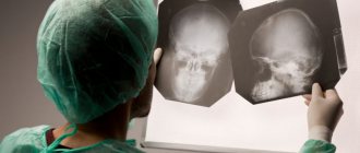
The primary diagnostic measure is examination and consultation with a neurologist. At the first suspicion of a disorder and to exclude similar diseases, a highly informative diagnostic method is prescribed - a computer scan. During screening, the presence of foci of accumulation of calcifications in intracerebral structures is revealed. An alternative method for examining the brain is magnetic resonance imaging, but when imaging tissue, calcium inclusions are less visible. But the accompanying atrophic processes appear better in the photographs.
The following are prescribed as additional parallel studies:
Laboratory analysis of blood solution for biochemistry. If natural blood electrolytes in the form of metal compounds remain relatively normal, this indicates the absence of disturbances in the body’s metabolism that affect the excessive accumulation of calcifications in the tissues.- Study of hormonal blood balance. If no hormonal disturbances were identified as a result of a clinical examination, then there is no reason to believe that calcification is possible.
- Ultrasound examination of the glandular structure of the thyroid gland. Degenerative changes in this structure are not observed in patients with Farah anomaly, but with hormonal imbalance, other pathologies can be identified that are provoking or related factors in the development of the phenomenon.
- TCD of the vascular network of the head brain. The technique allows you to determine the dynamics of blood circulation in the organ. If ischemia develops, it is likely that calcification is the underlying cause.
- PCR. This technique allows you to find pathogenic elements that cause infection of the lining of the brain (bacteria, viruses, fungi). Chronic inflammation can lead to subsequent calcium deposition in the affected areas.
Etiology
The main etiological aspects of atherosclerosis in the arteries of the lower extremities do not differ significantly from the mechanisms of formation of atherosclerosis in any other localization. The main importance is given to lipid metabolism disorders. Against the background of increased cholesterol levels in the blood, cholesterol infiltrates the vascular wall. In this case, the predominance of low-density lipoproteins (LDL) is most important. An indicator reflecting the balance between the level of atherogenic and antiatherogenic lipids is called the atherogenic index (coefficient) and is an important indicator of predisposition to the development of atherosclerosis.
Another important etiological factor is damage to the vascular wall - smoking, hypertension, immunological disorders, etc.
The course of OASNK is significantly complicated by the presence of concomitant diabetes mellitus and atrial fibrillation.
How to deal with pathology?
Since the main source of Farah's disease has not yet been determined, therapeutic manipulations are reduced to reducing symptomatic manifestations. Drug therapy is prescribed, aimed at improving metabolism and metabolic processes. Drugs are prescribed individually by the treating specialist. If the main symptoms are parkinsonian changes, medications taken for Parkinson's disease are prescribed. Modern anticonvulsants are used to reduce the frequency of epileptic seizures. An additional supportive effect is achieved by resorting to systematic courses of exercise therapy, water therapy and mental activity training.
Pathomorphology
The main changes develop in the intima of the arteries. There are 5 morphological stages of atherosclerosis:
- Prelipidic – characterized by increased endothelial permeability, destruction of the basement membrane, destruction of elastic and collagen fibers.
- Stage of lipoidosis - focal infiltration of the arterial intima with lipids occurs.
- Stage of liposclerosis - a fibrous plaque forms in the intima of the artery.
- Stage of atheromatosis - destruction of the plaque occurs with the formation of an ulcer.
- Stage of atherocalcinosis – plaque calcification occurs.
Based on the type of damage to the vascular bed, segmental and diffuse atherosclerosis are distinguished. In the first case, the process develops in a limited area of the vessel from single plaques to complete occlusion of the lumen. This type is more favorable in terms of the potential for performing bypass reconstructive operations on blood vessels. The diffuse type involves widespread atherosclerotic lesions predominantly in the distal bed, leaving no “window” for the surgeon to apply a shunt or prosthesis. The destiny of such patients is conservative therapy in order to delay the onset of gangrene as much as possible.
Forecast of the course and preventive recommendations
The disease is a chronic form of degenerative processes occurring in the brain, from which it is not yet possible to completely recover. If symptomatic treatment is started in time, the patient’s satisfactory condition can be maintained for many years. The asymptomatic course of the pathology has virtually no effect on the health and well-being of the patient. The anomaly in such cases is discovered by chance when diagnosing another disease using computed tomography of the brain.
There are no preventive measures to prevent Farah disease, since the etiology of the anomaly is not clear.
Diagnosis of astigmatism
In ophthalmology
Several methods are used to diagnose
astigmatism
. The most revealing is refractometry: an analysis of the refractive power of the eye, which allows us to identify refractive errors. Another popular method is visometry, a familiar vision test using tables. It is performed first without correction, and then with correction, when cylindrical lenses with different refractive powers are placed in front of the eye. Skiascopy, or the shadow test method, also helps identify problems: the patient is allowed to “try on” spherical and cylindrical lenses, and the doctor observes the movements of the shadows on the pupillary area.
The following may also be prescribed:
- Ultrasound of the eyeballs;
- computer keratotopography;
- ophthalmometry;
- autorefractometry and other methods.