Definition
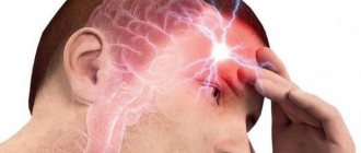
Benign paroxysmal positional vertigo (BPPV) occurs in repeated episodes, often lasting less than one minute. Attacks are triggered by changes in head position: turning, throwing back, as well as changes in body position, including lying down, even in sleep. Between attacks, autonomic disturbances (nausea, rarely vomiting, fluctuations in blood pressure, sweating) and imbalance may persist, so patients may describe constant dizziness.
Over time, the severity of attacks usually decreases. The word “benign” means that the disease goes away on its own, without treatment, without causing lasting harm to the patient.
BPPV is the most common type of dizziness. Attacks most often develop in older women. However, the disease can occur at any age.
Attacks of BPPV, in most cases, are associated with separation, destruction or increase in size of the otoliths.
Otoliths (otoconia) are layered pebbles consisting primarily of calcium carbonate crystals, like mother of pearl or nacre. They are immersed in a jelly-like layer that envelops the hairs of sensitive cells on the surface of the macula (macula) of the spherical and eleptic sacs of the vestibular analyzer. The otoliths, the jelly-like layer and the hairs of sensory cells form the otolithic membrane.
The elliptic sac (uterus) connects to three semicircular tubules (SCC), located in three perpendicular planes: lateral, anterior and posterior. In their extensions at the junction with the uterus, there is also a sensitive area - the ampullar ridge, covered with a structure similar to the otolithic membrane - the cupula. Normally, the cupula separates the RCC and the utricle. It does not contain otoliths. The cupula provides the perception of angular accelerations of the head, responding to changes in pressure in the ampulla arising due to the inertia of the endolymph (the fluid that fills the RCC and the sacs of the vestibular analyzer).
Detached otoliths or fragments thereof may enter the ampullae of the RCC and irritate the cupular areas. This more common variant of BPPV is called canalithiasis.
Thanks to the balance between the formation and resorption of the layers that make up the otoliths, their renewal is ensured, as well as the resorption of detached otoliths. When the balance is disturbed, one of the otoliths becomes large (2-4 times larger than neighboring cells); the large mass leads to greater displacement compared to neighboring fixed otoliths, which is a source of irritation of the vestibular system. This variant of BPPV is called dome lithiasis; it is characterized by a longer course (several months) and lack of effect from vestibular maneuvers.
Asymmetrical signal input to the brain during unilateral stimulation of the vestibular apparatus disrupts the illusion of balance created by the interaction of the vestibular, visual and proprioceptive systems (receiving signals from muscles and ligaments, assessing the position of limb segments). There is a feeling of dizziness.
Sensitive cells of the vestibular analyzer send a signal of maximum intensity to the brain during the first second of stimulation, then the signal strength decreases exponentially, which underlies the short duration of BPPV symptoms.
The most common lesion is the posterior SCC (90%), less often the lateral one (8%), the remaining cases are caused by damage to the anterior SCC and combined damage to several tubules. Classic cases of BPPV due to posterior RCC involvement are idiopathic in 35% of cases, with previous traumatic brain injury (sometimes minor) and whiplash in the neck occurring in 15% of patients.
In other cases, BPPV is caused by other disorders: most often Meniere's disease (30%), vestibular neuronitis, surgical interventions on the organ of hearing, paranasal sinuses, herpetic lesions of the ear ganglion and circulatory disorders of the structures of the inner ear. Population studies have revealed a direct relationship between the likelihood of developing BPPV and age, female gender, migraine, giant cell arteritis, risk factors for cardiovascular complications - arterial hypertension and dyslipidemia, as well as a history of stroke, which confirms the importance of vascular causes in some cases.
Lindsay-Hemenway syndrome has been identified - acute dizziness, followed by the development of BPPV attacks and a decrease or complete disappearance of nystagmus in the caloric test due to circulatory disorders in the anterior vestibular artery system.
The diagnosis of BPPV is made based on the assessment of nystagmus during special maneuvers - techniques that cause angular accelerations of the patient's head.
What it is?
When you turn your head quickly or change your body position suddenly, you may experience an attack of dizziness called benign paroxysmal positional vertigo (BPPV). This disease can be characterized as follows :
- benign – without negative consequences for life and health;
- paroxysmal – paroxysmal, sudden;
- positional – manifested when changing the position of the head or body;
- dizziness is a key symptom of the disease.
Although dizziness occurs in most diseases, with BPPV it has characteristic signs, based on which a specialist can make an accurate diagnosis at the first examination.
BPPV differs from other types in that a person can cope with it quite effectively on their own with the help of special exercises.
Most often, this disease occurs in people over 50 years of age. It occurs much more often in women than in men.
Terms and Definitions
Dizziness is a feeling of uncertainty in determining one’s position in space, apparent rotation of surrounding objects or one’s own body, a feeling of instability, loss of balance, the ground moving away from under one’s feet.
Vestibulometry is a set of tests carried out to determine the functional state and level of damage to the vestibular analyzer.
Vestibular neuronitis is a selective lesion of the vestibular ganglion (Scarpa's ganglion) (vestibular nerve), presumably of inflammatory origin and manifested by an acute episode of intense dizziness, lasting from 2-3 hours to several days, accompanied by a balance disorder with preserved hearing.
Dizziness is a feeling of uncertainty in determining one’s position in space, apparent rotation of surrounding objects or one’s own body, a feeling of instability, loss of balance, the ground moving away from under one’s feet.
ICD-10 code
ICD-10 is the international classification of diseases, 10th revision , approved by the World Health Assembly in 1990. It was created to encode diseases and pathologies and combine them into a single system. This classification is used by doctors all over the world, since the alphanumeric code that denotes any diagnosis is universal and understandable to every medical professional, regardless of the level of proficiency in foreign languages.
ICD-10 consists of 22 disease classes. Each class consists of an alphanumeric code. Classes are divided into blocks, and those in turn are divided into headings and sub-headings. Thus, the final codes very accurately characterize various diseases.
According to ICD-10, BPPV is coded H81.1:
- Class VIII. H60-H95 . Diseases of the ear and mastoid process.
- Block H80-H83 . Diseases of the inner ear.
- Category H81 . Vestibular dysfunction.
- Subsection H81.1 . Benign paroxysmal vertigo.
The ICD is actively used in medical documentation. Alphanumeric codes, used instead of the verbal formulation of diseases, provide convenient data storage, analysis and retrieval. This classification contributes not only to the exchange of information between doctors, but also to the preservation of medical confidentiality.
Complications
Despite the benign course, in rare cases paroxysmal dizziness becomes more pronounced and progresses. This course is characterized by frequent severe attacks of disorientation, accompanied by severe loss of balance, vomiting and nausea.
Injuries resulting from falls are a common complication in the elderly. The pathological process does not affect other organs and does not affect the functioning of the ear. But many people develop various mental disorders: depression, anxiety disorder, manic-depressive psychosis and others.
Timely consultation with a doctor and treatment allows you to completely cure the disease and eliminate any complications that may arise.
Causes of BPPV
To analyze how benign positional vertigo occurs, it is necessary to understand the structure of the vestibular apparatus.
Three semicircular canals, which are located in the vestibule , a special organ in the inner ear, are responsible for recording human movements. In these channels there is an ampoule with a gelatin-like substrate - cupula. It is her movements that create a sense of body balance.
Also in the ampoule there is a certain amount of liquid, which contains calcareous formations - otoliths. They are formed throughout a person’s life, and as the body ages, they are destroyed and processed by special cells.
In some cases, otoliths are not destroyed and their parts float freely in the liquid, irritating the receptor apparatus. As a result, a feeling of dizziness appears, which disappears when the crystals settle in any area.
Depending on the location of the floating otoliths, 2 forms of benign paroxysmal positional vertigo can be distinguished:
- Cupulolithiasis - otoliths are attached to the cupula of one canal.
- Canalolithiasis – otoliths move freely in the canal cavity.
When formulating a diagnosis, the side where the lesion occurred and the semicircular canal (posterior, anterior, external) are also noted.
The following factors can cause the appearance of free otoliths::
- chronic migraines;
- head injuries;
- Meniere's disease;
- use of gentamicin antibiotics;
- alcohol intoxication;
- inner ear surgery;
- viral labyrinthitis;
- inflammatory processes in the ear canals;
- spasm of the labyrinthine artery.
Prevention
It is difficult to prevent the development of pathology, since the cause of its occurrence is not fully understood. Considering that in some cases it occurs after a head injury or previous inflammatory processes, it is important to lead a healthy and active lifestyle, strengthen the immune system with the help of good nutrition, exercise, vitamin therapy and hardening. It is important to follow safety rules when playing sports and in everyday life.
There are no preventive measures to prevent the development of an idiopathic disease. In this case, prevention is aimed at reducing the number of attacks and reducing their manifestation. To do this, it is recommended to perform a number of exercises designed specifically for the prevention of BPPV.
Symptoms
Sudden attacks of dizziness occur when there is a certain change in the position of the body or head (most often when getting up in the morning or when turning in bed).- The duration of such attacks is no more than 1 minute.
- When dizziness occurs, a person feels as if he is falling, rising, or swaying, and surrounding objects are spinning before his eyes.
- The appearance of nystagmus - involuntary movements of the eyeball.
- dizziness may be accompanied by sweating, pale skin, irregular heartbeat, nausea, and vomiting.
- The attacks are most severe in the morning. In the afternoon they practically do not occur.
- In the absence of provoking movements, there is no dizziness.
- Attacks can be either isolated or occur several times a day.
- A certain position of the head provokes the appearance of dizziness (for example, throwing the head back or lowering it down).
- Outside of attacks, the person feels well.
- The attacks are always the same and there are no additional symptoms such as headaches, tinnitus, or hearing loss.
Diagnostics
To make an accurate diagnosis, two diagnostic methods are used:
- physical examination;
- instrumental examination.
The most common method for detecting benign paroxysmal positional vertigo during physical examination is the Dix-Hallpike test.:
- the patient sits down and turns his head to the side by 45°;
- with the support of a doctor, he abruptly lies on his back so that his head hangs back 20-30°;
- the doctor observes the patient’s reaction and waits for nystagmus to appear;
- then the patient sits down again, turns his head 45° to the other side and the procedure is repeated;
- the doctor determines the side of the lesion by the presence of nystagmus and dizziness.
The following methods are used for instrumental examination::
- Blessing or Frenzel glasses, electrooculography and videooculography (to determine nystagmus);
- magnetic resonance imaging of the brain and spinal cord;
- X-ray examination of the cervical spine and base of the skull;
- electrocardiogram and ultrasound examination of the heart;
- test to determine blood glucose levels.
On our website you can find out why dizziness occurs:
- during smoking and after quitting;
- in older people;
- in children;
- in teenagers;
- after training;
- before, during and after meals;
- with osteochondrosis;
- with VSD;
- among women;
- after a stroke.
Treatment
If you complain of dizziness, you should contact a general practitioner who will conduct an examination and, if necessary, refer the patient for a consultation with an otolaryngologist, neurologist or vestibulologist.
Treatment is divided into two areas :
- medicinal;
- non-drug (physiotherapy).
Drug treatment is aimed at relieving symptoms and improving the patient’s well-being in the acute stage of attacks. The patient may be prescribed the following medications :
- antiemetics (Cerucal, Metoclopramide);
- herbal nootropics (Tanakan, Cinnarizine, Bilobil);
- vestibulolytic (Vestibo, Betaserc, Meclizine);
- vasodilators (Cavinton, Magurol);
- antihistamines (Dramina, Diphenhydramine).
The main treatment is the so-called positional maneuvers. They consist in a progressive change in the position of the patient’s body and head in space in a certain order. With the help of exercises, doctors try to move the otoliths to the part of the vestibular apparatus where they belong.
During the maneuvers, attacks of benign positional paroxysmal vertigo may occur. The patient can perform some maneuvers without assistance, others only with the participation of a doctor.
Non-drug therapy consists of performing special gymnastics. The most famous and most effective physiotherapeutic techniques are the Brandt-Daroff method and Epley-Simon gymnastics.
Their essence is as follows: a person consistently changes the position of the head and body, which promotes the movement of otoliths into an area of the vestibular apparatus from which they can no longer move and, accordingly, cause dizziness.
The effect of such gymnastics, provided it is carried out correctly, occurs quite quickly, after just a few sessions. In severe cases, surgery may be required. The recovery period after it is 1 week.
Exercises (maneuvers)
Brandt-Daroff
The patient can easily perform the maneuver independently.:
- Immediately after waking up in the morning, you need to take a sitting position. Your legs need to hang down.
- Then quickly lie on your side, bending your lower limbs. In this case, you need to turn your head 45 degrees up and remain in this position for at least half a minute.
- Next, sit back on the bed.
If benign positional paroxysmal vertigo occurs, it is necessary to wait until it ends in a horizontal position. Repeat the maneuver 5 times on each side.