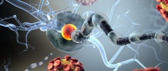
Multiple sclerosis is a disease that affects the central nervous system. Without proper treatment, this disease can lead to disability.
The causes of the disease have not yet been clarified. One thing is certain, people aged 25 to 50 are at risk. The age of the disease decreases every year.
Typically, the disease does not have specific symptoms. Each case of the disease manifests itself individually.
Often people simply do not pay attention to the first manifestations of the disease, and when they realize it, it may already be too late.
Here are the most common symptoms of multiple sclerosis:
- visual and speech impairment;
- weakness, memory and attention deteriorate;
- coordination is impaired;
- genitourinary system disorder.
When you first notice these warning signs, it is best to consult a doctor immediately!
Classification of multiple sclerosis
The following types of multiple sclerosis are distinguished:
- Primary progressive . The patient's condition worsens gradually. The attacks are weak or not expressed. The main symptoms are problems associated with impaired vision, speech, and urination. Motor activity is impaired.
- Relapsing-remitting . Seizures occur regularly. Symptoms are constantly changing. Most often, the patient experiences impaired thinking, coordination of movements, pain in the legs, eyes, and dizziness.
- Secondary progressive . It occurs without attacks and periods of remission. The patient feels severe weakness, coordination of movements is impaired, the muscles in the legs become dense and stiff, problems arise with the functioning of the bladder and intestines, mental activity is impaired, and severe depression occurs.
- Progressive-relapsing . Occurs least often. It manifests itself as periodic attacks, disruption of the musculoskeletal system, bladder, and intestines. The patient's coordination of movements is impaired and a depressive state occurs.
Causes of multiple sclerosis
Under the influence of a number of factors, the body’s immune defense is disrupted, an excess amount of T-lymphocytes enters the brain tissue and the inflammatory process begins.
As a result, the myelin sheath of the nerve is destroyed by its own immune system, which mistakes myelin antigens for foreign bodies and begins to destroy them.
As a result of the destruction of the myelin sheath, the transmission of nerve impulses is disrupted and multiple sclerosis develops.
There are a number of factors that can trigger the development of the disease. These include:
- past viral and bacterial infections;
- chronic intoxication of the body with gasoline, metals, solvents, chemicals, and so on;
- frequent stress, emotional overload;
- poor nutrition – the greatest danger is posed by animal proteins and fats, or rather their excessive consumption; if a person has been obese since youth, then the risk of developing multiple sclerosis doubles;
- previous surgical operations;
- physical stress;
- back and head injuries;
- genetic predisposition;
- high blood sugar;
- taking oral contraceptives;
- gender – the disease occurs more often in women than in men;
- race – representatives of the Caucasian race are most susceptible to multiple sclerosis.
Bee Honey
Bee honey is a natural product produced by honey bees from the nectar of flowering plants. It differs from nectar in its physicochemical and biological properties. Bees enrich nectar with enzymes, organic acids and other substances. Under the influence of enzymes, part of the sucrose in the nectar is broken down and converted into glucose and fructose.
Honey has the appearance of a thick, transparent, slightly colored, sweet aromatic liquid. The chemical composition of different varieties of honey is different and depends on the type of plant from which the nectar is collected, on soil and climatic conditions.
The main component of all types of honey are carbohydrates (glucose, fruit, sucrose) in the form of grape and fruit sugar. Honey also contains enzymes (invertase, catalase, acid phosphatase, etc.). Honey contains organic acids, vitamins, protein, coloring, antibiotic and mineral substances. Among the mineral substances, salts of calcium, sodium, magnesium, iron, sulfur, iodine, chlorine, and phosphorus were found. Honey also contains microelements (manganese, silicon, aluminum, boron, chromium, copper, nickel, lead, tin, zinc, octium, etc.), which play a huge role in the normal functioning of many organs and systems of the body. A constant admixture of honey is pollen, due to which the honey is enriched with vitamins and proteins.
Bee honey has high antibacterial, antimicrobial, immunostimulating, anti-inflammatory, analgesic, and antiallergic properties. Ingestion of honey improves metabolic processes in the body and has a calming effect on the nervous system. Under its influence, skin turgor improves, the function of the prostate and other sex glands is normalized, which serves as the basis for the use of honey in the complex treatment of patients suffering from MS. The most healing is honey collected from herbs, but honey from the flowers of buckwheat, rapeseed, mustard, leuzea, heather, lemongrass, clover, black elderberry, black currant, etc. also has high medicinal properties.
Symptoms of Multiple Sclerosis
The clinical picture may vary, depending on where exactly in the body the main lesion is located.
The following symptoms may indicate the development of multiple sclerosis:
- oculomotor disorders (double vision, strabismus);
- epileptic seizures;
- inflammation of the facial nerve (manifested by paresis of the facial muscles);
- neurotic disorders (impaired intelligence, fatigue, frequent mood swings, depression);
- dizziness;
- disturbance of urination (and sometimes defecation);
- the appearance of pathological reflexes, paresis of the limbs;
- sensitivity disorders (tingling, numbness of the skin);
- disruption of the cerebellum, which can be manifested by changes in handwriting, staggering while walking, speech impairment, and tremors of the limbs.
Bee venom
Bee venom (apitoxin) is a colorless liquid with a pungent odor, formed in the stinging apparatus of a working bee. Apitoxin has a complex composition: a protein complex, fat-like substances, mineral salts, amino acids, organic acids, enzymes, etc. The enzymes that make up bee venom affect the permeability of blood vessels and the condition of cell membranes. Bee venom, penetrating into the human body, has an analgesic, ganglion-blocking, anti-inflammatory, hyposensitizing, stimulating effect on the pituitary-adrenal cortex-gonadal system, enhances the production of corticotropin, etc.
Bee venom is introduced into the body of a patient with MS by direct injection of a bee sting (stinging), administration of ampoule preparations, electrophoresis, mechanical rubbing of ointments, inhalation or administration under the tongue. The most common of them are bee stings and ointment dosage forms containing bee venom.
Long-term observations have shown that bee venom, for established indications and taking into account individual tolerance, can be successfully used in the complex treatment of MS. Bee stings are carried out in the lumbar region by 2 to 6 bees daily or every other day. The course of treatment requires from 40 to 60 bee stings. Stings are carried out according to the instructions of the Russian Ministry of Health. It should be remembered that apitherapy for patients with allergic manifestations in the past should be carried out with great caution under the mandatory supervision of a doctor.
Recently, in the treatment of a number of diseases, including MS, bee venom is increasingly used using electrophoresis. The pharmaceutical industry produces a special tablet preparation “Apifor” for electrophoresis. The course includes 10 to 20 procedures, 10 to 15 minutes each.
The introduction of bee venom using ultrasound deserves special attention and wider use. Before starting apiultrasound therapy, the patient undergoes an examination to identify contraindications to the use of acoustic energy and bee venom. Treatment is carried out in cycles of 10 - 15 procedures using increasing doses. The duration of the procedure is 5-12 minutes. Continuous ultrasound - 0.5 - 1.5 W/cm2, depending on age and stage of the disease. An area of skin, usually in the lumbar or perineal area, is pre-lubricated with an ointment containing bee venom in a concentration of 150 mg/100 g. After this, the sonicator is stroked over it with light pressure in a circular or longitudinal motion. It comes into contact with the patient's skin due to the presence of a layer of contact medium.
Diagnosis of multiple sclerosis
The diagnosis is made on the basis of anamnesis, questioning and examination of the patient, and the clinical picture of the disease. Additional diagnostic methods can be used:
- blood test (general) - there is a decrease in the number of lymphocytes and leukocytes;
- blood test (biochemical) - there is a decrease in the level of cortisol, amino acids and proteins in the blood plasma, phospholipids and lipoproteins are increased;
- coagulogram - determine the rate of blood clotting;
- immunological study of cerebrospinal fluid and plasma - autoimmune and immunosuppressive components predominate;
- measurement of the electrical activity of the brain - for this purpose, a study of the patient’s visual, auditory and sensorimotor potential is usually carried out;
- SPEMS is the latest research method that uses an electromagnetic superposition scanner.
Treatment of multiple sclerosis (classical medicine)
If the disease is detected at an early stage and does not have many symptoms, this improves the prognosis.
The patient is hospitalized in a special medical center for the treatment of multiple sclerosis, where a treatment regimen is selected taking into account the stage of the disease, the presence of complications, and the individual characteristics of the body.
The main treatments for multiple sclerosis are:
- hormonal therapy - the patient is given large doses of corticosteroids;
- plasmapheresis – blood purification procedure;
- taking cytostatics;
- β-interferons - prescribed to prevent exacerbations, slow down the progression of the disease, prolong the period of working capacity and social adaptation of the patient;
- immunomodulators - prevent further destruction of myelin sheaths, reduce the severity and frequency of exacerbations, soften the course of the disease;
- symptomatic therapy - to alleviate or relieve the symptoms of multiple sclerosis, enterosorbents, antioxidants, muscle relaxants, nootropics, vascular therapy, vitamins B and E, and amino acids are prescribed.
During periods of remission, the patient is prescribed sanatorium treatment, massage, and exercise therapy. All thermal procedures are prohibited.
Complete recovery from multiple sclerosis is impossible, but it is quite possible to improve the patient’s quality of life and prolong periods of remission with adequately prescribed treatment.
Traditional medicine for multiple sclerosis
Traditional medicine has learned to treat multiple sclerosis quite effectively, but the disease is still considered incurable. Fighting a disease is a long process. Traditional medicine offers its own quite effective methods - infusions, decoctions, teas. It is worth using folk remedies together with taking pharmacological drugs to get the most effective result. Many patients maintain health and good spirits until old age.
Herbal infusions, decoctions
Various plants and herbal mixtures are used to combat the disease.
Larkspur reticulata
Grind the dried root of the plant well. For 1 tbsp. boiling water you will need 2 g of raw materials. Leave overnight, filter. Drink in 3 doses, 1 tbsp. l. It is also worth dripping 2 drops. infusion into the nose in the morning and immediately before bed, having previously sanitized the nasopharynx.
Duration of therapy is 2 months. Then they take a break of 2 weeks and continue treatment.
Hawthorn, rue, valerian
To prepare the medicine you will need the following ingredients:
- hawthorn leaves - 25 g;
- hawthorn flowers - 25 g;
- rue (herb) - 15 g;
- valerian (roots) - 10 g.
Mix all ingredients well, separate 1 tbsp. l. mixture and pour 200-250 ml of boiling water, leave for 50 minutes. Drink 200 ml immediately before bed.
Dandelion, wheatgrass, yarrow, soapwort
Ingredients:
- dandelion (roots) - 30 g;
- soapwort (roots) - 30 g;
- wheatgrass (roots) - 30 g;
- yarrow (herb) - 30 g.
Grind all components well and mix. Separate 1 tbsp. l. plants and steam in 1 tbsp. boiling water Leave in a closed container for 1 hour. Drink 1 tbsp. in the morning and before bed.
Immortelle, wormwood, mint, calendula and others
To prepare the infusion you will need the following plants:
- immortelle, wormwood, tansy, calendula, corn silk, dandelion (roots) - 1 part each;
- mint, chamomile (flowers), barberry (roots), fennel (fruits) - 2 parts each;
- celandine – 4 parts.
Dried ingredients are used . Grind everything, mix well and pour 1 tbsp. l. herbs 250 ml boiling water. Place in a water bath for half an hour, remove and leave. Filter the finished product and take a third of a glass in the morning.
Alcohol tinctures
Traditional medicine offers various recipes for alcohol tinctures:
- Propolis . To prepare the tincture you will need a solution of propolis (20%) and alcohol (70%). You can purchase a ready-made version. Take 20 drops. in three doses over 30 minutes. before meals. The duration of the course depends on the complexity of the disease - 1-3 months.
- Mordovnik . The medicinal plant stops the development of the disease. To prepare the tincture you will need unpeeled plant seeds (1 tbsp.) and 1 tbsp. vodka. Pour in the seeds and leave in the dark for 3 weeks, shaking the container periodically. The finished infusion is filtered. Drink 30 drops, diluting them in 50 g of water. The course is 2 months, the break is 10 days, after which the treatment is repeated.
- Clover . The dried heads of the plant are poured into a liter jar, then 0.5 liters of vodka are poured in and left for 2 weeks. Take 1 tbsp. l. in the evening before bed. The duration of therapy for multiple sclerosis is 1 month. Take a break of 2 weeks and continue treatment.
Honey against multiple sclerosis
There are several options for honey-based recipes:
- Honey and onions . Peeled onions are grated on a fine grater, and the juice is separated using gauze. The juice is poured into a separate container and honey is added in the same amount. Everything is mixed and stored in the refrigerator. The folk remedy should be taken in three doses per day, 15 ml each, preferably an hour before meals. It is allowed to take the medicine an hour after eating.
- Honey and woodlice (chickweed) . The plant is good at relieving pain from illness. To prepare the medicine, the crushed herb must be mixed with honey in equal quantities. Drink the finished product 15 ml in 5 divided doses per day.
Oils and fats for multiple sclerosis
Taking vegetable fats has a beneficial effect on the patient's health. They contain polyunsaturated fatty acids, valuable for the body, which are necessary for nerve fibers. Therefore, traditional medicine offers an abundance of recipes based on vegetable, flaxseed oils, fish oil and other ingredients.
To prepare the medicine you will need garlic, oil, lemon. Chop 1 head of garlic well, transfer to a jar, pour in 220 ml of sunflower oil. Place the contents in the refrigerator and leave for a day. Drink with lemon juice half an hour before meals. Dose – 1 tsp. medicine and lemon juice.
Massage Oil
To treat pathology, massage the body with St. John's wort flower oil. To make oil, the upper flowers of the plant are kneaded well, placed in a jar, filling it 2/3 full. Then pour in heated sunflower oil, cover with gauze, and expose to the sun.
After 5 weeks, when the oil turns dark red, the remedy is ready for use. Before use, the oil is filtered.
The effect of coronavirus on the nervous system and methods of brain recovery
What is SARS-CoV-2?
Coronavirus disease 2021 (COVID-19), the disease caused by the novel severe acute respiratory syndrome Coronavirus 2 (SARS-CoV-2), is a viral pandemic. As a member of the coronavirus family, SARS-CoV-2 shares 77.2% amino acid identity, 72.8% sequence identity, and structural similarity to severe acute respiratory syndrome coronavirus (SARS-CoV). With its high affinity receptor-binding domain of angiotensin-converting enzyme-2 (ACE-2), SARS-CoV-2 enters human cells in the same way as SARS-CoV. It has been confirmed by numerous studies that SARS-CoV-2 can also attack the central nervous system (CNS).
Patient complaints during and after coronavirus infection
Patients who had coronavirus infection noted that at the time of its manifestation there were headaches, changes in vision, loss of hearing, sense of smell and taste, impaired mobility, numbness of the limbs, tremors, fatigue (weakness), muscle pain (myalgia), memory loss, changes moods.
Clinical picture of COVID-19 infection
During SARS-CoV-2 infection, nearly 70% of patients experience mental disorders and neurological symptoms, including: mood changes (42%), fatigue (67%), headache (25%), vision changes (67%), myalgia (15%), impaired mobility *67%), memory loss (14%), loss of taste (67%), numbness of extremities (67%), tremors (4%), loss of smell (65%) and hearing loss (2 %). Other common symptoms include fever (88%), cough, 57%), and gastrointestinal discomfort (14%). Immediately after coronavirus infection, 55% of patients still have neurological symptoms. The number of patients experiencing fatigue and mood changes is, however, significantly reduced compared to the number of patients experiencing the complaints noted above - symptoms of the onset of the disease. And yet, 78% of patients can be classified as a mild type of COVID-19, 20% of patients can be classified as a severe type, and 2% can be classified as a critical type.
Neurological disorders after coronavirus infection
Neurological disorders can be found in at least 55% of patients with COVID-19. Approximately 40% of patients with coronavirus infection COVID-19 report neurological symptoms such as dizziness, headache and impaired consciousness during hospitalization. In addition to frequent olfactory and gustatory disturbances in mild to moderate COVID-19 patients reported from 12 European hospitals, cases of various neurological diseases have also been reported, including encephalitis, stroke, microhemorrhage, reversible posterior cerebral hemorrhage, encephalopathy and cerebral venous disease. embolism Additionally, it has been documented that SARS-CoV-2 specific RNA was detected in the cerebrospinal fluid (CSF) of a COVID-19 patient. In my opinion, after this viral infection, it is necessary to study the long-term effects of SARS-CoV-2 infection on the central nervous system, especially on structures that are easily attacked by the virus and structures that highly express ACE-2. During SARS-CoV-2 infection, 41 of 68% of patients have neurological symptoms, and more than 50% of recovered patients continue to have symptoms even after 3 months. It is interesting to note that gray matter in the hippocampus (a key brain structure in memory organization) and the cingulate cortex (an important part of the limbic system) were negatively associated with loss of smell during infection and memory loss after 3 months, supporting the hypothesis of neurogenesis in these regions. Tremor was found to be negatively associated with FA_WM scores both acutely and at 3-month follow-up, indicating disruption of WM fibers in both hemispheres, possibly resulting from the cytokine storm induced by SARS-CoV-2.
Pathological processes
According to research, coronaviruses can cause demyelination, neurodegeneration and cellular aging, which accelerates brain aging and exacerbates neurodegenerative pathologies, such as Alzheimer's disease or Parkinson's disease, multiple sclerosis and amyotrophic lateral sclerosis, etc.
Brain changes after coronavirus infection
Patients with COVID-19 have statistically significantly higher bilateral gray matter volumes (GMV) in the olfactory cortex, hippocampus, insula, left Rolandic operculum, left Heschl's gyrus and right cingulate gyrus, as well as an overall decrease in MD, AD, RD, accompanied by an increase in FA . in white matter, especially AD in right CR, EC and SFF and MD in SFF compared with non-COVID-19 volunteers. Global GMV, GMV in the left Rolandic operculum, right cingulate cortex, bilateral hippocampus, left Heschl gyrus, and Global MD of WM were found to be correlated with memory loss GMV in the right cingulate cortex and left hippocampus were found to be associated with olfactory loss. MD-GM, total GMV, and right cingulate GMV were correlated with LDH levels. In the gray matter of COVID-19 patients, the left insula, bilateral cingulate cortex, right precuneus, and right thalamus showed significantly lower MD values. In white matter, regional mean MD, AD, and RD values are generally lower in the COVID-19 group, and regional mean FA values are generally higher. Mean MD values of the right superior fronto-occipital fasciculus (SFF) in COVID-19 and AD values in the right corona radialis (CR), right external capsule (EC) and right SFF are significantly lower in COVID-19
MD GM values in the right thalamus and AD WM values in the right EC were positively correlated with visual changes. GMV in the right cingulate cortex and hippocampus are negatively correlated with loss of smell. Overall, recovered patients with COVID-19 are more likely to have enlargement of the olfactory cortex, hippocampus, insula, Heschl's gyrus, Rolandic operculum and cingulate cortex, as well as an overall decrease in MD, AD, RD accompanied by increased FA in white matter. especially AD in right CR, EC and SFF and MD in SFF. Global GMV, GMV in the left Rolandic operculum, right cingulate cortex, bilateral hippocampus, left Heschl's gyrus, and Global MD of WM correlated with memory loss. GMVs in the right cingulate cortex and left hippocampus are associated with loss of smell.
Overall, lower diffusion parameters (MD, AD, RD) and higher FA values were found in white matter from the COVID-19 cohort. In addition, the diffusion coefficient (MD and AD) of the right CR, EC and SFF is significantly reduced. The corona radiata (CR), consisting of a massive bundle of projection fibers, connects the cortex to the brainstem and thalamus in an afferent and efferent manner. The EC and SFF are a series of association fibers connecting the frontal, parietal, and temporal cortices. White matter is not the primary target of the neurotropic virus; however, connective fibers may act as a conduit for intracranial transmission of the virus. In contrast to the increased MD with compressed fiber volume in hydrocephalus, increased white matter fibers, decreased MD values, and increased FA suggest greater fiber alignment and limited freedom of diffusion, suggesting a possible intrinsic repair process (eg, remyelination) that occurs after infection.
Interhemispheric asymmetry
It is interesting to note that all white matter diffusion abnormalities are limited to the right hemisphere, without the asymmetrical symptoms reported by COVID-19 patients. The predominance of abnormal white matter diffusivity may be due to differences in blood volume in the hemispheres. The predominance of the right side in odor perception is indicated by numerous studies of olfactory function, which have not been fully studied. Changes in diffusion on the right side may be related to odor perception on the right side, which requires further research.
The study results showed possible disruption of the microstructural and functional integrity of the brain during the recovery phases of COVID-19, indicating the long-term consequences of SARS-CoV-2.
Brain changes after COVID-19 captured using voxel morphometry
VBM analysis shows significantly higher GMV in the left Rolandic operculum, bilateral olfactory cortex, bilateral insula, bilateral hippocampi, right cingulate cortex, and left Heschl's gyrus in COVID-19 patients.
Lactate dehydrogenase as an indicator of brain atrophy
Lactate dehydrogenase is one of the key enzymes of the glycolytic pathway, highly expressed in cells of the kidneys, heart, liver and brain. . Elevated lactate dehydrogenase concentrations are observed in patients with encephalitis, ischemic stroke, and head trauma. Higher serum lactate dehydrogenase concentrations always follow tissue destruction and are strongly associated with worsening disease and poor outcome. Decreased total brain gray matter volume (GMV) in patients with elevated lactate dehydrogenase levels may indicate brain atrophy due to a severe inflammatory response.
Hemostasis system for COVID-19
Because hemostatic abnormalities, including disseminated intravascular coagulation and severe inflammatory response, have been frequently observed in patients with COVID-19, some people may be predisposed to cerebrovascular events caused by infection and inappropriate treatment. Ischemic changes are known to be accompanied by lower FA values and higher MD values in ischemic lesions.
Angiotensin converting enzyme
It is important to investigate the relationship between abnormal anatomical brain regions and ACE-2 distribution. It is clarified that SARS-CoV-2 enters the host cell by attaching to ACE-2 through the Spike glycoprotein. Therefore, greater expression of ACE-2 may cause more severe abnormalities in brain structures. The distribution of ACE2 was nonequivalent throughout the brain and was most frequently expressed in gray matter, followed by the spinal cord, hippocampus, basal ganglia, limbic system, and frontal cortex. Study results showed that various components of the limbic system during coronavirus infection were affected by structures that have possible high expression of ACE-2, which overlapped with putative ACE-2-rich regions
How are we dealing with the consequences of COVID-19?
In our clinic, we have been conducting MRI 3 tests with diffusion tensor imaging (tractography) for patients with mental and neurological disorders for more than 3 years. The results of this study show the state of the connections (bundles of fibers) between brain structures and provide an answer to the question: why did these connections suffer (inflammation, loss of membranes of neuron processes - demyelination, compression, deterioration of blood circulation, etc.) Magnetic resonance In vivo tomography (MRI) can image cerebral structures non-invasively. Possible microstructural damage to the CNS can be detected using structural MRI and diffusion tensor imaging (DTI). Axial diffusivity (AD), radial diffusivity (RD), mean diffusivity (MD), and fractional anisotropy (FA) can be calculated using track-based spatial statistics (TBSS). Along with volumetric analysis, DTI is widely used in large-scale neuroradiological studies to detect microstructural changes in patients with cerebral viral infections, human immunodeficiency virus (HIV) and herpes simplex virus (HSV), etc. The more severe the case of COVID-19, the higher the MD value of bilateral cingulate gyri. The gyrus singularis typically plays an important role in attention, motivation, decision making, learning and cost-benefit calculations, as well as monitoring conflicts and errors, which often occur in limbic encephalitis. Thus, dysregulation of the cytokine response has been suggested in severe cases
Diffusion tensor image calculations
To calculate quantitative parameters including gray matter volume (GMV), white matter volume (WMV), FA, MD, RD and AD values for different brain regions, an atlas-based approach to image processing of brain structures must be used. DTI data sets are first processed using DSI Studio software ( https://dsi-studio.labsolver.org/ ) to generate FA, MD, RD, and AD maps for each fiber bundle. When assessing brain connections, we evaluate at least 12 fiber bundles, and if necessary, we can integrate the results of diffusion tensor imaging and functional MRI data (changes in brain activity during solving certain tasks). The researcher needs to maintain tight registration between the quantitative DTI and 3D-T1WI maps using the MATLAB-based SPM12 ( https://www.fil.ion.ucl.ac.uk/spm/ ) software (MathWorks, Natick, MA, USA. ). 3D-T1WI needs to be segmented and non-linearly normalized into MNI space using the CAT12 toolbox ( https://www.neuro.uni-jena.de/cat/ ) in SPM12 to obtain tissue probability maps and normalized quantitative DTI maps. Next, we need to combine automated anatomical labeling atlas-3 (AAL-3) and JHU DTI-based white matter (WM) atlas to extract quantitative parameters in GM and WM, respectively. Finally, the brain must be divided into 65 (AAL-3) plus 36 (JHU DTI-based WM atlas) anatomical regions. Regional brain volumes and diffusion indices, including FA, MD, RD and AD WM and GM, respectively, can be extracted by averaging values from voxels with a partial volume greater than 90% of the corresponding tissue. Estimated total GMV and WMV are further normalized by intracranial volume correction
Loss of smell and brain damage
Significant increased volumes were observed in the bilateral olfactory cortex, hippocampus, insula, left Heschl's gyrus, left Rolandic operculum, and right cingulate gyrus. All the structures mentioned above belong to the central olfactory system. Among them, the olfactory cortex, also called the piriform cortex, directly receives axonal projections from the olfactory bulb (OB), called part of the "primary olfactory cortex". Other structures are the cortical targets of the primary olfactory cortex in the bilateral limbic lobe, temporal cortex, which have been called "secondary olfactory cortex." The literature has reported that frequent loss of smell during upper respiratory tract infections led to loss of stimulation and subsequent loss of volume in the acute phase, while after recovery of smell, gray matter volumes in the central olfactory system subsequently increased (grey matter of the olfactory system - GMV central of the olfactory system as a whole were smaller in patients with persistent olfactory loss of function compared with patients without olfactory problems.
Several possible routes of SARS-CoV-2 invasion have been identified, including hematogenous, lymphatic, and neuro-retrograde, but the exact route remains unknown. Gray matter volumetric changes in the central olfactory system suggest that SARS-CoV-2 may enter the CNS via the OB (olfactory bulb)-mediated retrograde neuronal pathway. Two reasons are hypothesized to play a role in the increase in GMV: neurogenesis and functional compensation.
Neurogenesis after COVID-19
It is well known that adult neurogenesis is limited to two regions: the subventricular zone (SVZ) and the subgranular layer of the dentate gyrus of the hippocampus. Following coronavirus infection, neuroblasts from the SVZ migrate along the rostral migratory stream, first enter the olfactory cortex, and finally replace interneurons (e.g., periglomerular cells, granule cells) in the olfactory bulb. Therefore, an increase in the number of neurons may have led to an increase in GMV in the olfactory system. In order to compensate for the impaired sense of smell, the increased functional involvement of brain regions will be hypertrophy, which has been proven by experimental studies in sensory deprivation models to have enlarged neurons and increased the number of dendritic spines.
Other studies after coronavirus infection
We also conduct tests such as a routine electroencephalogram, cognitive evoked potentials and neuropsychological examination for each patient who has suffered a coronavirus infection. A detailed immunological examination of the patient is important. To avoid cross-infection, patients cannot undergo MRI during the acute phase, but we are still wondering whether there are any microstructural changes during the convalescent phase of COVID-19 and whether any indicators remain to suggest a likely intracranial infection and route of infection .
Treatment of the effects of coronavirus infections on the nervous system
Given the olfactory and gustatory dysfunction in patients with COVID-19 and evidence of invasion of the olfactory epithelium by SARS-CoV, the olfactory gyrus can be considered the first functional area in the CNS infected by SARS-CoV-2. Hence, it is extremely important to administer neuropeptides (Semax) as early as possible after recovering from the coronavirus infection. In our clinic (Mental Health LLC) we do this using the correct dosage of nasal electrophoresis. In addition, we have developed special meta-cognitive training to restore the memory of patients who have suffered from COVID-19. Our experience also shows the effectiveness of the antiviral drug amantadine and DEHA supplements for the treatment of neuropsychiatric consequences of COVID-19
Prevention of multiple sclerosis
There is no specific prevention of the disease. Following these recommendations will help reduce the likelihood of developing multiple sclerosis:
- rejection of bad habits;
- use of personal protective equipment when working in hazardous industries;
- constant physical activity (not exhausting, but moderate);
- proper nutrition, avoidance of fast food and other junk foods (especially fatty and salty), introducing foods rich in fiber into the diet, as well as fresh fruits and vegetables;
- refusal of overeating, strict diets, weight control;
- avoiding stress;
- normalization of work and rest regimes;
- refusal to take oral contraceptives and other hormonal drugs (if possible);
- hardening the body, strengthening the immune defense;
- preventing the body from overheating;
- Regular preventive examinations will help identify possible diseases at an early stage, which will significantly facilitate subsequent treatment and improve the prognosis.
Principles of treatment
Low cholesterol diet. It is recommended to completely or partially avoid eating the following foods: brains, fatty meat, kidneys and liver, lard, animal by-products, egg yolks.- Systematic aerobic physical activity at least 3 times a week: running, cycling, race walking, swimming, rollerblading, fitness or yoga classes, etc.
- Normalization of body weight. One of the main measures to eliminate sclerosis is to reduce excess weight to normal levels.
- Maintain blood pressure within reference values and stop using tobacco products and alcohol. It has been noted that with frequent changes in the diameter of blood vessels, the risk of sclerosis increases.
- A suitable regime of wakefulness and rest, which will help eliminate neuropsychic stress.
Read here how to treat multiple sclerosis, and also how to live with this disease - follow the link.
Where to buy traditional medicine for multiple sclerosis?
The online store “Russian Roots” offers natural products based on the knowledge of traditional medicine, as well as ingredients for the preparation of medicinal products. Delivery is available in Moscow, the region and all regions of the country. In Moscow, you can purchase goods in one of the herbal pharmacies in the chain. The products have all the necessary documentation.
Attention! All materials published on our website are protected by copyright. When re-publishing, attribution and a link to the original source are required.


