A special place in the development of the individual and the human collective is occupied by the ability to transmit, receive and process sound signals. The ability to recognize and work with a complex sign system has made a person not just a highly developed organism, but a fully functional personality. Initially exchanging simple sounds, society eventually learned to convey complexly constructed verbal sentences. It is thanks to the presence of the temporal lobe that the implementation of the most complex mental function - speech - is possible.
- Location
- Functions
- Fields
- Symptoms of the lesion
Location
The temporal lobe is part of the telencephalon and is included in the structure of the cortex. It is located on both hemispheres of the brain on the sides below, in close contact with neighboring areas - the frontal and parietal lobes. This area of the cortex has the most pronounced boundary lines. The upper part of the temple is slightly convex, and the lower part is concave. The temporal lobe is separated from all the others by a groove called the lateral (lateral). The close location of the temporal and frontal lobes is not accidental: speech develops in parallel with thinking (frontal cortex), and these two functions are closely interconnected, since the ability to formulate and express oneself clearly (speech) is ensured by the degree of development of mental functions.
The convolutions of the temporal lobe are located parallel to the grooves that limit the area. Anatomically, there are 3 gyri: superior, middle and inferior. However, the superior cerebral fold includes 3 more small convolutions located in the sulcus itself. This group of small structures is called Heschl's convolutions. The inferior gyrus of the temple borders the transverse medullary fissure. On the lower part of the temporal lobe, in addition to the inferior gyrus, additional structures are also distinguished: the hippocampal peduncles, the lateral occipitotemporal gyrus.
Organic brain damage
Symptoms of organic brain damage in children
The main sign of organic brain damage in children is psychoorganic syndrome. This condition is expressed in a violation of three aspects of brain function at once.
- Problems with memory - the child does not remember new information well and loses some of what has already been learned (partial amnesia). Moreover, with OPM, unreal (invented) memories may appear.
- Decreased intelligence - such children have difficulty concentrating, their thinking is impaired, and they have difficulty oriented in space.
- Affective disorders and reduced neurodynamics - children with organic brain lesions constantly experience weakness, dizziness and headaches, they are susceptible to depression and irritability. Children often display inappropriate emotions and “out-of-control” behavior.
- Delayed speech and intellectual development is another symptom of organic brain damage, which is characterized by impaired cognitive activity. This condition is not congenital, like mental retardation, but acquired. The functions of a child's damaged brain begin to disintegrate. Sometimes the developmental delay is so severe that the child cannot learn to care for himself.
There are also a number of focal symptoms , which depend on the area of the brain in which the disorders are localized.
Frontal lobe – facial and ocular muscles are paralyzed, the sense of smell is impaired and words are difficult to pronounce, difficulty performing purposeful movements, strange behavior on the verge of euphoria.
The parietal lobe is a violation of sensitivity, the inability to perform purposeful, meaningful actions, as well as the inability to learn reading and counting. Seizures with convulsions are common.
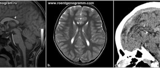
Temporal lobe - impaired sense of smell and hearing, problems with taste, there are hallucinations, emotionally unstable mood, partial or complete misunderstanding of speech.
Occipital lobe – visual impairment up to blindness, problems with coordination of movements and balance, hallucinations, convulsions during seizures.
Naturally, therapy and correction of APM should be prescribed based on the cause of its occurrence and development. For example, doctors recommend eliminating lesions caused by infection with the help of antibiotics, antiviral drugs and immunostimulants. If APM has developed as a result of a tumor, then, first of all, care must be taken to remove it. You can't argue with that.
Official medicine suggests treating ischemic diseases of the brain with nootropic drugs , as well as using decongestant and vascular therapy.
It is imperative to remember that drug therapy cannot but have side, sometimes harmful, consequences .
Eliminating the consequences is often more difficult and longer than the main violation.
But even in the case of successful drug therapy, you CANNOT do without psychological and correctional therapy.
If your child:
- very picky and capricious in choosing food
- reacts sharply to weather changes
- he gets carsick
- he's inattentive
- speaks poorly
- restless
- cries often
- clumsy
- lazy
Do you think that all these are character traits? In fact, these are neurological problems that may be associated with organic brain damage. If these violations are ignored now, then in the future they can cause serious behavioral disorders, speech delays and general development, and subsequently difficulties in learning at school.
The fact is that at an early age the consequences of many neurological disorders can be easily corrected and forgotten about them forever. But in adolescence, small problems often turn into big ones and are already very difficult to cope with.
Therefore, it is better not to postpone a visit to a neurologist and neuropsychologist.
It is especially important to conduct neuropsychological diagnostics in children before entering grade 1, to identify and correct neuropsychological disorders in a timely manner so that children do not develop “school failure”, which will affect not only the learning process, but also the psychological state of the child.
For example, in the temporal lobe of the left hemisphere there are areas “responsible” for the sound analysis of speech. If neuropsychological tests that check the state of this area of the brain are performed incorrectly, it is possible not only to diagnose its immaturity, but also to predict the occurrence of certain types of errors in speaking, writing, reading and remembering information.
Assigned functions
The functionality of the temporal cortex is insignificant, however, it is highly specialized. The functions of the temporal lobe of the brain are associated with the perception, analysis and synthesis of speech, the perception of auditory information, and partly gustatory and olfactory information. Also, the location of one part of the seahorse determines another function - memory, namely its mechanical component. One area has a special purpose: Wernicke's center (sensory speech area) - located on the back of the superior temporal gyrus. This zone is responsible for the perception and comprehension of oral and written speech.
What matters is the functional asymmetry of the brain, that is, the location of the dominant areas of the cortex on the surface of the brain. This specificity of the central nervous system did not bypass the temporal lobe.
The left temporal lobe is responsible for the following functions (it should be noted that the list of tasks is based on the fact that the left hemisphere is dominant):
- Understanding audio information (music, words and speech);
- Short-term memory;
- Choice of words during a conversation;
- Synthesis of visual information with auditory information;
There is an interesting phenomenon here - synesthesia . Only 0.05% of the population has this phenomenon. The essence of the phenomenon is the ability to see the qualitative parameters of sounds in a different color spectrum. Physiologically, this is explained by the process of irradiation (spread of action potential), when the excitation of an overly irritated area of the cortex passes to the neighboring part of the brain. As a rule, famous musicians (Rimsky-Korsakov, Franz Liszt) possessed and still possess this ability. - The connection between music and emotions;
The right temporal lobe of the brain is responsible for the following functions and abilities:
- Recognition of facial expressions;
- Identification of speech intonation;
- Musical tones and rhythm;
- Memorizing and fixing visual data.
In addition to recognizing speech intonation, the non-dominant lobe also analyzes it and subsequently integrates images into the general emotional attitude towards the interlocutor. It is this part of the brain that allows a person to know whether his conversation partner is happy with him or wants to get rid of him as soon as possible.
Insula
Recent neuroimaging studies of the insula have led to renewed interest in the role of this region in normal conditions and in the development of pathology.
In this article, the authors provide brief information about the anatomical and histological features of the insula of the human brain. The following describes the physiological functions of the insula and highlights its long-underestimated involvement in the pathogenesis of psychiatric and neurological disorders. In conclusion, the authors propose various techniques that will allow us to better study the role of the insula within both basic and clinical neuroscience. Glossary
Agranular region (cortex): an area of the neocortex with relatively indistinguishable layers II and III and the absence of layer IV.
Central executive network: A system of neurons in the brain, including the dorsolateral prefrontal cortex and posterior parietal cortex, that are responsible for high-order cognitive functions such as attention and working memory.
Cognitive resources: a set of mental abilities and resources related to cognitive activity (attention, memory, working memory, thinking, etc.).
Passive Brain Network: A system of neurons including the ventromedial prefrontal cortex and posterior cingulate cortex responsible for self-perception processes such as autobiographical memory processing and introspection.
Granger causality analysis: an approach for studying causal interactions between neuronal activities in successive series of fMRI scans. The basis of causality analysis is the property of causes to precede effects. Subtle statistical and predictive analysis allows us to answer the question of the cause-and-effect relationship between the activation of different areas.
Granule region (cortex): A region of the neocortex with six layers, including a well-defined layer IV, which contains many stellate granule neurons receiving thalamocortical afferents.
Valences (stimulus values): positive (attractive) or negative (repulsive) values of stimuli that underlie certain behavior. For example, the expectation of pleasure from specific types of behavior such as eating food or quenching thirst will have a positive stimulus value.
Interoception: The sensing and integration of autonomic, hormonal, visceral and immunological signals associated with the maintenance of homeostasis, which together provide information about the physiological state of the body. The progression of neuronal processing from the posterior to the anterior insula is represented as follows: the posterior insula is responsible for the primary (objective) projections of interoceptive signals, while the anterior insula carries out their secondary representation and integration with emotional, cognitive and motivational signals.
Neuronal projection of the state of the body: the process of topographically mapping the state of the body in the central nervous system, especially in the upper part of the brain stem and cerebral cortex, including the insula. For example, body responses caused by thermal and visceral stimuli are displayed in areas of the insula. Changes in these areas are constantly monitored and regulated in order to maintain the body's physiological parameters within optimal values.
Stimulus priority network: A system of neurons in the brain, including the anterior insula and anterior cingulate cortex, responsible for identifying salient stimuli and coordinating cognitive resources such as attention and working memory between the central executive network and the brain's passive mode network.
Self: conscious perception of one's own existence. Subjective sensations superimposed on constantly updated information about the objective state of the body allow one to become aware of one’s physical “I.”
Subjective sensations: conscious sensations of body states caused by internal signals (for example, thirst, shortness of breath, lack of oxygen, touch, itching, stimulation of the penis, sexual arousal, coolness, warmth, exercise, palpitations, wine tasting, distension of the bladder, stomach and etc.)
Trends
- Thanks to recent brain imaging studies, the insula is again being considered as an important brain region not only in the physiological but also in the pathological context of clinical research. The anterior lobule of the insula plays a key role in maintaining states of subjective sensations. It may also regulate the involvement of sensations in cognitive and motivational processes.
- It is very important to look at mental states through the prism of insula functions. To overcome the limitations of human brain imaging, much work needs to be done on statistical processing of human brain imaging data.
- Given recent technological advances in preclinical studies in rodents, it is reasonable to expect increased understanding of the causal role of the insula in higher nervous activity. Such understanding consists of information at various levels: from genes, molecules, cells and neural networks to physiology and behavior.
Introduction: Time to Focus on the Insula
The insula of the human brain was first described as an “island” of the cortex by Johann Christian Reil in 1796 (insula from Latin - island). Since then, the island was forgotten for a long time. Interest in it returned in 1994, when Antonio Damasio formulated the “somatic marker hypothesis,” which states that rational thinking is inseparable from feelings and emotions, which are a reflection of the state of the body [1]. Recent neuroimaging studies of the human brain have indicated the importance of the insula in many diseases of this organ [2, 3]. The purpose of this article is to shed light on the role of the insula, and especially the association of insular dysfunction with psychiatric and neurological disorders. To achieve this goal, the authors briefly describe the anatomical and histological features of the human insula. Attention is then focused on the physiological functions of the islet and its role in pathologies. Finally, promising strategies are suggested to better understand the role of the insula in normal and pathological brain function.
Anatomy and histology of the human insula
The insular cortex is located bilaterally in humans - in the depths of the lateral (Sylvian) fissure, separating the temporal lobe from the parietal and frontal lobes, at the bottom of the lateral fossa of the cerebrum (Fig. 1) [4]. Simply put, the insular cortex can be divided into anterior and posterior lobules, with each section having its own cytoarchitectonic features, its own configuration of connections and, therefore, performing different functions [4-7]. The posterior, granular (see glossary) regions of the insula, in addition to afferentation from association areas of the frontal, occipital, and temporal lobes, receive ascending sensory inputs from the spinal cord and brainstem through the thalamus. Thus, somatosensory, vestibular and motor signals are integrated in these regions. The anterior (agranular) regions have reciprocal connections with limbic structures such as the anterior cingulate cortex, ventromedial prefrontal cortex, amygdala, and ventral striatum. The anterior lobule of the insula is involved in the integration of information coming from visceral and other autonomic systems into the emotional, cognitive and motivational components of higher nervous activity.
Figure 1. Anatomy of the human insula.
The insular cortex is located bilaterally in the depths of the Sylvian fissure, which separates the temporal lobe from the frontal and parietal lobes. The insula is covered by parts of the frontal, parietal and temporal lobes, which together form the operculum. The perimeter of the islet is limited by a circular insular groove; the deep central sulcus of the insula divides the insula into anterior and posterior parts. The anterior lobule of the insula has three short gyri, and the posterior lobule has two long ones. Taking into account the cytoarchitectonics, the islet can be clearly divided into anterior agranular and posterior granular sections with the presence of a transitional dysgranular region between them.
The anterior insula is one of the most differentiated regions of the human neocortex relative to other primates [8]. Functionally and anatomically, this zone is closely connected with the anterior part of the cingulate gyrus and, therefore, the anterior part of the insula can be conditionally considered the “sensitive area of the limbic system”, associated with the anterior cingulate gyrus - the “motor area of the limbic system” [9, 10]. Interestingly, the anterior insula is significantly similar to the anterior cingulate cortex with a special structure of layer 5 pyramidal neurons, namely a high density of fusiform neurons called von Economo neurons [11]. Although the function of von Economo neurons in this region has not yet been clearly established, there is strong evidence that these neurons with large diameter axons are involved in enhancing rapid, long-term integration of information [12].
Physiological functions of the human insula
Countless sensory functions of the insular cortex are united by the concept of “interoception” [13]. Interoception is the neuronal representation (projection) of body parameters important for maintaining homeostasis. It is believed that interoception gradually becomes more complex as signals move in a caudo-rostral direction [14]: first, primary (objective) signals arrive in the posterior lobule of the insula, where low-order sensory stimuli are processed. This information is then transmitted to the anterior insula, where these secondary signals are integrated with emotional, cognitive and motivational signals collected from other cortical and subcortical areas such as the amygdala, anterior cingulate cortex, dorsolateral prefrontal cortex and ventral striatum ( Fig. 2).
The anterior insula plays a key role in maintaining subjective sensations [15]. It is well known that primary signals from receptors of various sensory organs are projected to specific areas of the primary sensory cortex, such as in the primary visual cortex [16]. Similarly, the posterior insula is the primary sensory cortex for primary interoceptive signals, and each of these signals has its own specific region within the posterior insula [13, 17]. It is important that such caudo-rostral switching of signals allows you to consciously perceive interoceptive signals (Due to the fact that objective signals from interoceptors are integrated with information about subjective parameters of the psyche. - Ed.) [14], therefore the anterior part of the insula is a neuronal projection of subjective sensations [13-15]. The subjective sensations arising in the insula can also be the perception of one’s “I”: a number of researchers suggest that the interoceptive representation in the anterior part of the insula provides our awareness of the parameters of the body as sentient (intelligent) beings, which ultimately can be the basis of self-awareness [10, 14, 18].
Figure 2. Interoceptive information and its integration with emotional, cognitive, and motivational signals from multiple cortical and subcortical areas.
Interoceptive information about constantly changing body parameters arrives at the posterior insula through ascending afferents from specific pathways in the spinal cord and brainstem and switches in the thalamus. This information is projected rostrally to the anterior insula, where it is integrated with emotional, motivational, and cognitive signals from the cortical and subcortical regions of the brain. Thus, the anterior lobule gives rise to unique subjective sensations. In addition, because of its location at the intersection of multiple intracortical pathways associated with high-order cognitive and motivational processes, the anterior insula regulates the involvement of subjective sensations in cognitive and motivational processes.
The insula has been shown to play an important role in the formation of consciousness [19, 20]. There is a number of evidence indicating that sensations arising with the participation of the insula influence consciousness [20, 21]: they determine the relative importance (salience) of competent stimuli, as a result of which the priority for the allocation of cognitive resources is set for these stimuli. We pay attention to and remember vivid events associated with feelings of joy and sadness, pleasure and pain [22, 23]. Sensations also influence the processes of forming conclusions and consolidating beliefs [22]. In general, the anterior insula identifies relevant information based on subjective sensations, and therefore helps cognitive processes select information for further processing [20, 21].
The insula also plays an important role in the formation of motivation, especially in explicit motivation [24, 25]. Explicit motivation is a conscious, subjective desire to change behavior, while implicit motivation involves an unconscious change in behavior. Research agrees that the insula determines the valence of stimuli based on the subjective feelings evoked by these stimuli [24–26]. Rewarding stimuli cause a feeling of pleasure that. in turn, leads to the emergence of a desire for appropriate actions, while aversive stimuli cause pain, which creates a feeling of disgust and sets avoidance behavior. In this context, sensations arising in the insula mediate human behavior [22].
The need for dynamic interaction between sensation and behavior and motivation explains the unique anatomical location of the anterior insula. The anterior lobule of the insula plays a key role in the formation of subjective sensations. In addition, it is connected to the dorsolateral and ventromedial regions of the prefrontal cortex. Recent studies have shown that significant information from the anterior insula is collected in the dorsolateral prefrontal cortex, resulting in control of attention and working memory [20], while the ventromedial prefrontal cortex, based on subjective feelings, receives information from the anterior insula about the results of previous behavioral experience taking into account the current situation, and then sets goals for making decisions about further actions [27, 28].
All of these functions performed by the anterior insula are very similar to those performed by the amygdala. The amygdala itself plays a key role in the process of processing emotions, but the functionality of the insula and the amygdala is still somewhat different: the work of the amygdala is associated with automatic (implicit) responses, while the anterior lobule of the insula is responsible for subjective (explicit) experience (t i.e. subjective sensations) [22]. Therefore, the amygdala belongs to the system of impulsive responses, and the anterior insula belongs to the analytical system [29]. Thus, in addition to performing the functions of an interoception center, the anterior lobe of the insula is also a “switchboard” in the regulation of cognitive processes and motivation.
Pathogenetic role of the insula in psychiatric disorders and neurological diseases
In shaping human behavior, sensations dynamically interact with consciousness and motivation; Dysfunction of these interactions underlies many mental disorders. Indeed, recent extensive meta-analyses of structural and functional imaging studies of the central nervous system have confirmed that the insula is the “common core” that is affected in many mental disorders [2, 3]. In the course of genomic studies, scientists learned about the high polygenicity of mental disorders [30]. Moreover, these studies also demonstrated the pleiotropy of genetic risk factors, which somewhat undermined the position of existing diagnostic classifications in terms of their biological correctness. Although the traditional classification approach is still applicable in clinical practice, where speed and reliability are highly valued, in the field of neuroscience it is becoming increasingly important to understand mental disorders in the context of functions associated with the corresponding neural networks of the brain [31]. As stated above, the insula plays a role in the processing of subjective sensations and emotions. In addition, it ensures the integrity of cognitive and motivational processes, connecting with each other the areas of the prefrontal cortex responsible for their formation: dorsolateral and ventromedial, respectively. Therefore, dysfunction of the insula is reflected not only in the aspect of emotions, but also affects cognitive and motivational processes in a wide range of psychiatric disorders. In some psychiatric disorders, insular dysfunction leads to distortion of subjective sensations.
Structural studies (neuroimaging using voxel morphometry) have shown a significant decrease in insular gray matter volume in patients with major depressive disorder [32, 33]. Imaging studies using fMRI (functional magnetic resonance imaging) have found that insular activity is significantly increased during emotional processing [34, 35], while in major depressive disorder, insular activity in a resting state paradigm (No Task) approx. transl.) decreases [36, 37]. Structural and functional abnormalities of the insula, including altered developmental pathways and decreased gray matter volume, are also observed in patients with bipolar disorder. [38-40]. At the same time, no specific changes in fMRI have been identified in bipolar disorder [41, 42]. In addition to affective disorders, inadequate processing of subjective sensations by the insula may underlie the development of many other psychiatric disorders accompanied by disturbances in the emotional sphere. For example, structural and functional deficits in the insula are associated with anxiety disorders [43, 44], disturbances in emotional processing in schizophrenia [45], abnormalities in the processing of social emotions such as empathy for pain in psychopathy [46], and distorted body image in anorexia nervosa [47]. Pathological changes in the insula are also involved in neurological diseases: in Huntington's disease and multiple sclerosis, impaired processing of facial expressions occurs [48], and in Alzheimer's disease, the sense of self is lost [49]. Insula dysfunction also underlies cognitive impairment in a wide range of mental disorders.
Functional imaging using Granger causality analysis has shown a decrease in the strength of causal influences of the insula priority network on the central executive network and the passive mode network of the brain in patients with schizophrenia [50, 51]. The insula mediates dynamic switching between the central executive network and the passive mode network, facilitating access to cognitive resources such as attention and working memory when a priority stimulus occurs [20]. Thus, changes in the strength of connections in these networks underlie the cognitive impairment that occurs in some forms of schizophrenia [51]. Hypofunction of the insular network has also been found in patients with autism spectrum disorders [52], which may fit into the concept of common genetic and biological risk factors for autism spectrum disorders and some forms of schizophrenia [53]. Insular pathology also contributes to the consolidation of false beliefs when delusional ideas arise [54, 55].
Insula dysfunction underlies motivational deficits, for example, in drug addiction. A number of meta-analyses of functional imaging studies have shown that drug-related cues induce a burst of activity in the insula in addicted individuals [56, 57]. Based on the physiological role of the insula in motivation discussed above, it is likely that drug-related pleasure sensations may influence the motivational significance of drug-related stimuli, which in turn will influence decision making. Motivation deficits in patients with anhedonia may also be associated with insula dysfunction. New evidence suggests that the deficits in decisiveness observed in patients with anhedonia may be associated with structural and functional changes in the insula [58, 59]. At the molecular level, a correlation was found between the strength of the dopaminergic response in the insula (bilaterally) and the willingness to expend energy to obtain rewards [60, 61].
Towards a better understanding of the physiological and pathological roles of the insula.
Brain imaging studies in humans have provided insight into the physiological functions of the insula and its role in pathology. However, it is much more difficult to infer cause-and-effect relationships between different phenomena using neuroimaging alone. Data obtained from human brain imaging are limited in both spatial and temporal resolution. In addition, the physiological basis of functional neuroimaging (for example, the BOLD signal - a change in the level of the MR signal with a local change in the degree of blood oxygenation) has only been partially studied. To overcome these limitations, significant efforts are required to statistically process neuroimaging data. For example, Granger causality analysis can be useful for studying the cause-and-effect relationships that exist in neural networks [62]. In addition, recent advances in non-invasive brain stimulation technologies, such as transcranial magnetic stimulation, may help scientists study new properties and connections of the insula without violating ethical research standards [63].
Animal studies provide an excellent opportunity to explore the causal roles of the insula by extrapolating observations from human studies. In addition, animal experiments are useful to overcome spatial and temporal limitations relative to brain imaging studies conducted in humans. Knowledge of comparative functional anatomy is important for correctly adapting animal observations to humans. Considering the similar cytoarchitectonics and set of connections, we can talk about some degree of homology between human and rodent islets [64, 65]. Animal experiments allow us to identify causal relationships by allowing us to perform direct invasive interventions on the brain without violating the basic ethical standards associated with human research.
Recent technological advances in preclinical studies in mice have allowed neuroscientists to formulate a complex picture of the architecture of neural networks and their activity in various behavioral situations. First of all, thanks to genetic modification technologies, such as specific labeling of cells of neural networks using Cre recombination [66], highly accurate anatomical and genetic identification of links and connections of neural networks has become possible. The anatomical and genetic “coordinates” of the elements and connections of neural networks can then be compared with data on their activity and/or changes in their activity in certain situations. This will provide a complete picture of the functions of neural networks in relation to behavior. Today, tools are available to determine the representation of functions in the brain, such as visualization of neuronal activity in vivo in freely behaving mice using miniature microscopes to record activity [67], and opto-/chemogenetic approaches to control neuronal activity [68, 69] (Fig. 3, main figure). Clinically detectable molecular markers of biological networks affected by diseases can be detected using modern techniques such as next-generation sequencing and single-cell analysis [70, 71]. The pathophysiological roles of these markers in psychiatric disorders can then be studied at multiple levels—cellular, neural, physiological, and behavioral—using relevant animal models such as transgenic and knockout/knockin mutant mice. In summary, the authors have high hopes for the use of translational and back-translational approaches to correctly interpret clinical and preclinical data and achieve a comprehensive understanding of the structure and function of the insula (see Open Issues).
Figure 3
New technologies for non-invasive brain stimulation and active statistical processing of imaging data can help study the functionality of the insula while maintaining the ethical principles of human research. Due to the similar cytoarchitecture and connections of the islet in rodents and humans, it can be argued that this region is somewhat homologous in different species. Experiments on rodents allow for invasive interventions on the brain, which will allow us to further understand the structure of the insula. Recent technological advances in experimental studies using mice provide a complex picture of the architecture of neural circuits and their activity in specific behavioral situations. Anatomically and genetically correct identification of neural network elements can be verified by measuring their activity or by interfering with cell activity in appropriate situations. Active application of translation and reverse translation between clinical and preclinical studies can improve understanding of the causal role of the insula in higher neural activity at all levels—from genes, molecules, cells, and neural circuits to physiology and behavior. Abbreviations: ACC, anterior cingulate cortex; PDO - anterior insula, CIS - central executive network, DLPFC - dorsolateral prefrontal cortex, SPRM - passive mode network, PCC - posterior cingulate cortex, PTC - posterior parietal cortex, SPS - stimulus priority network, VMPFC - ventromedial prefrontal cortex.
Unresolved issues
- What is the neuronal substrate of subjective sensations and other key features/functions of the insula at the microscopic level (i.e., neurons or glia, synapses or transmitters)?
- How do specific neural networks and pathways regulate the involvement of sensations processed by the insula in cognitive and motivational processes in relevant behavioral situations?
- How do genetic factors associated with psychiatric disorders affect insula function? How are these insular dysfunctions involved in the function of each brain region?
- What technical limitations of human brain imaging must we overcome to understand the causal functions of the insula?
- How can animal studies using advanced techniques such as in vivo imaging of neuronal activity, opto/chemogenetic approaches, genetic engineering, and artificial neural network modeling improve our understanding of the causal roles of the insula in brain function and pathology?
- What clinical observations and questions may further guide animal research? How can clinical and preclinical research complement each other?
What fields are included?
Brodmann fields are territorial boundaries of the structural organization of different parts of the cerebral cortex. The area of the temporal lobe includes fields 42, 41 and 22. A lesion of field 42 entails a violation in the recognition of sounds. Auditory hallucinations indicate damage to the 22nd field, and with organic damage to the 41st field, full-fledged cortical deafness occurs (the same Wernicke's Aphasia).