Normal physiology: lecture notes
2. Mechanisms for conducting excitation along the nerve fiber. Laws for the conduction of excitation along nerve fibers
The mechanism for conducting excitation along nerve fibers depends on their type. There are two types of nerve fibers: myelinated and unmyelinated.
Metabolic processes in unmyelinated fibers do not provide rapid compensation for energy expenditure. The spread of excitation will occur with gradual attenuation - with decrement. Decremental behavior of excitation is characteristic of a low-organized nervous system. Excitation propagates due to small circular currents that arise into the fiber or into the surrounding liquid. A potential difference arises between excited and unexcited areas, which contributes to the emergence of circular currents. The current will spread from the “+” charge to the “-”. At the point where the circular current exits, the permeability of the plasma membrane for Na ions increases, resulting in depolarization of the membrane. A potential difference again arises between the newly excited area and the neighboring unexcited one, which leads to the emergence of circular currents. The excitation gradually covers neighboring areas of the axial cylinder and thus spreads to the end of the axon.
In myelin fibers, thanks to the perfection of metabolism, excitation passes without fading, without decrement. Due to the large radius of the nerve fiber due to the myelin sheath, electric current can enter and exit the fiber only in the area of interception. When stimulation is applied, depolarization occurs in the area of interception A, and the neighboring interception B is polarized at this time. Between the interceptions, a potential difference arises, and circular currents appear. Due to circular currents, other interceptions are excited, while the excitation spreads saltatory, jumpwise from one interception to another. The saltatory method of propagation of excitation is economical, and the speed of propagation of excitation is much higher (70–120 m/s) than along unmyelinated nerve fibers (0.5–2 m/s).
There are three laws for the conduction of stimulation along a nerve fiber.
Law of anatomical and physiological integrity.
Conduction of impulses along a nerve fiber is possible only if its integrity is not compromised. If the physiological properties of the nerve fiber are disrupted by cooling, the use of various drugs, compression, as well as cuts and damage to the anatomical integrity, it will be impossible to conduct a nerve impulse through it.
Law of isolated conduction of excitation.
There are a number of features of the spread of excitation in peripheral, pulpal and non-pulpate nerve fibers.
In peripheral nerve fibers, excitation is transmitted only along the nerve fiber, but is not transmitted to neighboring ones, which are located in the same nerve trunk.
In the pulpy nerve fibers, the myelin sheath plays the role of an insulator. Due to myelin, the resistivity increases and the electrical capacitance of the sheath decreases.
In non-pulp nerve fibers, excitation is transmitted in isolation. This is explained by the fact that the resistance of the fluid that fills the intercellular gaps is significantly lower than the resistance of the nerve fiber membrane. Therefore, the current that arises between the depolarized area and the unpolarized one passes through the intercellular gaps and does not enter neighboring nerve fibers.
The law of two-way conduction of excitation.
The nerve fiber conducts nerve impulses in two directions - centripetal and centrifugal.
In a living organism, excitation is carried out only in one direction. Bilateral conductivity of the nerve fiber is limited in the body by the place where the impulse originates and the valve property of synapses, which consists in the possibility of excitation in only one direction.
Nervous tissue
Nervous tissue is the main tissue that forms the nervous system and creates the conditions for the implementation of its many functions. Nervous tissue is of ectodermal origin; it is not customary to divide nervous tissue into any types of tissue. It has two main properties: excitability and conductivity.
Neuron
The structural and functional unit of nervous tissue is a neuron (from ancient Greek νεῦρον - fiber, nerve) - a cell with one long process - axon (Greek axis - axis), and one / several short ones - dendrites (Greek dendros - tree ).
I hasten to inform you that the idea that a short process of a neuron is always a dendrite, and a long process is always an axon, is fundamentally wrong. From a physiological point of view, it is more correct to give the following definitions: dendrite - a process of a neuron along which a nerve impulse moves to the body of a neuron, axon - a process of a neuron along which an impulse moves from the body of a neuron.
Neurons have 4 properties:
- Reception (lat. receptio - acceptance) - capable of perceiving incoming signals (dendrites)
- In response to signals, they are able to switch to a state of excitation or inhibition
- Conduction of excitation (from the dendrite to the neuron body, then to the end of the axon)
- Transmission of a signal to other objects - a neuron or an effector organ
In physiology, an effector organ (from the Latin efferes - efferent) is often called an executive organ or a target organ (muscles, glands). The effector organ carries out certain “orders” of the central nervous system (CNS) or endocrine glands
The processes of neurons conduct nerve impulses and transmit them to other neurons, effectors, due to which muscles contract or relax, and the secretion of glands increases or decreases.
Myelin sheath
Nerve fibers are divided into myelinated and unmyelinated. A nerve fiber is one or more processes of neurons (can be either axons or dendrites) with a surrounding sheath.
Unmyelinated nerve fibers are found predominantly in the autonomic nervous system (conduction speed 1-2 m/s). Myelin - form the white matter of the brain and spinal cord, nerve fibers of the somatic nervous system (5-120 m/s).
In myelinated nerve fibers, the processes of neurons are covered with a myelin sheath (70-75% composed of lipids (fats)), which ensures isolated transmission of the nerve impulse along the nerve. If there were no myelin sheath (imagine!), nerve impulses would propagate chaotically, and when we wanted to move our arm, our leg would move along with our arm.
There is a disease in which one’s own antibodies destroy the myelin sheath of the nerve fibers of the brain and spinal cord (such malfunctions of the body also occur). This disease - multiple sclerosis, as it progresses, leads to the destruction of not only the myelin sheath, but also the nerves - which means muscle atrophy occurs and the person gradually becomes immobilized.
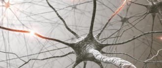
The myelin layer is represented by several layers of glial cell membrane (lemmocyte, Schwann cell), which twist around the axial cylinder (neuron process). This twisting is clearly visible in the picture of a healthy nerve, just above 
The myelin layer of the fiber sheath is regularly interrupted at the junction of neighboring lemmocytes - nodes of Ranvier. The myelin sheath provides isolated and faster conduction of excitation (saltatory type, Latin salto - I gallop, jump).
Neuroglia (Greek νεῦρον - fiber, nerve + γλία - glue)
You have already seen how important neurons are; their high specialization leads to the emergence of a special environment - neuroglia. Neuroglia (glial cells, gliocytes) are an auxiliary part of the nervous system that performs a number of important functions:
- Supporting - supports neurons in a certain position
- Regenerative (Latin regeneratio - rebirth) - in case of damage to nerve structures, neuroglia promotes regeneration
- Trophic (Greek trophe - nutrition) - with the help of neuroglia, neurons are nourished: neurons do not come into direct contact with blood
- Electrical insulating - lemmocytes (Schwann cells) curl around the processes of neurons and form the myelin sheath
- Barrier and protective - isolate neurons from the tissues of the internal environment of the body
- Some gliocytes secrete cerebrospinal fluid - cerebrospinal fluid (from the Latin liquor - liquid)
Neuroglia consists of different cells; there are tens of times more of them than neurons themselves. In the peripheral part of the nervous system, the myelin sheath, which we studied, is formed precisely from neuroglia - Schwann cells (lemmocytes). Between them, the nodes of Ranvier are clearly visible - areas devoid of the myelin sheath between two adjacent Schwann cells.
Classification of neurons
Neurons are functionally divided into sensory, motor and intercalary.
Sensory neurons are also called afferent, centripetal, sensory, perceptive - they perceive irritations, convert them into nerve impulses and transmit them to the central nervous system. A receptor is the terminal ending of sensory nerve fibers that perceive a stimulus.
Interneurons are also called intermediate, associative - they provide communication between sensory and motor neurons, transmit excitation to various parts of the central nervous system, and participate in information processing and the generation of commands.
Motor neurons are also called efferent, centrifugal, or motor neurons - they transmit a nerve impulse (excitation) to an effector (working organ). The simplest example of the interaction of neurons is the knee reflex (however, there is no interneuron in this diagram). We will study reflex arcs and their types in more detail in the section on the nervous system.
Synapse
In the diagram above, you probably noticed a new term - synapse (Greek sýnapsis - connection). A synapse is the point of contact between two neurons or between a neuron and an effector (target organ). At the synapse, the nerve impulse is “transformed” into a chemical one: special substances are released - neurotransmitters (the most famous is acetylcholine) into the synaptic cleft.
Let's look at the structure of a synapse in the diagram. It is made up of the presynaptic membrane of the axon, next to which there are vesicles (Latin vesicula - bubble) with a neurotransmitter inside (acetylcholine). If a nerve impulse reaches the terminal (end) of the axon, then the vesicles begin to merge with the presynaptic membrane: acetylcholine flows out into the synaptic cleft.
Once in the synaptic cleft, acetylcholine binds to receptors on the postsynaptic membrane, thus excitation (nerve impulse) is transmitted to another neuron. This is how the nervous system works: the electrical transmission path is replaced by a chemical one (at the synapse).
Poison curare
It is much more interesting to study any subject with examples, so I will try to please you with them as often as possible. I cannot hide the story about the poison curare, which the Indians have used for hunting since ancient times.
This poison blocks acetylcholine receptors on the postsynaptic membrane, and, as a result, the chemical transfer of excitation from one neuron to another becomes impossible. This leads to the fact that nerve impulses cease to flow to the effectors, including the respiratory muscles (intercostal muscles, diaphragm), as a result of which breathing stops and the death of the animal occurs.
Nerves and ganglia
When the processes of neurons (nerve fibers) come together, they form bundles of nerve fibers. Nerve bundles unite into nerves, which are covered with a connective tissue sheath. If the bodies of neurons are concentrated in one place outside the central nervous system, their clusters are called a nerve ganglion - or ganglion (from the ancient Greek γάγγλιον - node).
In the case of complex connections between nerve fibers, they speak of nerve plexuses. One of the most famous is the brachial plexus.
Nervous system diseases
Neurological diseases can develop anywhere in the nervous system: the clinical picture will depend on this. If the sensitive pathway is damaged, the patient ceases to feel pain, cold, heat and other irritants in the area of innervation of the affected nerve, while movements are fully preserved.
If the motor link is damaged, movement in the affected limb will be impossible: paralysis occurs, but sensitivity may remain.
There is a severe muscle disease - myasthenia gravis (from the ancient Greek μῦς - “muscle” and ἀσθένεια - “powerlessness, weakness”), in which one’s own antibodies destroy motor neurons (motor neurons).
Gradually, any muscle movements become more and more difficult for the patient, it becomes difficult to speak for a long time, and fatigue increases. A characteristic symptom is observed - drooping of the upper eyelid. The disease can lead to weakness of the diaphragm and breathing muscles, making breathing impossible.
© Bellevich Yuri Sergeevich 2018-2021
This article was written by Yuri Sergeevich Bellevich and is his intellectual property. Copying, distribution (including by copying to other sites and resources on the Internet) or any other use of information and objects without the prior consent of the copyright holder is punishable by law. To obtain article materials and permission to use them, please contact Yuri Bellevich
.
Structure and properties of nerve fibers.
Nerve fibers are processes of nerve cells (neurons) that have a membrane and are capable of conducting nerve impulses.
The main component of the nerve fiber is the process of the neuron, which forms, as it were, the axis of the fiber. Mostly this is an axon. The nerve process is surrounded by a membrane of complex structure, together with which it forms a fiber. The thickness of the nerve fiber in the human body, as a rule, does not exceed 30 micrometers.
Nerve fibers are divided into pulpy (myelinated) and non-myelinated (non-myelinated). The former have a myelin sheath covering the axon, the latter lack a myelin sheath.
Myelin fibers predominate in both the peripheral and central nervous systems. Nerve fibers lacking myelin are located predominantly in the sympathetic division of the autonomic nervous system. At the point where the nerve fiber departs from the cell and in the area of its transition into the final branches, the nerve fibers can be devoid of any membranes, and then they are called bare axial cylinders.
Depending on the nature of the signal carried through them, nerve fibers are divided into motor autonomic, sensory and motor somatic.
The structure of nerve fibers:
Myelinated nerve fiber contains the following elements (structures):
1) an axial cylinder located in the very center of the nerve fiber,
2) the myelin sheath covering the axial cylinder,
3) Schwann shell.
The axial cylinder consists of neurofibrils. The pulpy membrane contains a large amount of lipoid substances known as myelin. Myelin ensures the speed of nerve impulses. The myelin sheath does not cover the entire axial cylinder, forming gaps called nodes of Ranvier. In the area of the nodes of Ranvier, the axial cylinder of the nerve fiber is adjacent to the superior Schwann membrane.
The fiber space located between two nodes of Ranvier is called a fiber segment. In each such segment, the nucleus of the Schwann membrane can be seen on stained preparations. It lies approximately in the middle of the segment and is surrounded by the protoplasm of the Schwann cell, the loops of which contain myelin. Between the nodes of Ranvier, the myelin sheath is also not continuous. In its thickness, so-called Schmidt-Lanterman notches are found, running in an oblique direction.
Schwann membrane cells, as well as neurons with processes, develop from the ectoderm. They cover the axial cylinder of the nerve fiber of the peripheral nervous system, similar to how glial cells cover the nerve fiber in the central nervous system. As a result, they may be called peripheral glial cells.
In the central nervous system, nerve fibers do not have Schwann sheaths. The role of Schwann cells here is performed by elements of oligodendroglia. An unmyelinated (unmyelinated) nerve fiber is devoid of a myelin sheath and consists only of an axial cylinder and a Schwann sheath.
Function of nerve fibers.
The main function of nerve fibers is the transmission of nerve impulses. Currently, two types of nerve transmission have been studied: pulsed and non-pulse. Impulse transmission is provided by electrolyte and neurotransmitter mechanisms. The speed of nerve impulse transmission in myelinated fibers is much higher than in nonmyelinated fibers. In its implementation, the most important role is played by myelin. This substance is capable of isolating a nerve impulse, as a result of which signal transmission along the nerve fiber occurs spasmodically, from one node of Ranvier to another. Pulseless transmission is carried out by axoplasmic current along special axon microtubules containing trophogens - substances that have a trophic effect on the innervated organ.