Cysts of the pineal gland (epiphysis) are common, asymptomatic, and in most cases are an incidental finding. Cysts, especially large and atypical-looking ones, are difficult to distinguish from cystic tumors, so patients with suspicious changes should undergo long-term observation. On MRI or MRI, a pineal gland cyst appears as a single-chamber fluid formation with the density of cerebrospinal fluid or with a signal intensity similar to that of cerebrospinal fluid. Peripheral contrast enhancement is typical for most cysts, and rim-shaped calcifications are found in 25% of cases.
Get an MRI of the pineal gland in St. Petersburg
Symptoms of a pineal gland cyst
The vast majority of pineal gland cysts are small in size (in 80% of cases less than 1 cm) and are characterized by an asymptomatic course. Cysts that cause symptoms mainly occur in women in the second half of life. Larger cysts can cause a volumetric effect on the quadrigeminal plate, leading to compression of the superior colliculus and the occurrence of Parinaud's syndrome. When the cerebral aqueduct located between the third and fourth ventricles is compressed, obstructive hydrocephalus may develop. Rarely, if the cyst bleeds, it can quickly increase in size. This condition is called pineal cyst apoplexy.
A pineal gland cyst can cause headaches, blurred vision, and the inability to look up or down. Rare symptoms: ataxia, emotional disorders, disturbances in mental activity, dizziness, sleep disturbances, nausea, hormonal imbalance (early puberty, secondary parkinsonism).
↑ Diseases associated with impaired sexual differentiation
Among the diseases associated with impaired sexual differentiation, in which pathological changes in the organ of vision are observed, we will consider the following: Shereshevsky-Turner syndrome, trisomy X, Klinefelter syndrome.
Diseases associated with impaired sexual differentiation are based on chromosomal abnormalities
.
Shereshevsky-Turner syndrome
caused by monosomy on chromosome X (karyotype XO). With this anomaly, newborns experience lymphatic swelling of the dorsal surfaces of the feet, legs, and neck. The swelling goes away after a few months; folds of skin remain on the neck, running from the occipital bone to the shoulders; these folds give a sphinx-like expression to the face. Other characteristic signs of Shereshevsky-Turner syndrome are short stature, infantility, hypoplasia of the female genital organs, and low-set ears. Qualitative skeletal abnormalities are noted (diffuse osteoporosis, shortening of the metacarpal bones, high hard palate, etc.) A low hairline on the back of the neck, age spots, and telangiectasia are often observed. With this syndrome, there may be congenital heart defects, large vessels, kidney malformations (absence of one kidney, anomalies of the pelvis and ureters, horseshoe kidney). Mental development is normal.
Of the eye syndromes with Shereshevsky-Turner syndrome,
epicanthus, drooping upper eyelid (Fig. 70),
Rice. 70.
Congenital bilateral ptosis. Epicanthus.
color anomaly, strabismus, exophthalmos, cataract, choroidal coloboma (Fig. 71),
Rice. 71.
Coloboma of the choroid.
lack of pigment in the retina, glaucoma and some other changes.
The domestic literature provides a description of eye symptoms
in 29 people with this disease [Kaplan A.I. et al., 1969]. One patient had facial asymmetry with the location of the eye sockets at different levels and a wide nasal bridge, 10 had epicanthus, 12 had ptosis of the upper eyelid, 3 had a Mongoloid eye shape, 10 had strabismus, 4 had nystagmus, and 8 had pigment deposition on the lens capsule. One patient each had a coloboma of the iris and choroid, decoloration of the optic nerve head, a decrease in visual acuity to 0.06 and a narrowing of the boundaries of the visual field by 15-20°. 5 people had a color anomaly (protanomaly and deuteranomaly). The above changes in the organ of vision occurred either in isolation or in various combinations.
Klinefelter's syndrome
. There are chromatin-positive and chromatin-negative Klinefelter syndromes. In chromatin-positive Klinefelter syndrome, the sex chromosome set is XXV. Carriers of this syndrome are characterized by tall stature, a eunuchoid physique, often with gynecomastia, infertility. Quite often, mental retardation is noted with this syndrome.
Various changes in the organ of vision have been described
, color anomaly, hypertelorism, epicanthus, ptosis, ametropia, strabismus, coloboma of the iris, choroid, congenital cataract, pigmentary degeneration of the retina, optic nerve atrophy are found in this syndrome. A.I. Kaplan and co-authors, in a study of 17 patients with Klinefelter syndrome, revealed the following changes: 3 had epicanthus, 2 had a mongoloid eye shape, 3 had nystagmus, 1 had an irregularly shaped pupil, 4 had unilateral nystagmus, 4 had clouding on the lens capsule, 6 had a decrease in visual acuity, 2 had a narrowing of the visual field by 15°. These changes occurred either in isolation or in various combinations. No color anomalies were noted in any case.
Trisomy X
.
In this disease, the chromosome set (karyotype) contains three X chromosomes. Patients have
normal growth and sexual development may be normal. The offspring of women with trisomy X are healthy. Along with this, with normal sexual development and regular menstruation, it is noted that the latter disappear after a few years and secondary amenorrhea occurs. Mental development with trisomy X may not be impaired, but with this anomaly the incidence of mental defects is increased. A contributing factor in the occurrence of trisomy X in newborns is the advanced age of the mother. In total, a little more than 50 cases of trisomy X have been described, in which pathological changes in the organ of vision were observed. These include bilateral optic atrophy, chorioretinitis, and unilateral corneal opacification. Due to the small number of observations, it is difficult to express an opinion on the specificity of these changes.
Structure
The wall of a pineal cyst consists of three concentric layers:
- The inner layer is represented by fibrillary glial tissue, often with inclusions of hemosiderin
- The middle layer is the parenchyma of the pineal gland with or without calcifications
- The thin outer layer consists of connective (fibrous) tissue
Hormonal changes are thought to play a role in the formation of cysts, as they are most often found in young women. With age, cysts first increase in size and then “shrink.” In men, these formations remain in a stable state for a long time. Cysts usually contain proteinaceous fluid that differs from cerebrospinal fluid on CT scans; Sometimes blood is found in them. The walls of the cyst intensively accumulate contrast.
Location
The pineal gland is the only brain structure in the midline that is an unpaired organ.
The pineal gland is located in the epithalamus, the supratuberous region of the midbrain (the quadrigeminal region), near its center, between the two hemispheres.
Location of the pineal gland
The pineal gland is located between the laterally (laterally) located thalamus and the leash commissurra - a strip of nerve fibers, one of the structures of the commissural system, anatomically connecting the hemispheres of the brain. The pineal gland is located in the groove where the two halves of the thalamus are connected.
The pineal gland is located in front of the cerebellum and is attached to the first ventricle of the brain. Located behind the third ventricle, it is washed by cerebrospinal fluid, which enters through a small pineal-shaped depression of the third ventricle, protruding from the stalk of the gland.
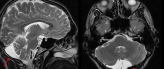
What does a pineal gland cyst look like on CT and MRI?
Computed tomography visualizes a fluid-dense mass with well-defined edges and peripheral calcifications found in 25% of patients. In many cases, there is also a peripheral accumulation of contrast in the cyst in the form of a thin and smooth “rim”. The cyst disrupts the course of the internal veins of the brain, displacing them upward.
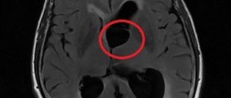
Pineal cyst with a typical hyperintense signal on MRI (T2 WI) (marked with a blue arrow). Left: axial tomogram, right – sagittal
The following signs are observed on MRI:
- T1 VI
- Typical iso- or hypointense signal compared to brain parenchyma
- The signal is usually uniform
In 55–60% of cases, the signal is hyperintense compared to the cerebrospinal fluid
- High intensity signal
- High intensity signal that is often not completely suppressed
- There is no diffusion restriction
- Approximately 60% of cysts accumulate contrast
The influence of pineal gland hormones on the human body
The most important function of the pineal gland in the brain is to produce the hormones melatonin, serotonin, histamine and norepinephrine. The healthy functioning of the organ in childhood ensures the child’s intellectual development and his ability to learn.
The effect of hormones on the body as a whole:
- Supports stress resistance of the central nervous system;
- Control blood pressure;
- Regulate sleep starting from embryonic development;
- Supports carbohydrate and fat metabolism in the body.
Malfunction of the pineal gland affects the biological rhythms of the body: insomnia, shallow sleep and feeling tired in the morning.
Causes of pineal gland dysfunction:
- Cystic formation in the epiphysis of the brain;
- Degeneration of cellular composition with age-related changes in the body;
- Amyloidosis;
- Accumulation of calcifications;
- Neoplasm in the pineal gland;
- Blockage of a vessel providing blood supply to an organ by a thrombus;
- Stroke;
- Inflammation and lack of blood supply (with intoxication, infectious diseases, hemorrhagic diathesis);
- Hypertension.
Each hormone produced by the pineal gland is responsible for certain conditions of the human body.
Serotonin
The hormone is the main neurotransmitter belonging to biogenic amines. Once in the bloodstream, there is an uplift in mood and a surge of strength, which is why you can come across the name “happiness hormone”.
Serotonin is synthesized from tryptophan; the body obtains the amino acid when processing food. The hormone is synthesized in the pineal gland of the brain. The intake of light carbohydrates from food (confectionery) and sufficient sunlight increase the production of serotonin, which directly affects a person’s mood.
Melatonin
The hormone is formed from serotonin and acts on the entire cellular composition of the body, helping to maintain their tone. It is often called the “sleep hormone” because most of it is synthesized during nighttime sleep.
Melatonin regulates the biological clock in the body and activates cell renewal and growth, hence its second name – “youth hormone”.
The hormone is produced only in conditions of complete darkness and helps restore the body, relieving the feeling of fatigue and toning the entire body. Age-related changes affect this hormone - the production of melatonin decreases, which leads to damage to hormonal DNA and withering of the body.
Functions of the hormone in the body:
- Stimulates memory function;
- Reduces pain;
- Prevents obesity;
- Helps produce immune cells;
- Controls blood pressure;
- Regulates the sleep-wake cycle.
Norepinephrine
The hormone is a biogenic amine and is synthesized from tyrosine (an amino acid), which the body receives from food. The process of norepinephrine synthesis occurs in the pineal gland of the brain and adrenal glands.
The hormone improves the functioning of brain structures and promotes the absorption of glucose in muscle fibers.
Norepinephrine begins its work in stressful situations, increasing heart rate and increasing blood pressure, which is why it is called the “stress hormone.”
Histamine
The hormone is an organic compound formed from histidine (the chemical structure of a protein) and is a mediator of allergic reactions.
Participates in metabolic processes and the body’s immune defense when an allergen penetrates, which is why it received the name “alarm hormone.” Massive production of this hormone sharply reduces blood pressure, causing anaphylactic shock.
We recommend watching an educational film and learning more about the pineal gland.
How to distinguish a pineal cyst from a tumor?
A pineal cyst, especially one with nodular contrast enhancement, cannot be differentiated from a cystic pineocytoma based on imaging techniques alone. Other formations may also occur in the pineal area: papillary tumor, germinoma, embryonal cancer, choriocarcinoma, teratoma, epidermoid cyst, arachnoid cyst, vein of Galen aneurysm, metastases.
Although such cases are rare, it is always best to have the results examined by an experienced neuroradiologist. Rechecking CT or MRI images will help you more accurately understand whether a tumor is suspected. Today this can be done remotely using Second Medical Opinion services, such as the National Teleradiological Network.
Possible contraindications
Magnetic resonance imaging is considered absolutely safe for the body only if the patient has no contraindications. Therefore, before making an appointment for an MRI, you need to visit a doctor and check if you have one of the serious contraindications:
- The presence of a pacemaker or metal structures in the body that cannot be removed independently;
- Diseases of the central nervous system or serious psychological disorders in which the patient is unable to control his body and lie still;
- The patient's weight is over 250 kg.
- Severe fear of confined spaces.
If the doctor recommended undergoing magnetic resonance imaging with contrast, you need to make sure there is no pregnancy, lactation, renal failure, kidney problems or allergies to the components included in the drug.
Treatment of pineal cyst
In almost all cases there is no need for treatment; in most cases, small cysts do not require follow-up studies. Depending on the size and location of the cyst, as well as symptoms, treatment may consist of stereotactic removal of the cyst, aspiration of its contents, creation of a connection with the cerebrospinal fluid spaces, and shunting. If the cyst recurs after treatment, radiation therapy is possible.
Get an MRI of the pineal gland in St. Petersburg
Conclusion
Remember that health is a gift that can be easily lost! In order for the pineal gland to cope with its assigned functions, you should strictly monitor your daily routine, diet, get enough sleep, and lead a healthy active lifestyle.
You shouldn’t change your habits suddenly; the main thing is to gradually bring them back to normal. These are excellent preventive measures that will prevent pathological changes in this mysterious gland, which is responsible not only for physiology, but also for intelligence.
How to properly prepare for the procedure
To obtain clear and informative images, it is recommended to follow a few simple rules:
- Do not eat for 7-8 hours before the examination if an MRI with contrast is planned;
- Do not drink a lot of liquid immediately before the procedure, so as not to experience the urge to urinate;
- Tell your doctor if the test worries you. If you are experiencing severe panic, your doctor may recommend taking a sedative;
- Take with you a change of clothes without metal fittings;
- To avoid wasting time in the diagnostic room, remove all metal-based jewelry and accessories in advance.
What else do you need to know?
If you are about to undergo magnetic resonance imaging for the first time, we recommend that you familiarize yourself with some features:
- If loud noises scare you, be sure to take earplugs or special noise-isolating headphones with you. The tomograph makes a loud noise while taking images.
- There is lighting and ventilation inside the tomograph.
- A microphone and speakerphone are provided to communicate with a specialist.
- If you feel unwell, you can use the panic button, which will stop the research process.
- It is important to remain completely still. Even minimal body movements can lead to blurry pictures.