What is arachnoiditis?
Dear Alla!
Unfortunately, after head injuries, post-traumatic arachnoiditis of the brain can develop.
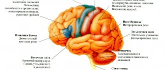
Arachnoiditis of the brain is a disease characterized by inflammation of the arachnoid membrane of the brain. The disease accounts for about 4 - 5% of all organic diseases of the nervous system. A chronic inflammatory productive process occurs in the arachnoid and choroid of the brain, which involves the choroid plexuses of the ventricles, the subepidermal layer, the molecular layer of the cerebral cortex (cerebral cortex), and the epidermis. The brain is surrounded by three meninges: on the outside the dura mater, then the arachnoid mater, then the pia mater.
Elimination of the disease
The disease cerebral arachnoiditis must be treated over a long period of time, in courses. To eliminate the source of infection, doctors prescribe antibiotics to their patients. The following tools are also used:
- anti-inflammatory;
- absorbable;
- hyposensitizing;
- dehydration, etc.
When intracranial pressure increases, diuretics (for example, Furosemide, Mannitol) and decongestants are needed. If patients experience seizures, doctors prescribe antiepileptic drugs. If necessary, symptomatic medications are used.
Pathogenesis of arachnoiditis
With injuries and bruises of the brain, subarachnoid hemorrhages and damage to the soft meninges occur, as a result of which tissue decay products enter the subarachnoid space. Subsequently, the arachnoid membrane becomes denser and thickens. Adhesions and adhesions appear between the arachnoid membrane and the pia mater. Then the circulation of cerebral fluid is disrupted, cyst-like expansions are formed, and the ventricles of the brain enlarge.
General cerebral symptoms
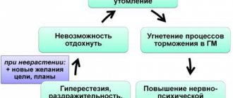
Cerebral arachnoiditis is characterized by certain clinical manifestations. First of all, the disease makes itself felt by general cerebral symptoms. Headache is common. It is strongest in the morning. In some people it is accompanied by nausea and vomiting.
Headache may worsen with tension, straining, or awkward movements. In addition to it, people with cerebral arachnoiditis report dizziness. In patients, memory deteriorates, irritability appears, fatigue quickly sets in, sleep is disturbed, and general weakness is observed.
Treatment of post-traumatic arachnoiditis in Saratov, Russia
Sarklinik provides treatment for arachnoiditis in Saratov , Russia, treatment of post-traumatic arachnoiditis using hardware and non-hardware techniques. With complex treatment, a significant improvement in the condition is possible. Sarklinik knows how to effectively treat arachnoiditis. Consultations, comprehensive medical care.
In any case, it is necessary to carry out differential diagnosis and establish a true diagnosis, and only then carry out treatment. Differential diagnosis is varied, it is necessary to exclude various diagnoses: somatoform autonomic dysfunction, vegetative-vascular dystonia, cerebral vascular atherosclerosis, arterial hypertension, hypertension, cerebral aneurysm, age-related changes, stress, auditory neuritis, neurosis, depressive neurosis, cervical osteochondrosis spine, anemia, diseases of the cardiovascular system, arrhythmia, kidney disease, brain tumors, diseases of the endocrine system, taking a number of medications, consequences of brain injuries, concussions, otosclerosis, labyrinthitis, tumors of the tympanic cavity, Meniere's disease, acute trauma, exostoses , fistula, autoimmune diseases, exposure to loud noises at work or at home, the effects of ototoxic drugs, temporomandibular joint diseases.
Of course, it is necessary to conduct an MRI of the brain, REG, EEG, MRI of the cervical spine, vascular angiography, audiogram, CBC, TAM, glycated hemoglobin, cholesterol, lipoproteins (lipoproteins).
Tinnitus (tinnitus) and noise in the head make it very difficult to live a full life, so you should definitely consult a doctor.
Sign up for a consultation. There are contraindications. Specialist consultation is required.
Related posts:
Resistance index in fibroids, blood flow intensity: results
Psychogenic erectile dysfunction, erection disappears, treatment in Saratov
Why do I cum very quickly, I get very excited at the sight of a naked girl
Lost sensitivity to sexual intercourse, treatment in Saratov, Russia
Unprotected sexual intercourse, discomfort in the testicle, spot, odor, short intercourse, yellow clots in semen
Comments ()
- Anamnestic information: 1) etiological risk factors; 2) development gradually, gradually. At first, often astheno-neurotic symptoms, irritative (epileptic seizures), then hypertensive (headache, etc.). Gradual deterioration of the condition, sometimes temporary improvement.
- Neurological examination: general cerebral and local symptoms in varying proportions depending on the predominant localization of the process.
1) General cerebral symptoms.
It is based on a violation of liquorodynamics (cerebrospinal fluid circulation), most pronounced during obliteration of the foramina of Magendie and Luschka, much less often due to chorioependimitis. Clinical symptoms are caused by intracranial hypertension (96%) or CSF hypotension (4%). Hypertensive syndrome is more common and most pronounced in arachnoiditis of the posterior cranial fossa, when 50% of patients have congestive optic discs against the background of focal symptoms. Headache (in 80% of cases), often morning, bursting, pain when moving the eyeballs, physical stress, straining, coughing, nausea, vomiting. Also include: non-systemic dizziness, tinnitus, hearing loss, autonomic dysfunction, increased sensory excitability (intolerance to bright light, loud sounds, etc.), weather dependence. Neurasthenic manifestations (general weakness, fatigue, irritability, sleep disturbance) are common. Liquorodynamic crises (acutely occurring dyscirculatory disorders), manifested by increased cerebral symptoms. It is customary to isolate the lungs (short-term increase in headache, moderate dizziness, nausea); moderate severity (more severe headache, poor general health, vomiting) and severe crises. The latter last from several hours to 1-2 days and are manifested by severe headache, vomiting, general weakness, and impaired adaptation to external influences. Autonomic-visceral disorders are also common. Depending on the frequency, there are rare (1-2 times a month or less), medium frequency (3-4 times a month) and frequent (more than 4 times a month) crises.
2) Local or focal symptoms. They are determined by the predominant localization of organic changes in the membranes of the brain and adjacent structures. In general, focal symptoms are characterized by a predominance of irritation rather than loss. The exception is opticochiasmatic arachnoiditis.
- convexital arachnoiditis (in 25% of patients, more often of traumatic etiology). The predominance of local symptoms is characteristic (depending on the damage to the area of the central gyri, parietal, temporal). In the cystic form of arachnoiditis, usually mild or moderately expressed motor and sensory disturbances (pyramidal insufficiency, mild hemi- or monoparesis, hemihypesthesia). Epileptic seizures are typical (in 35% of patients), often the first manifestation of the disease. Secondary generalized partial seizures (Jacksonian) are common, sometimes with transient postparoxysmal neurological deficit (Todd's palsy). Polymorphism of seizures is characteristic due to the frequent temporal localization of the process: simple and complex (psychomotor), partial with secondary generalization, primary generalized, their combination. EEG reveals epileptic activity in 40-80% of patients (according to various sources), including peak-slow wave complexes. General cerebral symptoms are moderate, severe headaches, crisis states more often during exacerbation;
- basilar arachnoiditis (in 27% of patients) can be widespread or localized mainly in the anterior, middle cranial fossa, in the interpeduncular or optic-chiasmatic cistern. With significant spread, many cranial nerves at the base of the brain (I, III-VI pairs) are involved in the adhesive process, which determines the clinical picture of the disease. Pyramidal insufficiency is also possible. General cerebral symptoms are mild and moderate. Mental disorders (fatigue, decreased memory, attention, mental performance) are more often observed with arachnoiditis of the anterior cranial fossa.
The most distinct symptoms are in the case of opticochiasmatic arachnoiditis. Currently, it is rarely diagnosed, since in the vast majority of cases, chiasmatic syndrome is caused by other causes (see “Differential diagnosis”). More often it develops after a viral infection (flu), trauma, or against the background of sinusitis. General cerebral symptoms are mild or absent. Initial symptoms are the appearance of a grid before the eyes, a progressive decrease in visual acuity, often immediately bilateral, over 3-6 months. In the fundus there is first neuritis, then atrophy of the optic discs. The field of view is concentric, less often bitemporal narrowing, unilateral or bilateral central scotomas. Endocrine metabolic disorders are possible due to the involvement of the hypothalamus in the process;
- arachnoiditis of the posterior cranial fossa (in 23% of patients), usually post-infectious, otogenic. A severe, pseudotumorous course is often encountered due to severe disturbances in cerebrospinal fluid dynamics, damage to the caudal cranial nerves, and cerebellar symptoms. With predominant localization in the region of the cerebellopontine angle, damage to the VIII pair of cranial nerves (tinnitus, hearing loss, dizziness) appears earlier. Subsequently, the facial nerve suffers, cerebellar insufficiency and pyramidal symptoms are detected. The first symptom of the disease may be facial pain as a consequence of trigeminal neuropathy. Arachnoiditis of the cistern magna and adjacent formations is characterized by the greatest severity of hypertensive syndrome due to obliteration of the cerebrospinal fluid ducts and impaired outflow of the cerebrospinal fluid. Severe crises are frequent, cerebellar symptoms are pronounced. Complication is the development of a syringomyelitic cyst. Due to the serious condition of patients, it is sometimes necessary to resort to surgical intervention.
- Data from additional studies. The need for adequate assessment due to the difficulties and frequent errors in diagnosing arachnoiditis. It is incorrect to judge true (actual) arachnoiditis only on the basis of morphological changes detected on PEG, CT, MRI studies without taking into account the characteristics of the clinical picture, the course of the disease (progression of the first symptoms, the appearance of new symptoms). Therefore, it is necessary to objectify complaints indicating hypertensive syndrome, visual impairment, frequency and severity of crises, and epileptic seizures.
Only a comparison of clinical data and the results of additional studies provides grounds for diagnosing true current arachnoiditis and distinguishing it from residual stable conditions:
- Craniogram. Symptoms that are sometimes detected (digital indentations, porosity of the dorsum sella usually indicate a past increase in intracranial pressure (long-standing traumatic brain injury, infection) and cannot in themselves indicate current arachnoiditis;
- echo-EG allows you to obtain information about the presence and severity of hydrocephalus, but does not allow you to judge its nature (normotensive, hypertensive) and cause;
- Lumbar puncture. CSF pressure is increased to varying degrees in half of the patients. With exacerbation of arachnoiditis of the posterior cranial fossa, basal water levels can reach 250-400 mm of water. Art. lying down. A decrease in pressure occurs with chorioependimitis. The number of cells (up to several dozen in 1 μl) and protein (up to 0.6 g/l) increases in the case of active process, in other patients it is normal, and the protein content is even below 0.2 g/l;
- PEG has important diagnostic value. Signs of adhesive arachnoiditis, cicatricial changes, internal or external hydrocephalus, and an atrophic process are revealed. At the same time, PEG data can be the basis for the diagnosis of true arachnoiditis only if the clinical features and course of the disease are taken into account. They are often evidence of residual changes in the subarachnoid space and brain substance after injury, neuroinfection, and occur in patients with epilepsy without clinical signs of arachnoiditis;
- CT and MRI make it possible to objectify the adhesive and atrophic process, the presence of hydrocephalus and its nature (aresorptive, occlusive), cystic cavities, and exclude space-occupying formations. CT cisternography allows us to identify direct signs of changes in the configuration of the subarachnoid spaces and cisterns. However, the information obtained has diagnostic value only with an adequate assessment of the lumbar puncture data (especially the state of liquor pressure) and the clinical picture of the disease as a whole;
- EEG reveals foci of irritation in convexital arachnoiditis, epileptic activity (in 78% of patients with epileptic seizures). These data only indirectly indicate the possibility of an adhesive process and do not in themselves constitute a basis for diagnosis;
- immunological studies, determination of the content of serotonin and other neurotransmitters in the blood and cerebrospinal fluid are important for judging the activity of the inflammatory meningeal process, the degree of involvement of the brain substance in it;
- ophthalmological examination is used for diagnostic purposes not only for opto-chiasmal, but also diffuse arachnoiditis due to the frequent involvement of the visual pathways in the pathological process;
- otolaryngological diagnosis is important primarily for determining the etiology of arachnoiditis;
- experimental psychological research allows us to objectively assess the state of mental functions and the degree of asthenia of the patient.
Causes of disability
It was said above that cerebral arachnoiditis can lead to disability. Thus, the disease provokes a limitation of life activity, that is, patients completely or partially lose the opportunity or ability to carry out the main components of everyday life. This happens for the following reasons:
- Convulsive seizures. Sick people periodically lose control over their behavior. In this regard, life activity is limited and ability to work is impaired.
- Deterioration of visual functions. In people suffering from cerebral arachnoiditis, visual acuity decreases and the field of vision narrows. They cannot work with small parts or perform their professional duties that require eye strain. Some people constantly need help from people around them due to blindness.
- Disturbances of cerebrospinal fluid dynamics in the disease cerebral arachnoiditis. The consequences are the manifestation of hypertensive syndrome with repeated crises. Crises are accompanied by dizziness and disorientation.
- Neurasthenia and concomitant vegetative dystonia. People's endurance to climatic factors decreases, and the ability to endure prolonged physical and mental stress is lost. Patients react negatively to loud sounds and too bright light.