Medical certificate
The main components of the brain are 4 ventricles. They are represented by small cavities. They constantly produce and circulate cerebrospinal fluid. Its main purpose is to transport nutrients to cells and remove waste products. In addition, cerebrospinal fluid plays a kind of shock-absorbing role during head impacts.
Normally, each ventricle contains about 150 ml of cerebrospinal fluid. It is completely updated three times a day. An increase in its volume provokes an increase in the size of the ventricles and a change in their shape. When the lateral C-shaped curvature disappears, the cavity becomes rounded. In this case, they speak of the initial stage of lateroventriculoasymmetry of the brain.
What it is? Such a terrible diagnosis refers to a disorder characterized by an asymmetric change in the size of the ventricles. To a small extent, such changes are not considered a pathology. However, their noticeable progression can lead to severe brain disorders. Enlarged ventricles begin to put pressure on surrounding elements and structures, causing disruption of the functioning of neurons. In some cases, death of nerve cells is observed.
Symptoms of ventricular dilatation
The principle of the development of the disease has general features, without taking into account the characteristic causes. Fluid collects in the ventricles, they enlarge and begin to put pressure on the brain tissue. The pressure in these cavities increases.
Patients experience: headaches, nausea, vomiting, a feeling as if the eyes are being squeezed out, problems with hearing and vision, apathy, fatigue, dizziness, memory deterioration, fainting, coma, disorientation, problems with coordination of movements, enuresis. , depressive state. All these are symptoms of dilatation of the lateral ventricles.
Reasons for the development of the anomaly
The pathology can either be congenital or be the result of other health problems. In infants, lateroventriculoasymmetry is more common than in adults. Approximately every 500th newborn is diagnosed with an increase in the size of the ventricles, and in 80% of cases it is caused by congenital abnormalities in brain development. The remaining 20% are due to birth injuries. A child may develop intrauterine anomalies due to problems with the woman’s health during pregnancy. We are talking about such diseases as toxoplasmosis, rubella and mumps.
The causes of lateroventriculoasymmetry of the brain in adults are somewhat different. An increase in cerebrospinal fluid and, as a consequence, the size of the ventricles can be provoked by:
- skull injuries;
- previous neuroinfections (for example, meningitis);
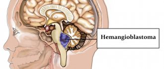
- tumors, cysts, hemangiomas;
- thrombosis of cerebral vessels.
Accurate determination of the cause of the outflow disturbance and excessive production of cerebrospinal fluid allows you to prescribe effective treatment.
How does hydrocephalus of the brain manifest?
The main signs of hydrocephalus are directly related to the deterioration of the patient’s general health. Symptoms can be pronounced or unexpressed. Common consequences of cerebral hydrocephalus are headaches, nausea, and vision problems. Secondary symptoms: the occurrence of convulsive syndrome, tinnitus, impaired coordination of movement and fear of light.
Even moderately severe hydrocephalus, located in the cerebral ventricles, can lead to impaired sensitivity of the limbs and even paralysis. Convulsive seizures also appear, which in turn can be symmetrical or asymmetrical. Ventricular hydrocephalus is a rather dangerous disease. In newborns, the consequences of hydrocephalus are retardation in physical and mental development.
Clinical picture
Moderate lateroventriculoasymmetry of the brain does not affect the human condition. We can talk about the development of a pathological process in the presence of pronounced changes in the lateral ventricles. In this case, the patient feels the symptoms of the primary disease, which provoked lateroventriculoasymmetry. This may involve problems with sensation, memory, coordination or thinking. Typically the patient complains of:
- pain in the head, a feeling of squeezing and bursting;
- nausea and vomiting, especially in the morning;
- dizziness;
- state of anxiety.
The patient is constantly accompanied by apathy and drowsiness. Such symptoms of lateroventriculoasymmetry of the brain indicate that the pathology is progressing. As a result of impaired outflow of cerebrospinal fluid, pressure on the brain increases, which leads to:
- oculomotor disorders;
- development of dementia;
- blurred vision;
- urinary incontinence;
- impaired motor function (gait becomes unsteady).
If treatment for the anomaly is not started in a timely manner, it can become chronic.
Headache and venous congestion
Venous stagnation (or disturbances of venous outflow from the cranial cavity) is a syndrome that often occurs with headaches of various types. Unfortunately, it is a common situation when venous outflow disorders are not taken into account in the treatment of tension headaches and migraines; as a result, patients suffer for years.
Even though venous outflow disorders are often described during head studies (MRI, vascular ultrasound), they are practically not taken into account in combination with other signs and complaints of the patient.
Manifestations of venous stagnation
Venous pressure inside the cranial cavity normally changes constantly - it increases with straining, coughing, sneezing, and decreases with rest. If venous outflow becomes difficult, characteristic complaints may appear:
- Heaviness in the head and pain in the morning.
- Swelling of the face (especially the eyelids) after sleep.
- Bursting headache.
Disturbances of venous outflow and nature of pain
Disturbances in venous outflow can change the nature of pain in patients with tension-type headaches. For example, pressing pain in the temples is accompanied by heaviness in the head after sleep.
There are even situations when disturbances in venous outflow are the main and only cause of headaches, and tension headaches come later.
The situation is no easier for migraine patients. In addition to the migraine itself, many also experience tension in the neck muscles and impaired venous outflow. In such patients, headaches almost never stop.
In practice, it turns out that an incorrect diagnosis (without taking into account venous outflow disorders) leads to long-term and ineffective treatment of headaches.
Difficulties in diagnosis
How can a doctor suspect the presence of a “venous factor”? After all, as we see, complaints with venous stagnation are not so specific. Only on the basis of symptoms such as swelling of the face and heaviness in the head in the morning, a correct diagnosis cannot be made.
The patient should be asked about changes in pain patterns and headache frequency. Since the tone of the veins can change with changes in atmospheric pressure, weather or cycle phase in women, all this must be taken into account.
Venous congestion can be confirmed using the following methods:
– Consultation with an ophthalmologist - dilated veins will be visible in the fundus. – Ultrasound examination of blood vessels - a decrease in the speed of blood flow through the veins will be noted. – Magnetic resonance imaging in phlebographic mode - dilated venous sinuses and veins will be noticeable.
However, the conclusion “impaired venous outflow” in itself does not carry much value - what is more important is the full clinical picture with the specifics of your complaints.
How to treat?
With regard to drugs for disrupting venous outflow from the cranial cavity, the situation is complex. There are currently no effective drugs with proven action against veins inside the skull. In practice, vascular drugs (for example, Cavinton) and metabolic drugs (Mexidol) are often prescribed, which may cause some improvement (although their effect has not been proven). Sometimes venotonics (for example, Detralex) are prescribed with a positive effect
Among other methods of treating headaches with impaired venous outflow, the methods of manual therapy, massage, and physiotherapy (if there is tension in the neck muscles or the effect of osteochondrosis on the blood vessels) have proven themselves to work well in practice.
Timely detection of venous outflow disorders and consultation with a neurologist can protect against progression and chronicity of pain.
Be healthy!
Maria Meshcherina
Photo istockphoto.com
Course of the disease in children
The manifestation of the pathological process in young patients depends on age. Children are usually capricious and do not eat well. Their gaze is directed downward, but their eyes are excessively open. In children, the bones of the skull are flexible and plastic. Therefore, disorders can manifest themselves in an increase in head size. Pulsation of the fontanel and swelling of the veins are often observed.
School-age children also suffer from lateroventriculoasymmetry of the brain. Only a few know what kind of disease this is. Its development may be preceded by congenital disorders of brain formation or failures at the genetic level. At this age the disorder manifests itself:
- divergence of seams between the bones of the skull;
- slow development;
- overweight;
- sudden mood swings;
- impaired ability to move the eyeballs;
- problems with mental activity;
- decreased attachment to loved ones.
In adolescents, this disease is a consequence of cranial injuries or previous infections. It is accompanied by severe headaches, vomiting and nausea, and convulsions. Psychosis cannot be ruled out.
In newborns
Ventriculomegaly refers to the dilatation of the lateral ventricles in infants who have recently been born. The first signs of the disease can be detected at 17 weeks of pregnancy. More often the disease develops in premature infants. If the cause of the deviation does not appear clearly, it means that after some time the size of the ventricles will return to normal.
Why does this disease develop in newborns:
- Infectious processes in the mother's body during pregnancy.
- Asphyxia as a result of the umbilical cord entangling the neck.
- Injuries received during childbirth.
- Hydrocephalus.
- Hemorrhages.
- Lack of oxygen.
- Bad heredity.
- Mothers over 35 years of age are more likely to develop the disease.
Asymmetry is formed in children taking into account their age category. A baby with an illness is constantly capricious, reacts inhibited, often refuses breastfeeding, and his gaze is directed downwards.
If the baby has an isolated disease and does not have any abnormalities or genetic abnormalities, the prognosis is often favorable. This is only possible in a situation where the newborn has been treated for a month using the correct therapeutic technique.
Diagnostic methods
Prescribing treatment and explaining what lateroventriculoasymmetry of the brain is is the task of a specialist. If symptoms of the disorder appear, you should immediately consult a therapist. After studying the clinical picture and complaints, the doctor will refer you to a neurologist. It is this specialized specialist who can confirm the preliminary diagnosis. For this purpose, he prescribes a comprehensive examination to the patient, which consists of the following activities:
- CT, MRI. These methods are considered the most informative. Using tomography, you can assess the condition of the ventricular system and determine the causes of impaired outflow of cerebrospinal fluid.
- Echoencephaloscopy.
- Electroencephalography.
- Fundus examination.
If the listed options for diagnosing lateroventriculoasymmetry of the brain give conflicting results, a lumbar puncture is prescribed. The results of cerebrospinal fluid analysis can accurately determine the causes of deformation of the lateral ventricles.
Drug therapy
Treatment of lateroventriculoasymmetry of the brain with drugs is prescribed only when the clinical picture is severe. The main direction of such therapy is the elimination of the underlying disease, which provoked a change in the size and shape of the ventricles. For this purpose, drugs from the following groups are recommended:
- Diuretics.
- Neuroprotectors.
- Vascular agents.
- Nootropic drugs.
- Sedative medications.
- Anti-inflammatory drugs.
All medications, as well as their dosage, are selected individually.
The need for surgery
When conservative therapy is ineffective, surgery is prescribed. A puncture is performed at the site of the greatest accumulation of cerebrospinal fluid. If the cause of compression of the cerebrospinal fluid outflow tract is a tumor, it must be removed.
In modern medical practice, ventricular bypass surgery has become widespread in the treatment of lateroventriculoasymmetry. During the procedure, the doctor installs a shunt, with the help of which cerebrospinal fluid begins to flow into the bladder. After the operation, the patient can return to the usual rhythm of life. Intracranial pressure gradually decreases, and other symptoms of illness disappear. Unfortunately, in half of the cases, shunting loses its effectiveness over time. The development of complications, including inflammation of soft tissues and the addition of a secondary infection, cannot be ruled out. In this case, a repeat operation with replacement of the shunt is required.
Consequences of the disease
A common question that almost all patients diagnosed with “lateroventriculoasymmetry of the brain” ask specialists: is it dangerous? It is not possible to answer this unequivocally.
If there is an unexpressed form of the disease, when changes in the lateral ventricles do not cause discomfort to the patient, specific therapy is not required. Simply observing the anomaly is enough. In case of progressive disease, treatment is necessary. It can be either medicinal or surgical. If you ignore the symptoms of the disorder and do not seek help from a doctor, the likelihood of complications increases. In addition to impaired consciousness and motor function, such a patient is at risk of coma.
The main types of hydrocephalus
At the initial consultation, the neurosurgeon will listen to the patient’s complaints, but will be able to make an accurate diagnosis only as a result of a comprehensive diagnosis. Special studies make it possible to determine what stage of the disease we are talking about - advanced or mild hydrocephalus.
There are quite a large number of varieties of dropsy:
- by origin, acquired and congenital diseases are distinguished;
- according to the characteristics of the course, hydrocephalus can be closed (occlusive), open (non-occlusive) and mixed;
- by location - internal and external hydrocephalus;
- according to clinical signs - acute, subacute, chronic degrees of hydrocephalus.
The dynamics of pathology development is also important. The most dangerous is considered progressive hydrocephalus, in which the number of symptoms constantly increases. The mild type is considered a regressive disease. With stable hydrocephalus, significant changes in the human body, as a rule, do not occur.