Magnetic resonance imaging is recognized as one of the most informative neuroimaging research methods. During the procedure, detailed layer-by-layer images of the brain are obtained, allowing one to assess the organ’s compliance with standards.
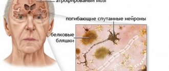
MRI is relevant in the diagnosis of post-traumatic conditions, inflammatory, tumor, and neurodegenerative changes. One of the most severe disorders detected by tomography is brain atrophy. This is a chronic progressive disease accompanied by the death of neurons, a decrease in the volume and density of brain structures.
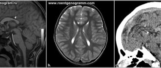
Atrophy of the frontal and temporal lobes on MRI scans
The pathology is based on organic changes in the nervous tissue. The progression of atrophy leads to a decrease in the functionality of the central nervous system. In fact, this means a gradual decline in the work (involution) of the brain until vital processes stop. The prognosis is unfavorable, since the changes are irreversible. Prospects depend on the etiology, form, rate of progression of the disease and the quality of medical care provided.
Using MRI, it is possible to identify and localize these processes in the early stages of development.
General information
Natural aging processes affect the state of the central nervous system. All older people experience memory deterioration and a decreased ability to learn, remember, and process information. In elderly and senile people, brain atrophy occurs (0.1-0.3% of volume is lost per year), associated with age-related physiological processes, but cognitive function does not quickly reach the level of dementia .
Therefore, in patients with a normal level of consciousness, the term “infectious changes” in the brain is often used. Physiological atrophy begins to develop at the age of 40-60 years and reaches severity by 75 years. In psychoneurological diseases ( Alzheimer's disease , Pick's disease , multiple sclerosis , Lewy body disease ), brain atrophy and, accordingly, neurological disorders progress quickly, leading patients to disability. With these diseases, from 0.5 to 1.4% of brain volume is lost per year. Atrophic changes in the brain, what is it? This is a decrease in the volume and density of brain tissue due to the death of neurons, which occurs for various reasons. Typically, atrophy is the outcome of long-term processes in which organic, irreversible changes in the parenchyma occur and nerve connections are destroyed.
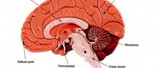
In this case, atrophy of the cerebral cortex, subcortical structures (visual thalamus, limbic system, basal ganglia, hypothalamus) and atrophic changes in the cerebellum are observed to varying degrees. Cortical (or external) atrophy is observed in all patients with secondary atrophy. The changes can be generalized (the entire brain is affected) or local (specific areas of the brain are affected).
In some diseases, the white matter of the brain predominantly atrophies, while in others, the gray matter atrophies. Thus, in multiple sclerosis , atrophic changes in the gray matter occur in the early stages; they develop faster than white matter atrophy and are associated with cognitive impairment. As the volume of the cortex decreases, neurological changes progress. Women get sick more often than men and the first signs of pathology appear at 55 years of age. In this regard, it is important to carry out effective treatment that would reduce the rate of atrophic changes. This pathology cannot be cured and sooner or later ends in dementia . The rate of atrophy determines the speed of onset and severity of disability.
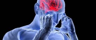
The larger the lesion, the worse the manifestation
As for the consequences that threaten the death of brain cells, the rule is relevant here: the larger the damage, the worse the manifestation.
And also in the absence of treatment, a person’s condition worsens faster. As a result, convulsions, loss of muscle function, or respiratory depression may occur. The simultaneous manifestation of such consequences can lead the patient into coma or stupor.
Nothing good can be expected here, since such a process can no longer be stopped, and when a significant part of the cells for life dies, death occurs.
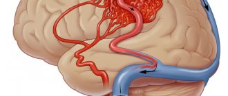
Pathogenesis
The development of atrophic processes in most cases is based on deterioration of blood supply. The cerebral cortex is gray matter. The white matter is located under the gray matter. The subcortical substance is formed by the thalamus, caudate and lenticular nuclei, and basal ganglia. It was found that the subcortical substance and the white matter of the hemispheres, located around the ventricles of the brain, are more affected by chronic cerebral ischemia than the gray matter. When cerebral vessels are damaged, the supply to the frontal subcortical areas is disrupted. Large areas of infarction develop in the white matter, in which axons and oligodendrocytes .
Damages to the cerebellum cause a decrease in its function: muscle contractions are discoordinated and it becomes impossible to perform normal movements in the limbs. The coordinated function of the muscles of the speech apparatus is disrupted, resulting in speech becoming slow and intermittent. Ataxia of the respiratory muscles is manifested by jerky breathing. Hypotonia of the tongue muscles causes soft consonants to be pronounced firmly.
What disorders affect the brain?
Subatrophic changes are precursors to global neuronal death. At this stage, it is necessary to promptly identify brain disease and prevent the rapid development of atrophic processes.
For example, with hydrocephalus of the brain in adults, the voids free from destruction are filled with the resulting cerebrospinal fluid. This type of disease is difficult to diagnose, but with proper therapy it will be possible to delay the development of the disease.
You may be interested in: Upper eyelid blepharoplasty: description of the procedure, pros and cons
Changes in the cortex and subcortex are caused by thromophilia and atherosclerosis, which without treatment lead to hypoxia and insufficient blood supply. As a result, neurons die in the back of the head and parietal part, so treatment is necessary to improve blood circulation.
Classification
Cerebral atrophy occurs:
- Primary. It is rare and is caused by genetic disorders and congenital brain abnormalities. It appears at any age, progresses quickly and cannot be corrected.
- Secondary. It is associated with the impact of adverse factors on the brain, among which are vascular pathology, traumatic brain injury, exposure to radiation, toxic effects, metabolic disorders, and bad habits. With secondary atrophy, eliminating the cause to some extent slows down the development of the disease, and adequate treatment ensures a stable condition for many years and a relatively good quality of life.
- General (the entire volume of the brain parenchyma decreases and the volume of the ventricles and subarachnoid spaces increases).
- Local (the volume of some brain structures decreases).
Depending on the location, there are:
- Cortical atrophy (the main symptom is the involution of the cortex in the temporal and frontal lobes).
- Generalized (changes in all parts of the brain).
- Multisystem (foci develop in several areas).
Depending on the stage:
- Stage I. Organic changes are minimal, but mild neurological disorders are noted. Emotional lability, memory impairment, and decreased concentration are noted.
- Stage II. Severe atrophic manifestations, which are manifested by hearing, speech and vision impairments. Behavior changes, a person commits illogical actions that he cannot explain and quickly forgets about them.
- Stage III. Cortical atrophy is sharply expressed and multiple foci are noted in the hemispheres. The patient cannot care for himself and needs outside help. Personal degradation is expressed to the maximum. This is how a person lives for many years until the destruction of important centers occurs and death occurs.
Cortical cerebral atrophy
This form is manifested by loss of intelligence and behavioral disturbances. The cerebral cortex (gray matter layer) is associated with higher cortical functions: perception, memory, imagination, thinking and speech. It has a complex six-layer structure and the neurons of each layer differ in function. Cortical areas, and accordingly, functions are divided into sensory, associative and motor.
Cortical atrophy occurs with the death of neurons in the frontal lobe cortex. The frontal association area is involved in higher mental functions. It controls behavior, logical thinking, object recognition and speech understanding. After damage to the frontal cortex apathy , there is no critical attitude towards oneself and one’s actions, the patient cannot use past experience, and his behavior becomes inadequate and unpredictable, he commits unmotivated actions. Cognitive (cognitive) abilities are gradually lost: the patient loses the ability to perceive, perceive information, analyze and remember it, and think. In general, cortical atrophy of the brain is characterized by progressive personality degradation, which occurs in stages.
Grade 1 cortical cerebral atrophy is the initial stage of the disease, in which there are no symptoms, but this conclusion can be given with MRI even in young people. At the same time, a person lives a full life and is active in his professional activities, but may periodically experience headaches , irritability and be emotionally unstable. Factors causing cerebral atrophy of the 1st degree may be various intoxications, previous traumatic brain injuries and deterioration of blood supply to the brain with osteochondrosis of the cervical spine .
Generalized atrophy
This is a common atrophy characterized by uniform death of areas of the entire brain. This form is characteristic of extensive ischemic and post-traumatic conditions, as well as neurodegenerative diseases - Alzheimer's disease , Parkinson's disease , Pick's disease and frontotemporal dementia. The progression of neurological symptoms depends on the degree of atrophy of the cortex and subcortical gray matter. Decreased cognitive function is associated with damage to the white matter and parietal cortex.
In this regard, generalized cerebral atrophy of the 2nd degree is characterized by a noticeable decrease in the patient’s ability to think and analyze. The level of critical thinking and evaluation of one's actions is reduced. The patient's habits, speech and handwriting also change. The person loses emotional connection and communication abilities. Neurological disorders - movements and coordination of movements - are more pronounced.
The third degree is characterized by degeneration of gray and white matter. The patient cannot control his behavior, and also needs care and observation, since there is a deterioration in hand motor skills and coordination of movements. The patient cannot use basic household items and is unable to make decisions independently.
Local degeneration is associated with:
- vascular diseases;
- alcohol abuse (atrophy of the cerebellar vermis is noted);
- multiple sclerosis;
- drug use;
- brain injury;
- infection of the central nervous system.
In the early stages of multiple sclerosis, the disease is characterized by degeneration of subcortical gray matter, most pronounced in the thalamus. After this, damage to the cortex develops (the central gyri are more affected), over time, destruction of the white matter is noted, and only then the spinal cord. In multiple sclerosis, not only general atrophy is observed, but also local atrophy - structures that have gray matter are affected: the cerebellum , basal ganglia and thalamus . In multiple sclerosis, gray matter atrophy predominates over changes in white matter, and it determines the degree of disability of patients.
Cerebellar atrophy
The cerebellum is located below the hemispheres and above the brain stem. This formation coordinates movements and their precision, receiving information from the cortex and basal ganglia about the position of the legs. Cerebellar lesions are characteristic of multiple sclerosis , Friedreich's ataxia , olivopontocerebellar degeneration , Pierre-Marie's ataxia . Ataxia (meaning disorder) is a characteristic feature of lesions of this brain formation.
Friedreich's ataxia appears at 10-20 years of age. Patients develop weakness in the legs, falls, staggering and uncertainty when walking, handwriting, speech and hearing are impaired. Muscle atrophy gradually increases (at first it is expressed in the legs, and over time it also covers the arms), deep sensitivity is impaired, cataracts develop, the optic nerve atrophies, and dementia develops. Computed tomography for this disease is ineffective, since a weak degree of atrophy of the hemispheres and cerebellum, expansion of the cisterns, and ventricles is detected in the later stages. Olivopontocerebellar atrophy is considered a form of multiple system atrophy.
Multiple system brain atrophy
This is a variant of a degenerative progressive pathology of the brain, which affects the basal ganglia, cerebellum and centers that are responsible for autonomic reactions. Clinically, multiple system atrophy is manifested by parkinsonism , cerebellar ataxia and autonomic disorders . The disease appears at the age of 50-60, progresses rapidly and leads to death. The muscles become stiff, the patient has difficulty moving, coordination and function of internal organs are impaired.
Depending on the predominant syndrome, the following forms of the disease are distinguished:
- Striatonigral degeneration or parkinsonian form . With it, degenerative changes are most pronounced in the striatum (belongs to the basal ganglia) and the substantia nigra (part of the extrapyramidal system, which plays a role in motor function). The leading symptom is parkinsonism .
- Olivocerebellar atrophy or cerebellar form . The cerebellum, olives and pons are affected. In the clinic, cerebellar syndrome predominates, manifested by impaired coordination and inability to maintain balance.
- Shy-Drage syndrome . In this form, the leading ones are autonomic failure and orthostatic hypotension - in patients, the pressure sharply decreases and a pre-fainting state appears when moving to a vertical position. Patients are also concerned about decreased sweating, darkening of the eyes, urinary disorders, impotence and unsteady gait.
Forms of multiple system atrophy
The danger of multiple lesions of brain structures is determined by a complex of pathological damage from the hemispheres, subcortical formations, cerebellum, spinal trunk, and white matter. Concomitant changes in the optic nerve lead to blindness, and in the trigeminal nerve – disruption of the innervation of the face.
Forms of multiple system atrophy:
- Olivopontocerebellar – damage to the cerebellum with impaired mobility;
- Striatonigral degeneration – muscle tremors with manifestations of Parkinsonism;
- Shy-Drager syndrome – vegetative-vascular dystonia, decreased blood pressure;
- Kugelberg-Welander amyotrophy is brain atrophy with muscle wasting and hyperplasia of connective tissue fibers.
Symptoms are determined by the predominant form of the lesion.
Causes
Cerebral atrophy in most cases is secondary in nature and it is worth noting the reasons that lead to this condition:
- Genetic predisposition.
- Chronic cerebral ischemia , causing diffuse damage to the white matter (cerebral atrophy), against which vascular dementia develops. It should be noted that cerebral atrophy in vascular dementia is not very pronounced when compared with Alzheimer's disease , and hippocampal atrophy in chronic cerebral ischemia is the same as in Alzheimer's disease.
- Arterial hypertension hypertensive encephalopathy develops . With this disease, MRI reveals multiple changes in the subcortical zones around the ventricles (leukoaraiosis), cortical atrophy and dilatation of the ventricles. The same changes are observed during physiological aging.
- Antiphospholipid syndrome.
- Central nervous system infections ( meningitis , polio , leptospirosis ).
- State of decortication in coma .
- Atherosclerosis general and cerebral. The condition worsens when atherosclerosis is combined with arterial hypertension.
- Brain tumors.
- Embolism of the cerebral arteries. The source of embolism is fragments of atherosclerotic plaques and blood clots from the heart.
- Atrial fibrillation . In patients with fibrillation, the brain matter as a whole, as well as individual parts, decreases. The frontal regions of the brain and hippocampus are primarily affected.
- Congenital anomalies of cerebral vessels, causing cerebrovascular disorders.
- Blood rheology disorders (increased viscosity, platelet aggregation, etc.).
- Elevated homocysteine . Homocysteine is a sulfur-containing amino acid, an increase in the level of which is associated with the development of vascular disorders of the brain and heart and neurodegenerative processes in the brain. Hyperhomocysteinemia is a risk factor for recurrent strokes and white matter changes. Patients with elevated homocysteine levels have greater hippocampal atrophy.
- Diabetes . Patients with diabetes mellitus are characterized by mild cerebral atrophy of the cortex, subcortical structures and leukoaraiosis . Even an increase in glucose to the upper limit is associated with atrophic changes in the hippocampus and amygdala complex.
- Multiple sclerosis . Already in the early stages, patients experience degenerative changes - neurons and axons die. Axonal damage is observed in active and chronic lesions of sclerosis. In the latter, axon density decreases to 80%. These processes, plus gliosis and demyelination, lead to a decrease in brain volume. Patients with relapsing-remitting sclerosis (it occurs with exacerbations and remissions) lose 0.5-1.3% of brain volume per year.
- Alzheimer's disease . MRI reveals atrophy of the temporal lobe (reduction in the volume of the gyri and widening of the sulci) and hippocampus, which are markers of this disease. The hippocampus plays an important role in memory formation (categorization of information and long-term memory). Any traumatic brain injury accelerates the development of Alzheimer's disease.
- Any intoxication of the body (drug, alcohol, drugs). With alcoholism, diffuse brain atrophy develops.
- Age-related changes.
- Hydrocephalus in children. This pathology is accompanied by structural changes in the brain: thinning of the cortex, reduction of white matter, vascular atrophy and atrophy of the basal ganglia and cerebellum.
- Traumatic brain injury . 3-4 weeks after the injury, minor hemorrhages resolve, the ventricles and subarachnoid space expand, and generalized brain atrophy develops, which is caused by prolonged intracranial hypertension.
- Long-term corticosteroid therapy can cause cerebral atrophy.
- Acute pain. Acute pain that persists for 3 months causes atrophic changes in the cerebral cortex.
- Physical inactivity . A risk factor for the appearance of atrophic processes and a decrease in cognitive cognitive functions is the limitation of physical activity.
How to stop or slow down cell death
In order to stop the disease, it is necessary to eliminate its causes. In most cases, this is very difficult to do, especially considering the fact that nerve cells are not restored - this is impossible.
If cell death was diagnosed at the initial stage, then it is possible to stop it or at least minimize the consequences for the brain with the help of vitamin complexes that strengthen cells and antioxidants that block the oxidation process. This treatment is aimed only at eliminating symptoms. Atrophy itself cannot be treated with modern drugs.
If we talk about the patient’s lifestyle, then all responsibility now falls on the shoulders of loved ones. They must provide the person with constant care. The patient needs to be surrounded with care, ensure comfort and absence of stressful situations.
The patient should not be spared from homework; on the contrary, it would be better if he went about his usual activities. As for inpatient treatment, this will only worsen the situation. When focusing on the problem, the patient worries more, which leads to the progression of cell death.
A calm and stable environment without changes can slow down the development of the disease, and in the best case, stop it. In addition, you can use antidepressants or tranquilizers, thereby avoiding outbursts of aggression.
Symptoms of brain atrophy
The initial symptoms of the disease reflect changes in the mental state of patients. Early symptoms are anxiety and depression . Patients become apathetic, lethargic, they are not interested or attracted to anything, they prefer to retire and constantly stay at home, their night sleep is disturbed. The mask of depression is irritability, dissatisfaction with everything and grumpiness, which are regarded as characteristics of older people.
If depression is a marker of internal atrophy, then anxiety disorders are characteristic of focal and external cerebral atrophy.
Cognitive disorders (memory, attention, concentration, ability to recognize objects and perform actions purposefully) are detected in all patients in the early stages of brain degeneration. Cognitive symptoms and depression are often regarded as senile forgetfulness and low mood. Impaired memory and attention first occur against the background of maintaining professional and everyday skills. As it progresses, social skills are gradually lost (the patient cannot independently go to the store, pharmacy, or visit the clinic, since he lacks purposefulness of actions) and household skills (he cannot simply take care of himself, eat and take a shower). Over time, sloppiness appears, the vocabulary becomes poor, the patient does not comprehend the speech and requests addressed to him. His movements are sweeping, his handwriting changes and his fine motor skills deteriorate.
In severe cases, the patient does not recognize his relatives and friends, cannot navigate the place and cannot answer questions. The specific behavior is that patients constantly repeat actions and words after others. The final stage of brain degeneration is marasmus - complete regression of mental abilities and destruction of personality. At the same time, infant reflexes are activated: a sucking reflex appears, the patient lies in the fetal position, recovers and urinates under himself. He responds to calls with inarticulate sounds.
Symptoms of cerebellar atrophy
Since the cerebellum regulates movements, if it is damaged, the following will be observed:
- Balance and muscle tone problems.
- Gait disturbances (wide-spaced legs, unsteadiness, difficulty turning).
- Fine motor disorders.
- Uncoordinated movements of arms and legs.
- Unsteadiness and frequent falls.
Patients are also worried about headaches , dizziness , attacks of nausea and vomiting, visual disturbances, pronunciation problems, scanned and slow speech, uncoordinated eye movements, and decreased hearing acuity. As it progresses, urinary incontinence , paresis and paralysis .
Treatment of Alzheimer's disease
Drug treatment
The doctor prescribes symptomatic therapy. It is important to alleviate the patient’s condition as much as possible, to help him retain memory and attention longer, and to increase life expectancy.
Drug treatments for Alzheimer's disease include:
- Cholinesterase inhibitors: rivastigmine, donepezil, haoantamine.
- NMDA-glutamate receptor blockers: akatinol-memantine.
The drugs are included in the list of vital drugs, their purchase is financed from the budget. When prescribed in the early and middle stages, medications help stop the progression of the disease.
To treat concomitant disorders, the doctor prescribes antipsychotics, antidepressants,
Social rehabilitation
It is important to maintain the patient’s usual lifestyle for as long as possible. Intellectual regression will be steady, but doing what you love and hanging out with friends will help slow it down. A person with Alzheimer's disease often lives “in the past,” so relatives are advised to discuss current events with him as much as possible and remind him of certain facts. If the patient was drawing or playing a musical instrument before diagnosis, let him continue. Active intellectual activity and movement stimulate the formation of new connections between neurons and use brain reserves, which helps resist dementia.
In many large cities, “schools for relatives” have been opened. The essence of the classes largely boils down to psychological support, but as part of the training they provide a lot of valuable information on caring for people with Alzheimer's disease. The attention of relatives is an important aspect of the social rehabilitation of each patient.
Some rules for caring for a person with dementia:
- As memory deteriorates, teach the patient to use simple paper notes. Everything in the room must be signed. Post detailed instructions for daily tasks, lists with meal times, etc. in visible places.
- Establish a clear daily routine. Breakfast, lunch, dinner, hygiene procedures - everything should be done at the same time. This contributes to the formation of “muscle” memory when a person acts automatically.
- Find something feasible for the patient to do. Many older people like to knit, read newspapers, and do crossword puzzles.
- Stay calm when communicating. You cannot shout at the patient, swear, or use physical force. With severe dementia, a person’s character and personality change greatly, but he does not behave this way on purpose, but because of his illness.
- Allow the patient to serve himself as much as possible. It is important for a person to feel his independence. You don't need to do all the household chores for him. The patient can wash the dishes or make the bed himself, even if it takes twice as long as usual.
- Make your surroundings safe. It is worth removing cutting and piercing objects from the room and leaving a minimum number of electrical appliances. As Alzheimer's disease progresses, patients tend to act rashly.
An integrated approach to treatment allows patients to maintain a fairly clear mind and the ability to self-care until old age. Life expectancy after diagnosis reaches 14 years or more. Relatives are advised to use the services of specialists, especially in the later stages of Alzheimer's disease. Professional care for a patient in a boarding house or hospice will significantly improve his well-being and allow the family to return to work and their normal lifestyle.
Tests and diagnostics
When examining a patient, psychodiagnostic examinations are used - tests, Schulte tables, MMSE mental status assessment scale, memorizing 10 words, etc.
- An MRI scan of the brain is also used. MRI is more effective at detecting local changes. Characteristic signs of atrophy are widening of the grooves (cortical atrophy) and enlargement of the ventricles. Based on this, the cerebroventricular index (the ratio of the size of the ventricles to the transverse diameter of the brain) is calculated. With this pathology, MRI reveals: an increase in CVI; expansion of subarachnoid spaces; white matter degeneration; decreased tissue density; reduction in shares in size. Based on the study, a quantitative assessment of atrophy is made. In generalized cortical atrophy, sulcal and ventricular widening is assessed in 13 different areas. Certain diseases are characterized by certain changes: with Pick's disease, atrophy is expressed in the frontal and temporal regions. In Huttington's disease , changes in the heads of the caudate nuclei. Parkinson's disease is accompanied by generalized atrophy and atrophy of the substantia nigra, and in Alzheimer's disease - atrophy of the hippocampus.
- The Fazekas scale evaluates quantitative white matter damage. The score on this scale has prognostic value. If the total score is 3, then the patient loses the ability to care for himself and live independently within a year: 0 - no leukoaraiosis; 1— multiple point lesions; 2 - moderate leukoaraiosis, with a tendency to merge; 3 - severe leukoaraiosis (“confluent”).
- Doppler ultrasound. Detects vascular patency.
- Electroencephalography (studies the degree of brain activity).
- Rheoencephalography (studies the state of blood circulation in the brain).
- Angiography (radiography of blood vessels with a contrast agent).
- Blood pressure monitoring and ECG monitoring if indicated.
- Ophthalmoscopy.
- Biochemical blood test.
Features of cortical atrophy
The death of cortical cells begins in the frontal lobes, where the functional centers for controlling movement and speech are located. Gradually, atrophy spreads to surrounding structures. In older people, the pathology leads to senile dementia.
Diffuse cortical changes are accompanied by microcirculation disorders and progressive clinical symptoms. Fine motor skills of the upper limbs and coordination of movements are impaired. The pathological complex leads to Alzheimer's disease and senile dementia.
MRI of cortical atrophy shows a decrease in the size of the frontal lobes. If there are changes on both sides, the functioning of the internal organs controlled by the frontal lobes is disrupted.
Congenital cortical atrophy of newborns is localized on one side. Symptoms are mild. With the help of rehabilitation procedures, it is possible to socialize the child.
Diet
Mediterranean diet
- Efficiency: from 2 kg in 7 days
- Terms: from 7 days
- Cost of products: 4000-6000 rubles for 7 days
Diet for Parkinson's disease
- Efficacy: therapeutic effect after a month
- Timing: constantly
- Cost of food: 1600-1700 rubles per week
In addition to drug maintenance treatment, it is important for patients with beginning signs of degenerative age-related processes to follow a diet. The Mediterranean diet has been proven to have a neuroprotective effect and prevent the development of cognitive impairment. Its features are a large amount of vegetables, fruits, fish (at least 3 times a week), which should replace meat, whole grain products and vegetable oils rich in ω-3 PUFAs (flaxseed, rapeseed) and ω-9 PUFAs (olive).
For problems with the cardiovascular system, the patient should receive 1 g of ω-3 PUFAs daily from fatty fish (salmon, herring, mackerel), nuts and flax oil. Mackerel has the highest omega-3 content (2299 mg per 100 g), followed by salmon (1966 mg) and herring (1571 mg). Depending on the type of fish, 50-80 g of it contain the daily requirement of 1 g of ω-3 PUFAs, and a handful of walnuts - 2.5 g of omega-3.
Flaxseed oil is a source of alpha-linolenic acid , from which eicosapentaenoic and decosahexaenoic acids are formed in the body. It is useful to consume not only flax oil, but also whole flax seeds, which contain fiber and phytoestrogens . Other sources of omega-3 include broccoli, melon, cauliflower, canola oil, beans, spinach, and Chinese cabbage.
In addition, it is better to replace omega-6 fatty acids (sunflower oil and soybean oil) with omega-9 - oleic acid found in olive oil. High doses of antioxidant vitamins (vitamins C and E) have been proven to be beneficial for the restoration of vascular endothelium. Flavonoids in red wine, red grapes and chocolate improve endothelial function in large arteries.
What does it look like?
At first, it is very difficult to notice anything suspicious, since externally only changes occur in the person’s character. The person becomes distracted, lethargic, sometimes aggressive and indifferent. After a short time, a person develops memory problems, decreased logic, loss of meaning in actions, and depleted vocabulary.
In addition, over time, the death of brain cells is accompanied by the following symptoms:
- constant aggression;
- selfishness;
- lack of self-control;
- frequent irritability;
- asociality;
- abstract thinking is lost;
- mental disorders;
- depression;
- lethargy.
Symptoms will vary depending on the location of the atrophy in the brain.
Prevention
There is no specific prevention, but following generally accepted recommendations for a healthy lifestyle can to some extent prevent or reduce the severity of degenerative changes in old age.
- Full sleep.
- Physical activity. Sufficient activity in old age has a positive effect on brain function and slows down age-related changes. Individuals with high activity exhibit less atrophy—less damage to white matter and greater volume of gray matter.
- Sufficient intellectual activity.
- Maintain a diet rich in antioxidants, vitamins and omega PUFAs. The results support the possibility of effective use of fish oil in reducing cognitive ability in the elderly and in Alzheimer's disease. Often cognitive impairment is associated with a deficiency of folic acid and vitamin B12 .
- Quitting the use of alcohol and drugs.
- Elimination of risk factors (high blood pressure and blood sugar levels, lipid metabolism disorders, anxiety, depression, sleep disturbances).
- Timely treatment of initial cerebrovascular disorders.
- Prevention of head injuries and infections of the nervous system.
- Genetic counseling for people with a family history of Alzheimer's disease .
- To prevent hydrocephalus in children, which causes brain atrophy, it is important to promptly identify and treat infections in pregnant women.
Congenital
These include:
- genetic factor;
- intrauterine infectious diseases;
- genetic mutations.
One of the genetic diseases that affects the cerebral cortex is Pick's disease. It is usually observed in middle-aged people, which manifests itself in the gradual damage of frontal and temporal neurons. The disease can develop rapidly and after 5-6 years leads to death.
Infection of a child during pregnancy also leads to the destruction of various organs, including the brain. For example, infection with toxoplasmosis in the early stages of gestation leads to damage to the nervous system of the fetus. After this, babies usually do not survive or are born with congenital abnormalities and mental retardation.
Forecast
The prognosis for cerebral atrophy is unfavorable, since this pathology is not treated, progresses and is associated at first with a decrease in the quality of life, and then with disability. Irreversible changes in the brain lead to a decrease in intelligence to the point of mental retardation. Cerebral atrophy does not always reduce life expectancy, but significantly worsens its quality. Life expectancy with cerebral atrophy depends on the degree of damage and the rate of progression. With rapid progression and involvement of vital centers, life processes may stop.
Life expectancy with multiple system atrophy is significantly reduced as bulbar palsy , a swallowing disorder, progresses rapidly. Dysphagia is complicated by aspiration of food and pneumonia , often leading to death. asphyxia occur .
The main stages of atrophic brain changes
The disease has five degrees of progression. Based on clinical symptoms, it is possible to verify nosologies starting from the second or third stage.
Degrees of cortical atrophy:
- There are no clinical symptoms, but the pathology progresses rapidly;
- 2nd degree – characterized by a decrease in communication skills, lack of an adequate response to critical remarks, and an increase in the number of conflicts with other people;
- Lack of behavior control, causeless anger;
- Loss of adequate perception of the situation;
- Elimination of the psycho-emotional component of behavioral reactions.
Identifying any symptom requires additional study of the structure of the brain.
List of sources
- Lokshina A. B., Zakharov V. V. Mild and moderate cognitive impairment in dyscirculatory encephalopathy. //Neurologist. magazine – 2006. – T.11, appendix.1. – P.57 – 64.
- Vorobyova O. V. Chronic cerebral ischemia: from pathogenesis to therapy (recommendations for an outpatient neurologist) // RMJ “Medical Review” No. 5, 2021. - P. 26-31.
- Golubev V.L., Vein A.M. Neurological syndromes: a guide for doctors. 6th ed. – M.: MEDpress-inform, 2021. – 736 p.
- Ekusheva E.V. Clinical portraits of “typical” patients in neurologist practice. Consilium Medicum. 2019; 21(9): 131-135.
- Odinak M.M., Voznyuk I.A. Neurometabolic therapy for pathologies of the nervous system. Emergency medicine. 2013; 3(50): 72-77.
Coma and stroke
Have you been struggling with HYPERTENSION for many years without success?
Head of the Institute: “You will be amazed at how easy it is to cure hypertension by taking it every day.
Stroke is a fairly serious disease that can lead to a number of tragic consequences. One of these is coma. A comatose state occurs in a patient due to apoplexy. After a stroke, a huge number of brain cells are affected. It is hemorrhage that can lead to a condition such as coma after a stroke.
Basic Prerequisites
There are several prerequisites for a person’s transition to this state, which is often called vegetative. The first of these is damage to the vast majority of cells in the cerebral cortex. Inaction during a stroke and ignoring the first symptoms of this disease will in any case lead to a coma.
The second prerequisite is serious injury or pressure on the center of the person’s brain. The central nervous system is significantly inhibited and cell activity decreases.
The risk of developing a coma largely depends on the prognosis of specialists. Hemorrhagic stroke is considered a very dangerous type of disease. Often, coma during a hemorrhagic stroke leads to the death of the patient.
A person who is comatose may open their eyes or move their pupils. This is a response to external stimuli. There are frequent cases of laughter, unusual grimaces, etc. in patients.
Causes
There are a certain number of reasons that can have a strong influence on the development of a condition such as coma during a stroke. Among these reasons are:
- Severe bleeding in the brain. This may be caused by increased pressure in a certain area of the organ.
- Ischemia. This term means poor circulation.
- Brain swelling. This disease can, in turn, be caused by a lack of oxygen, which is necessary for the normal functioning of cells, or by sudden hormonal changes in the patient’s body.
- Atheroma. This is a benign formation that can negatively affect the work and normal functioning of the walls of blood vessels.
- Intoxication. With this disease, the functioning of the patient’s excretory system is disrupted. Many breakdown products must be completely eliminated, as they can harm many internal organs. During a stroke, this function is greatly inhibited, which leads to intoxication.
- Lack of vitamins and nutrients necessary for life.
- Diseases of the circulatory system.
- Diseases associated with changes in the properties and functions of connective tissue.
- Inflammation of the capillaries.
A special cause of coma after this disease is thrombosis. Thrombosis is a disease characteristic of people:
- Suffering from arrhythmia.
- Having undergone surgery on the circulatory system and heart.
- Those who are overweight.
- Having bad habits (drug addiction, alcoholism, smoking).
- Using oral contraceptives.
Coma after hemorrhagic or ischemic stroke
Coma after a hemorrhagic stroke is simply inevitable in 90% of cases. This disease is very serious. It is accompanied by extensive bleeding in the body's brain. A hemorrhagic stroke of the brain can lead to swelling of this organ. With such indications, normal functioning of the body is simply impossible.
Our readers successfully use ReCardio to treat hypertension. Seeing how popular this product is, we decided to bring it to your attention. Read more here...
With ischemic stroke, the prognosis is more promising. A few days before the onset of a coma, the patient experiences stupor or is in the precoma stage. In this case, doctors can timely predict the onset of coma and take all necessary actions to save a person’s life. The patient may complain of severe dizziness, drowsiness and blurred vision.
Symptoms
The duration of the coma depends entirely on the prognosis and characteristics of the patient’s body. You can be in a comatose state for several weeks or for years. But timely detection of the disease can greatly alleviate its consequences.
Symptoms of the onset of coma include:
- A crooked smile.
- Barely audible, unintelligible speech.
- Facial asymmetry.
- Lethargy.
- Loss of coordination.
- Delirium.
- Cloudiness of mind.
- Weakness.
- Nausea and vomiting.
Degrees
Experts distinguish several degrees of coma. It is worth considering each of them separately.
1st degree. The patient experiences partial disturbances in the functioning of the central nervous system. This is expressed in the absence of a reaction to external influences, irritants and pain. The patient's consciousness becomes clouded. This prevents him from contacting other people. But the fundamental reflexes remain unchanged. The patient is able to respond to visual stimuli.
2nd degree. This degree is characterized by immersion in deep sleep. The possibility of contact with other people is completely excluded. The reaction to external stimuli is suppressed. The pupils are constricted. Spontaneous muscle contractions and limb movements may be observed. The receptors are insensitive. The chances of a positive outcome are rapidly decreasing.
3rd degree. Fundamental reflexes and reactions to external stimuli are completely absent. The pupils are motionless. There is no muscle tone. The patient's body temperature and blood pressure rapidly decrease. The patient defecates involuntarily. The chances of survival in most cases are zero.
4th degree. This degree is incomparable to life. There are disturbances in the functioning of the medulla oblongata. It is almost impossible to return to life after a 4th degree coma.
Artificial coma
Doctors resort to an induced coma during a stroke when the patient’s prognosis does not suggest other ways to save his health from life-threatening consequences. The patient is immersed in an unconscious state for a certain period of time. The main reasons for immersion in this state are increased intracranial pressure, cerebral edema or increased hemorrhage.
An artificial coma can in many cases replace anesthesia. In such cases, complex long-term brain operations may be performed.
It is worth noting that such a comatose state also has a lot of negative consequences. Artificial ventilation can lead to pneumonia, narrowing of the trachea, etc.
Brain atrophy leads to the death of all organ structures. In this case, all functions are disrupted, and the person becomes unable to take care of himself. The pathology usually affects older people, but also occurs in newborns. It is impossible to restore the functions of the organ with treatment. Therapy will only alleviate the course of atrophic changes.
Neuroleptics
Impaired coordination of movements, tremors, “restless” limbs... These are side effects that may accompany the first stage of treatment for schizophrenia. They also appeared in healthy adult volunteers who took part in a study of the side effects of the drug Haloperidol, usually prescribed to schizophrenics. Within 2 hours after the administration of this substance, volunteers developed problems with motor skills. Brain MRIs showed that they were associated with a decrease in the volume of gray matter in a region called the striatum, which is responsible for controlling movement.
But the effect of the drug was temporary - a few days after the experiment, the brain volume of the volunteers returned to its original level. According to scientists, this result may reassure people who are panicky that drugs will destroy their brain cells.
Dead neurons in the brain are not restored, therefore, when destroyed by the drug, a return to the original volume is impossible. Therefore, scientists believe that the reason for the reduction in volume is a temporary decrease in the number of synapses (functional connections between neurons). The BDNF protein, which is involved in synapses and disappears after the use of antipsychotic agents, is most likely responsible for this.
Treatment of pathology using traditional medicine
Brain atrophy is characterized by irreversible processes in the body that cannot be cured. However, with the help of traditional methods, it is possible to slow down the rate of development of pathological processes and improve the patient’s well-being. Herbal teas and tinctures are used with great success in the treatment of brain atrophy. Here are the best options for such fees:
- Take oregano, horsetail, motherwort and nettle in equal proportions. Pour boiling water into a thermos and leave overnight. Take 3 times a day;
- A tincture of young rye and chickweed, steamed with boiling water, can be drunk after meals in any quantity. This tea helps a lot after an injury.
- A tincture of viburnum, rosehip and barberry, steamed with boiling water and infused for 8 hours, is taken instead of tea in unlimited quantities, or with honey.
Preventive examinations, careful attitude towards one’s health, and strict adherence to the recommendations of specialists and the attending physician are very important for every person. This will help you maintain good health and live many more years of a happy and fulfilling life.
Forms of vascular dementia
The brain provides us with conscious existence, being responsible for mental, emotional, and adaptive processes. Its substance is strictly structured. Each of its departments is responsible for its functions.
However, any part of the brain can undergo destructive changes, as a result of which a certain type of activity is disrupted. Based on this, several forms of the disease are distinguished, differing not only in location, but also in the caliber of the affected vessels.
Dysmnestic or lacunar dementia occurs against the background of destruction of small-diameter vessels. As a result, multiple infarct foci appear in the thickness of the white and gray matter. This is the most classic variant of the course of the disease, in which all pathological manifestations are not clearly expressed. There is a measured decrease in intellectual abilities, mild memory impairment, and slight slowness of psychomotor skills.
The multi-infarction form is accompanied by damage to vessels of medium diameter, and usually develops in non-acute pathological processes. Its manifestations are insignificant and go unnoticed for a long time even by the patient himself. Its course is gradual. The disorders first progress and then freeze at a certain stage until the next micro-stroke. Among the symptoms of this type of disease, cognitive impairment comes to the fore. Neurological and emotional disorders gradually develop.
Subcortical vascular dementia is a disease of small vessels, against which atrophy of white matter cells occurs with the formation of ischemic areas. Scientists see the reason for this process in the accumulation of amyloid in the walls of the arteries, followed by its inflammation. The clinical picture of the disease is somewhat atypical. It can occur as Alzheimer's disease or as isolated dementia.
Autoimmune vasculitis, such as systemic lupus erythematosus and panarteritis, cause another form of the disease - cerebral vasculitis. It is expressed by dementia and confusion. As a rule, it affects patients over 50.
Mixed dementia combines two forms: vascular and atrophic, that is, Alzheimer's type. Therefore, in the picture of the disease one can observe symptoms of both vascular dementia and Alzheimer's disease, but the latter prevail over the former.
Characteristic signs of atrophy
How does the disease develop? The clinical picture is usually the same, despite different causes. The differences concern only the localization of lesions in the lobes or hemispheres, various zones of the cortex, subcortical substance, and cerebellum. For example, signs of cerebral atrophy appear mainly after 45 years, but early cases are also possible. Symptoms: decreased brain activity, poor vocabulary, lack of comprehension.
In general, signs of atrophy can be identified by the following manifestations:
- mental abnormalities appear in behavior;
- mental abilities decrease, memory deteriorates;
- motor activity is impaired.
Brain atrophy progresses with the following manifestations.
- Speech can become incoherent and meaningless; a person takes a long time to select words when describing.
- Mental abilities and the quality of thought processes quickly decline.
- There is no self-criticism.
- It is difficult for a person to take care of himself.
- Poor spatial orientation, the patient may get lost.
- Bad feeling.
Subsequently, a person cannot recognize ordinary things and use them. Elderly people experience a loss of ability to independently support their life activities. This is the last degree of atrophy - insanity, dementia, degradation of the physical and mental spheres are revealed. The patient is unable to perform simple actions.
The role of nutrition in the treatment of disease
Proper nutrition is very important to support the normal functioning of the brain and saturate its cells with all the necessary vitamins and minerals. A person diagnosed with cortical atrophy of the brain must necessarily include omega acids, unsaturated fats, and fat-soluble vitamins in their diet. It is necessary to remove everything fatty, fried, smoked and floury. Eating walnuts, fruits and vegetables has a good effect on brain nutrition. You should introduce fatty fish into your diet. Proper nutrition and a healthy lifestyle can stop the death of nerve cells and enable the patient to carry on with his usual life activities.