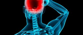
70% of adults have neck pain of varying frequency. At the same time, pain can not only be of a different nature, but also caused by a variety of diseases. Therefore, they are generally not observed in isolation, but are accompanied by other symptoms that help to correctly diagnose and select the optimal treatment tactics.
Types of neck pain
Neck pain can be short-term and limited to minor discomfort, or very acute, significantly interfering with everyday life. More often, people complain that they suddenly feel a shooting sensation, followed by persistent pain. It can be aching, tingling, pulsating, squeezing and of a different nature.
Neck pain is also called cervicalgia.
Discomfort usually increases during movements, for example, when turning or tilting the head, while maintaining a static, especially uncomfortable position for a long time, in particular when working at a computer for a long time. In this regard, a person is forced, often even unconsciously, to turn his whole body in order to spare his neck as much as possible.
So, it is clear that neck pain can be very different. Therefore, they are divided not only into acute (lasting less than 10 days) and chronic, but also into:
- vertebrogenic – occurring against the background of pathological changes in the cervical spine, including damage to the intervertebral discs, compression of the spinal roots and spinal cord;
- nonvertebrogenic - resulting from all other causes, including infectious diseases, inflammatory processes in the muscles, diseases of the thyroid gland, lymph nodes, etc.
Vertebrogenic pain is characterized by irradiation to the occipital region, shoulders, arms, up to the hands and fingers. Often, numbness, impaired sensitivity and mobility are observed, which indicates the development of neurological disorders, i.e., infringement by a particular anatomical structure of the nerve roots or spinal cord. Also in such situations, dizziness, attacks of loss of consciousness and headaches may occur, which indicates compression of the vertebral arteries, for example, by an intervertebral hernia.
It is necessary to make an urgent appointment with a doctor if the neck pain is acute or accompanied by dizziness, a feeling of numbness in the back of the head, or tinnitus.
Primary and secondary prevention
Primary prevention , which is aimed at preventing the development of the disease, comes down to treating concomitant pathologies, reducing the load on the cervical spine, alternating static and dynamic loads on the occipital muscles, as well as proper nutrition.
You should also beware of general and local hypothermia. It is recommended to use an orthopedic pillow for sleeping and wear a Shants collar to relieve the muscles of the cervical spine.
Secondary prevention involves preventing exacerbations of the underlying disease and involves:
- treatment and prevention of underlying pathology (osteochondrosis and other spinal diseases, myofascial syndrome, etc.);
- active lifestyle, avoidance of excessive physical activity, proper nutrition and the use of preventive courses of vitamin therapy;
- the use of preventive courses of physiotherapy, massage, reflexology, physical therapy.
Causes of neck pain
Sometimes it is difficult for patients to independently differentiate a sore throat from a pain in the neck, since it can be so diffuse that the sensations are blurred and it seems that the whole neck hurts. If this is accompanied by a feeling of soreness or scratching in the throat, an increase in body temperature, then this may indicate laryngitis, ARVI, sore throat, pharyngitis and other colds. Also, such symptoms are typical for:
- gastroesophageal reflux;
- Itsenko-Cushing syndrome;
- foreign bodies entering the throat, for example, fish bones;
- hypovitaminosis;
- formation of benign and malignant tumors in the throat.
But still, in most cases, people can differentiate problems with the throat from true pain in the neck, which can be observed both on the anterolateral surfaces, and behind and in front.
Localization of pain is an important diagnostic sign, which allows you to determine the source of pain at the examination stage, and subsequently thoroughly study the changes that have occurred using instrumental diagnostic methods.
Pain in the front of the neck: causes
The occurrence of unpleasant or even painful sensations directly in the area under the chin or diffuse pain along the entire front of the neck may indicate:
- thyroid diseases;
- inflammatory processes in muscles affecting ligaments and nerves;
- formation of cysts and abscesses;
- damage to the lymph nodes;
- angina pectoris.
That is, the list of possible causes is quite extensive and it is impossible to independently determine what exactly caused neck pain. In such situations, it is worth contacting a therapist for an examination. If necessary, the doctor will refer the patient to a specialist of the required profile.
Also, pain in the front of the neck can be accompanied by vertebral artery syndrome and radicular syndrome. In both cases, the cause is the pinching of blood vessels or nerves by a deformed intervertebral disc, edematous tissue, a displaced cervical vertebral body, or other structure. Our clinic has developed an integrated approach to solving this problem - experienced chiropractors, neurologists, rehabilitation specialists, endocrinologists will conduct an examination and prescribe the necessary treatment.
Pain in the back of the neck: causes
Due to the peculiarities of the modern lifestyle, people are very often bothered by pain in the back of the neck. Working at a computer, constant use of gadgets, low levels of physical activity and bad habits, all this causes pathological changes in the structure of the spine, which manifest themselves as neck pain. Therefore, if aching, nagging, sharp pain or shooting in the back of the neck occurs, first of all, consultation with a chiropractor or vertebrologist is required, since in the vast majority of cases this indicates diseases of the cervical spine, namely:
- osteochondrosis is a widespread disease accompanied by degenerative changes in the intervertebral discs, a decrease in their height and a decrease in strength;
- protrusion – a complication of osteochondrosis, in which the intervertebral disc protrudes (usually into the spinal canal, where the spinal cord and nerve roots pass), which creates a risk of pinching nerves and vertebral arteries;
- intervertebral hernia is a consequence of untreated protrusion, in which the internal contents of the intervertebral disc penetrate into the spinal canal, gradually increase and can seriously compress the nerves and spinal cord, and at the last stage of development, separate and “travel” along the spinal canal, causing serious and sometimes irreversible complications including paralysis;
- spondylosis is a complication of osteochondrosis, in which bone protrusions called osteophytes grow along the edges of the vertebral bodies, and the intervertebral discs become critically flattened, which over time leads to the fusion of adjacent vertebrae with each other and limitation of neck mobility;
- myofascial syndrome - a disease in which individual muscles spasm, which leads to attacks of acute pain during physical activity or when pressing on certain points in the neck area;
- vertebral compression fractures - most often occur in older people due to osteoporosis or are the result of a whiplash injury, for example in an accident, and pose a serious threat to human life and health;
- Ankylosing spondylitis is a systemic disease that affects almost all joints of the body and causes severe limitations in mobility due to the fusion of individual vertebrae, forming strong and immobile conglomerates.
Much less commonly, pain in the back of the neck is caused by spinal tuberculosis, osteomyelitis or Reiter's syndrome.
Side neck pain: causes
Most often, the pain in the lateral surfaces of the neck is burning or throbbing, and it can also be tingling in nature. They tend to radiate to the shoulder and ear, sometimes accompanied by the formation of secondary torticollis, as a result of which the head tilts to the affected side. This is typical for:
- pathologies of blood vessels, including atherosclerosis;
- muscle spasms, which can be provoked by heavy strain on the neck, sudden movement or hypothermia;
- formation of malignant tumors in the thyroid gland, pharynx and larynx.
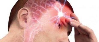
Manifestations of occipital neuralgia
At the beginning of the disease, periodic and then constant pain appears in the occipital part of the head, usually on one side, moving to the upper scalp. There is also pain in the upper neck. The pain is sharp, burning, often unbearable. Pain may appear or intensify when touching the back of the head or the top of the head. With ineffective treatment or its absence, the pain intensifies and becomes constant.
In addition, dizziness, blurred vision, and numbness of the skin on the back of the head and neck may occur. There is sleep disturbance, dizziness, nausea. The disease significantly worsens the patient's quality of life.
Diagnosis of the causes of neck pain
Since in the vast majority of cases, neck pain is caused by the development of pathologies of the cervical spine, if they appear, you should contact a chiropractor, vertebrologist or neurologist. Based on the complaints, examination data and neurological tests, the doctor will be able to determine whether the discomfort in the neck is really due to disorders in the spine or is caused by other diseases.
Nevertheless, it is precisely deviations from the norm in the condition of the spine that, in fact, are one of the reasons for the development of concomitant diseases of the internal organs. After all, changes in the position of the vertebrae, a decrease in the height of the intervertebral discs and other, even minor changes, lead to disruption of the transmission of nerve impulses from the spinal cord through the nerve roots along the nerves to the internal organs.
Since the spinal cord is distinguished by segmental innervation, i.e. each section is responsible for the correct functioning of a specific organ, if disorders occur in the cervical spine, it is the innervation of the ENT organs, vocal cords, thyroid and parathyroid glands, as well as neck muscles that may suffer , shoulders and forearms. Therefore, initially their work is disrupted, and over time, organic changes occur, that is, one or another disease develops, which can provoke pain of varying intensity and nature in the neck.
Therefore, diseases of internal organs are extremely rarely diagnosed and changes in the spine are not detected, and therefore consultation with a chiropractor is mandatory. But if the patient has signs of diseases of the thyroid gland, heart, ENT organs or others, he is additionally advised to obtain consultation from specialized specialists (endocrinologist, cardiologist, otolaryngologist, etc.).
Thus, diagnosing the causes of neck pain is always complex. This may include:
- general and biochemical blood test;
- determination of the level of thyroid hormones in the blood;
- Ultrasound with Dopplerography of the vessels of the neck, thyroid gland, salivary glands;
- X-ray of the cervical spine;
- electroneuromyography;
- CT;
- MRI.
For the purpose of diagnosing pathologies of the spine, MRI, i.e. magnetic resonance imaging, has the greatest information content. The method allows you to study the condition of the intervertebral discs down to the smallest detail, assess the quality of blood flow in the vertebral arteries, and detect signs of compression of the spinal cord or its roots. With its help, diseases are diagnosed at the earliest stages of development, which ensures the earliest possible start of treatment and its high effectiveness.
In our clinic, you can also learn in more detail about the composition of your body and the state of the vascular system, which is involved in the blood supply to internal organs, skeletal muscles, and the brain. Our experienced doctors will explain the data obtained to you in detail. Bioimpendansometry calculates the ratio of fat, muscle, bone and skeletal mass, total fluid in the body, and basal metabolic rate. The intensity of recommended physical activity depends on the state of muscle mass. Metabolic processes, in turn, affect the body's ability to recover. Based on the indicators of active cell mass, one can judge the level of physical activity and nutritional balance. This simple and quick test helps us identify disturbances in the endocrine system and take the necessary measures. In addition, it is also very important for us to know the condition of blood vessels for the prevention of diseases such as heart attacks, hypertension, heart failure, diabetes and much more. Angioscan allows you to determine such important indicators as the biological age of blood vessels, their stiffness, stress index (which indicates heart rate), and blood oxygen saturation. Such screening will be useful for men and women over 30, athletes, those undergoing long-term and severe treatment, as well as everyone who monitors their health.
In this case, body composition analysis gives us information that adipose tissue predominates in the body, and the bone-muscle component is in relative deficiency. These data will help the rehabilitation doctor competently draw up a physical activity plan, taking into account the individual characteristics of the patient.
Causes of occipital neuralgia
The disease develops as a result of compression of a nerve or its branches due to injury, inflammation or some other disease.
Causes of occipital neuralgia can be:
- injuries to the back of the head;
- psycho-emotional stress;
- viral infection;
- head muscle tension;
- osteocondritis of the spine;
- hypothermia;
- diseases of internal organs, such as diabetes and others.
Treatment for neck pain
For each patient, treatment tactics are developed strictly individually based on the diagnosis, the severity of pathological changes, the presence of concomitant diseases and other factors. Even patients with the same disorders may be prescribed different treatments, since even age and level of physical development influence it.
Nevertheless, the treatment of diseases that cause neck pain is always complex and includes both symptomatic and etiotropic therapy. In other words, it is aimed at eliminating both the symptoms of the disease and the causes of its occurrence. The main components of therapy are often:
- drug treatment;
- osteopathy;
- manual therapy;
- massage;
- physiotherapy (phonophoresis, carboxytherapy, ozone therapy, RF current pressure therapy);
- individual sessions with a rehabilitation specialist.
When diagnosing pathologies of the cervical spine, it is recommended to make some adjustments to your lifestyle and habits. Thus, office workers, drivers, seamstresses and representatives of other sedentary professions are recommended to stretch as often as possible and avoid long-term forced body positions, especially ones that require tension in the neck muscles.
It is also important to eat properly so that the body receives all the substances it needs. This will not only slow down the progression of pathology, but will also contribute to faster regeneration processes.
In case of severe pain, the chiropractor may recommend that the patient purchase a neck brace or Chance collar. Wearing it will help relieve the load on the cervical spine and avoid making thoughtless sudden movements, which will lead to a gradual fading of the pain syndrome. In our center you can select and purchase a bandage that suits you
Before visiting a doctor, in order to reduce pain, you can wrap your neck with a woolen scarf or take a painkiller tablet no later than 12 hours before your appointment.
Drug therapy
The nature of the prescribed drugs and their quantity can vary within very wide limits, which directly depends on the diagnosis and the presence of concomitant diseases. Therefore, we list only those groups of drugs that are prescribed to the vast majority of patients:
- NSAIDs are nonsteroidal anti-inflammatory drugs that also have analgesic properties (produced in various dosage forms, including tablets, ointments, injection solutions, etc.);
- corticosteroids are drugs with a powerful anti-inflammatory effect, which are used only in cases of severe inflammatory processes that cannot be eliminated by NSAIDs;
- muscle relaxants - medications that eliminate muscle spasms of various origins, which helps reduce the severity of pain;
- chondroprotectors are drugs that promote the restoration of the cartilage tissue that makes up the intervertebral discs, but are effective only in the initial stages of the development of osteochondrosis. To prevent diseases of the musculoskeletal system, we recommend to our patients the most effective drug Mermaids Marine Collagen;
- B vitamins - are used to improve the conductivity of bioelectric impulses from the spinal cord to the corresponding organs and restore their proper functioning;
- Vitamin D is a remedy responsible for the condition of bone tissue, as well as for higher brain functions, such as memory, memory, attention, and speech.
Patients may also be prescribed antibiotics, hormone replacement therapy, antihypertensive drugs, and others.
For particularly severe pain in the neck, blockades are used. The procedures involve the injection of anesthetic solutions with corticosteroids into the area where the nerves pass in the neck, which allows for very quick pain relief. But, in addition to the fact that they are effective only for pain caused by nerve damage, they require a high level of professionalism from medical workers, since when performing a blockade there is a risk of injuring the nerve, causing infection and causing serious complications.
Manual therapy
Manual therapy is one of the most effective ways to combat neck pain and the causes of its occurrence, but only if carried out correctly. It involves a direct impact on the spine and surrounding soft tissues. Do not confuse manual therapy with therapeutic massage, which only works the muscles of the back and neck.
Techniques and methods of manual therapy allow literally after the first session to achieve a significant improvement in well-being, and ultimately complete remission. This is achieved through:
- restoration of the normal axis of the spine by returning the vertebrae to their anatomically correct position;
- increasing the distance between the vertebral bodies and releasing compressed nerves and blood vessels, which helps eliminate signs of radicular syndrome, improve the functioning of internal organs and especially the brain;
- elimination of spasm of neck muscles and functional blocks, which improves mobility and reduces pain;
- improving blood circulation and tissue nutrition.
We do not have general standards of treatment; we apply an individual approach to each patient. You should trust manual therapy only to a highly qualified specialist. Otherwise, the use of inappropriate techniques can only harm the patient, aggravate the problem, or provoke the development of complications.
Physiotherapy
Physiotherapeutic procedures are used to increase the effectiveness of the therapy. They are indicated exclusively outside the acute period and have an analgesic, anti-inflammatory effect, and also improve microcirculation in soft tissues and help normalize muscle tone.
Typically, courses consisting of 8-15 procedures are prescribed. It can be:
- electrophoresis;
- UHF;
- ultrasound therapy;
- laser therapy;
- traction therapy (spinal traction);
- mud baths, etc.
The number of sessions of physiotherapeutic procedures and the frequency of their implementation are selected by the doctor individually. Some procedures can be combined with each other, while others should be performed strictly at a certain interval.
Exercise therapy
Physical therapy is one of the mandatory components of conservative therapy for neck pain. Since very often they occur against the background of muscle overstrain and the resulting degenerative-dystrophic processes in the intervertebral discs, it is important to pay due attention to exercise therapy and practice daily.
For each patient, a physical therapy program is developed separately by a rehabilitation physician. Using complexes presented on the Internet can be dangerous, since they do not take into account the individual characteristics of the patient, his age, the degree of neglect of the pathological process and the level of physical fitness of the patient. Therefore, to develop a physical therapy program, the help of a specialist is required. Moreover, the first few classes must be carried out under his supervision, so that the patient can understand how to perform each exercise and does not harm himself in the process of independent training. You should exercise at home in a calm environment, avoiding any sudden movements, and if pain occurs, you should consult a doctor.
Thus, neck pain is a widespread phenomenon. They can be caused by the development of various diseases, but most often the cause of discomfort is problems with the spine caused by a sedentary lifestyle. Therefore, you should not hesitate to consult a doctor. Indeed, in the early stages of the development of the disease, it can be easily dealt with using conservative methods. While in advanced cases, neck pain and the causes that cause them can be eliminated exclusively through surgery.
5 1 vote
Article rating
Why do teeth hurt when the trigeminal nerve is inflamed?
How is inflammation of the trigeminal nerve related to teeth? The connection here is the most direct. Firstly, severe pain during attacks “radiates” to the upper or lower jaw (depending on which nerve extension is inflamed), and sometimes to both jaws. This causes severe and unbearable toothache.
Secondly, those nerves that are found in the upper and lower teeth and gums are directly connected to the trigeminal nerve. The second branch of the trigeminal nerve innervates the upper teeth and gums, and the third, respectively, the lower jaw and gums.
The trigeminal nerve can also be damaged during a number of dental manipulations. Unsuccessful tooth extraction or improper filling, surgical intervention in the jaw structure that ended unsuccessfully, and injuries can cause trigeminal neuralgia.
It is very important to distinguish inflammation of the trigeminal nerve from other diseases of the oral cavity, in particular pulpitis - inflammation of the internal dental tissues. A characteristic sign of inflammation of the trigeminal nerve is that, despite severe pain, the sensitivity of the teeth does not increase when they come into contact with cold and hot foods, and hypothermia or heating does not provoke the onset of a new pain attack. Acute tooth sensitivity is a symptom of pulpitis.
When the trigeminal nerve is damaged in the jaw area, acute toothache, pain in the chin, ears and sometimes in the lower lip occurs. The pain may become increasingly intense, and often the patient continues to feel a dull ache after the attack.
A distinctive feature of modern human life is hypokinesia. Reduced muscle loads and prolonged forced positioning of the body and head lead to muscle detraining, weakening of their spring function, and increased load on the intervertebral discs, joints and ligaments. Diseases of the central nervous system of a vertebrogenic nature are currently second only to acute respiratory diseases in prevalence among the population. Spinal diseases occur in 60-90% of the population over 50 years of age. The number of young people suffering from this pathology is constantly increasing [9, 25, 30].
A normally functioning spine is a natural kinematic chain consisting of seven individual vertebrae articulated with each other by intervertebral discs, paired true joints and ligaments. The occurrence of ocular symptoms is facilitated by the anatomical features of the cervical region. This is the most mobile and most vulnerable part of the spine; normally it describes a regular arch with a forward convexity - lordosis. The two upper (rotational) vertebrae are significantly different from the remaining (flexor) vertebrae.
The first cervical vertebra - the atlas - is connected to the occipital bone by two joints and supports the skull. The second cervical vertebra has an odontoid process directed upward, with the atlas “strung” on it. Thanks to this unpaired joint between the atlas and the tooth of the second vertebra, the head turns to the sides. The remaining 5 cervical vertebrae are connected to each other by discs and processes. The muscular system also plays a huge role in ensuring the motor function of this part of the spine [10, 11, 20, 24, 44, 47, 48].
All cervical vertebrae (except VII) have openings in the transverse processes through which the vertebral arteries and accompanying veins and nerves pass. A.a. vertebralis
, being the first branch of the subclavian arteries, form the main artery. It constitutes the vertebrobasilar system that supplies the occipital lobes with the central link of the visual analyzer, the brain stem, the nuclei and conductors of the oculomotor, trochlear, abducens nerves and the system of the posterior longitudinal fasciculus, the fibers of which ensure the joint and simultaneous work of the oculomotor muscles [35, 37, 43 ].
The sympathetic nerve trunk extends from the base of the skull to the neck of the first rib, located behind the carotid arteries on the deep muscles of the neck. These are three sympathetic ganglia: the upper cervical, middle cervical and lower - cervicothoracic (stellate ganglion). The internal carotid nerve n. departs from the superior cervical ganglion . caroticus internus
, the lower cervical ganglion gives off the vertebral nerve
n.
vertebralis . These nerves accompany the arteries of the same name, forming plexuses around them, ascending into the cranial cavity, where they anastomose with each other and give branches to the vessels of the brain. From the branches of the sympathetic plexus of the carotid artery and the upper cervical ganglion, vasoconstrictors go to the glands of the head, including the lacrimal gland [8, 13, 24, 33, 36, 46].
Pupil dilatation center (Budge center, or centrum ciliospinale
) is located in the anterior horns of the spinal cord at the level from the 8th cervical to the 2nd thoracic segment.
Innervation of the muscle that dilates the pupil is provided by n.
sympaticus .
The anterior roots of these parts of the spinal cord pass through all three cervical sympathetic ganglia. In the ganglion cervicale,
the preganglionic neuron ends and a new postganglionic neuron begins, which reaches the plexus of the internal carotid artery [1, 3, 21, 42].
Sympathetic fibers enter the orbit in the form of the periarterial plexus of the ophthalmic artery, give off a root to the ciliary ganglion and, as part of short ciliary nerves, reach the eyeball. The cervical sympathetic nerves are connected to the smooth muscles of the orbit, narrow the lumen of the ophthalmic arterioles and dilate the pupil. The peripheral section of the parasympathetic pupillomotor pathway extends from the nucleus n. ocolomotorius
to the sphincter [1, 19, 41, 43].
The openings in the transverse processes of the cervical vertebrae are quite narrow, the neurovascular bundle is closely adjacent to the bone walls. Therefore, when you rotate your head, strain your neck muscles, or organic changes in the spine, neurovascular formations can be subject to trauma and deformation. The slightest displacement of the cervical vertebra causes a reflex spasm of the vertebral artery, which subsequently causes compression. Local (cervical sympathetic nodes and fibers) and general (by the type of reflex involvement of hypothalamic autonomic centers) autonomic formations are involved in the process. Pathological afferentation that occurs in the area of the vertebral nerve, through a large number of anastomoses, can spread to the periarterial plexuses of the carotid and ophthalmic arteries, which leads to pain in the orbit, eyeballs, and decreased blood circulation in the organ of vision [8, 10, 13, 22, 32 ].
The relationship between changes in the cervical region and some eye pathologies has been established during experimental and clinical studies. In 1866, a printed work by Prof. E.V. Adamyuk “On the influence of the sympathetic nerve on intraocular pressure”, in which glaucoma was considered as a secretory neurosis caused by irritation of the cervical sympathetic nodes [1].
When studying the regulatory role of the central nervous system and individual nerves affecting the eye, Soviet scientists obtained a lot of valuable data. In the 20s of the last century V.P. Roshchin confirmed the role of the sympathetic nervous system in the origin of glaucoma [17]. Further experimental studies conducted by L.S. Levina (1941), A.B. Desyatnikov (1953), I.F. Sinitsin (1971) showed that irritation or extirpation of the superior cervical sympathetic ganglion leads to fluctuations in intraocular pressure [2]. I.B. Kaplan and A.T. Gudneva (1980) in 32% of patients with intraocular pressure of 27-33 mm Hg, by treating the cervical spine, they achieved its reduction to normal levels [30]. A.M. Grinstein (1957), G.N. Grigoriev (1969), as well as D.I. Antonov (1970) pointed out the sometimes occurring attacks of unilateral loss of the visual field or part of it, combined with spasm of the retinal artery when the cervical sympathetic structures are damaged [2].
In the middle of the last century, works devoted to the clinic of visual disturbances in occlusions of the intra- and extracranial sections of the vertebral arteries also appeared in foreign literature: Synonds, Mackenzie (1957), Hoyt (1959), Minoretal (1959), Kearns (1960). Visual disorders have been associated with ischemia of the occipital lobe cortex, especially their poles and areas adjacent to the calcarine sulcus [2, 40].
The dependence of visual disturbances on the condition of the spine is indicated by changes in the fundus of the eye that appear after exposure to the muscles and ligaments of the cervical spine. During the Bertschi stretch or during neck extension, some subjects observed changes in the fundus: dilation of large veins, narrowing of arterial trunks, or dilation of arteries with unchanged vein diameter [30].
G.P. Smolyakova et al. (1988) found that when the superior cervical sympathetic nodes are irritated, changes characteristic of age-related macular degeneration appear. E.S. Abdulaeva (2002) recorded changes in blood flow in the siphon of the internal carotid and vertebral arteries in patients with non-exudative forms of macular degeneration [14].
Swiss ophthalmologist Johann Friedrich Horner in 1869 described anterior cervical sympathetic syndrome as changes in the face on the side where there is insufficiency of sympathetic innervation caused by a pathological process in the neck or chest. In France and Italy, the name of physiologist Claude Bernard is also associated with this pathology [1, 39].
Bernard-Horner syndrome can be congenital, acquired, or iatrogenic. The reasons for the development of such a symptom complex are numerous: surgical manipulations on the sympathetic nodes, tumor and inflammatory processes of the cervical spine, spinal cord, upper segments of the chest, syringomyelia, multiple sclerosis. Cases of the appearance of Horner's syndrome in individuals weakened after acute infections, herpetic diseases, and scleroderma have been described; with improvement in general condition, anterior cervical sympathetic syndrome completely disappeared in these patients [1, 21, 27].
The Bernard-Horner triad is a set of symptoms: ptosis, miosis, enophthalmos. Miosis is formed due to the shutdown of m. dilatator pupillae
, a narrow palpebral fissure is associated with sympathetic ptosis, switching off the Müller muscle, and enophthalmos is a consequence of switching off the Landström orbital muscle. Of the three main symptoms, miosis and ptosis are always the most prominent [1].
Horner's syndrome is often complicated by a number of other painful conditions: hypotension, discoloration of the iris, cataracts, lacrimation, dilation of the conjunctival and retinal vessels. Sometimes all this is accompanied by sweating and hyperemia of the skin of the corresponding half of the face [1, 22]. In children, the lack of sympathetic innervation prevents the pigmentation of melanocytes located in the stroma of the iris, so Horner's syndrome can lead to heterochromia [1, 38].
With anterior cervical sympathetic syndrome, pupillary reactions to light and reactions to near installation proceed in the same way as normal, or may even be somewhat more vibrant, but to a lesser extent. The return to the initial state occurs more slowly [1, 18, 42].
The painful pupillary reaction, in contrast to the light reaction and the reaction to near installation, in Horner's syndrome decreases significantly, becoming noticeable only with strong painful stimulation [1, 8].
Drooping of the upper eyelid may also be associated with damage to n. oculomotorius
. In such cases, insufficient innervation of the sphincter causes pupil dilation. In real clinical work, such ptosis is quite easy to distinguish. In addition to mydriasis, when the oculomotor nerve is damaged, ptosis is more pronounced and can completely close the entire eye [1, 40, 42].
Posterior cervical sympathetic syndrome was described by the French neurologist J. Barre in 1925, later supplemented in more detail by the Chinese physician J. Lieo in 1928. Synonyms of the Bare-Lieu symptom complex are: vertebral artery syndrome, cervical migraine, spinal nerve syndrome, sympathetic vertebral nerve neuritis [5, 11, 15, 32].
The disease occurs if compression of the vertebral artery or its sympathetic plexus occurs due to osteochondrosis, trauma or tumor in the neck. But the first place in the pathogenesis of the development of vertebral artery syndrome belongs to the growth of the uncinate processes in the direction of the intervertebral foramina and the narrowing of the latter. Another reason is lesions and changes in the vertebral arteries themselves: occlusion, deformation, anomalies of origin, location and entry. Posterior cervical sympathetic syndrome leads to various manifestations of vertebrobasilar insufficiency. There are functional and organic stages [2, 9, 15, 16, 45].
The functional stage of vertebral artery syndrome is characterized by three groups of symptoms: headache, cochleovestibular and visual disorders [5, 19, 21, 23].
In most patients, pain, starting in the cervico-occipital region, spreads anteriorly to the mastoid-temporal, parietal and frontal regions with irradiation into the eye, accompanied by vestibular (dizziness, loss of balance), auditory (hearing loss and tinnitus), autonomic (sweating , feeling of heat, hot flashes, chills) disorders. Laryngeal and pharyngeal symptoms (dysphonia, dysphagia, pharyngeal paresthesia) may occur [5, 7, 12, 25, 27].
Painful sensations appear as a result of irritation of pain sensitivity receptors in the fibrous and muscular tissues of the spine, in the capsule of the intervertebral joints, ligaments, and tendons of the neck muscles. Palpation often reveals pain in the area of paravertebral points, spinous processes, and along the vessels of the cervical region. When palpating the muscles, one can find Cornelius and Müller's nodes, pain and tension in the anterior scalene muscle. Pain can spread to the shoulder girdle and chest, and radiate to the heart [5, 8, 10, 12, 44].
Visual disturbances were included by J. Barre (1926) in the description of posterior cervical sympathetic syndrome as an obligate symptom. Upon careful questioning, they are detected in almost all patients with vertebrobasilar insufficiency of any etiology. Ophthalmological symptoms of vertebral artery syndrome are described somewhat differently by different authors. In this work we have attempted to summarize the descriptions found in various literature sources. Ocular symptoms can be transient or persistent [5, 7, 25, 27].
Transient include photopsia. Patients complain of the appearance before their eyes of “black flies”, “soot”, “sparks”, “lightning”, multi-colored and golden dots that seem to flicker and oscillate. Photopsia in cases of cerebral circulatory disorders are point-like, their occurrence is not related to the light source, they continue even with the eyes closed [2, 12, 28, 29].
More complex photopsia may also appear, for example in the form of “white shiny rings”, often shiny zigzag lines, in the form of a moving stream of multi-colored (red, yellow and green) cubes. In all cases, visual disturbances are short-term and last only a few seconds [2, 11, 13, 21].
Blurred vision of objects, a feeling of “veil” or “fog” before the eyes is noted, according to the literature, in approximately half of patients with vertebral artery syndrome. With posterior cervical sympathetic syndrome, visual disturbances appearing along with pain in the cervical-occipital region are described: flickering scotomas, fog and darkening before the eyes, photophobia, lacrimation, a feeling of sand behind the eyelids, nystagmus, changes in pressure in the retinal arteries or the tone of the fundus vessels. Sometimes there is redness of the conjunctiva and the development of neurodystrophic keratitis [1, 5, 29, 31].
Such patients turn to an ophthalmologist with complaints of discomfort in the eyes, especially when working at close range for a long time. There may be a feeling of “fullness” in the eye socket, “twisting” of the eyeballs, which intensifies with strained vision, while reading, or eye movements. Patients with posterior cervical sympathetic syndrome may tell their ophthalmologist that they are experiencing double vision. The examination reveals a decrease in acuity and a narrowing of the visual field [6, 13, 30, 31, 34].
In patients with diseases of the cervical spine, blurred vision and blurred images are often aggravated by a sudden change in head position. At the same time, the general condition may worsen: headache, dizziness, and increased blood pressure may appear or worsen [4, 6, 7, 11, 35, 45, 48].
Sometimes eye symptoms and dizziness are provoked by throwing the head back, and in some cases they appear after excessive physical or emotional stress. They also often occur against a background of fatigue: during prolonged walking over rough terrain, physical work, situations involving holding one's breath, or develop after fainting. At the end of such an attack, vision can be completely restored [6, 13, 30, 31, 34].
With vertebrobasilar insufficiency, short-term disturbances of higher cortical functions are possible, such as various types of visual agnosia with impaired optical-spatial perception. They appear as a result of ischemia in the distal cortical branches of the posterior cerebral artery [2, 4, 7].
The appearance of bilaterally intermittent symptoms: darkening of the eyes, narrowing of visual fields, dysarthria, dysphagia or dizziness - confirms the presence of vertebrobasilar insufficiency. A person may lose vision for a few minutes or report short-term loss of peripheral vision due to dizziness. In such a situation, vomiting and instability when walking may occur [4, 6, 10, 12, 22].
The organic stage is characterized by transient and persistent circulatory disorders in the brain and spinal cord. In this case, visual symptoms include: decreased vision, double vision, ptosis. The eyeballs may be completely motionless or make floating movements, strabismus and anisocoria appear. In hemorrhagic stroke, unilateral mydriasis is determined on the side of the hemorrhage, along with the abduction of the eyes towards the lesion - “the patient looks at the paralyzed limbs” [4, 6, 8-10, 12, 22].
Vertebrogenic pathological conditions are a problem that is addressed by neurologists, orthopedists, neurosurgeons, etc. The close connection between the pathology of the brain and the eye is due to the anatomical and functional unity of their blood circulation. Therefore, situations are possible when ocular symptoms become one of the earliest cerebral manifestations of a disease of the cervical spine. Ocular changes may occur long before the development of other symptoms of vertebrobasilar insufficiency [4, 7, 13].
In cases where patients do not associate the pathology of the organ of vision with an uncomfortable posture during sleep or at the workplace, turning and tilting the head, it seems to them that it arises as if “by itself”, and it is to this that the ophthalmologist directs his main attention, while the root cause of the disease may go unnoticed [4, 7, 11, 13, 26, 42].
In conclusion, the authors of the article express the hope that the presented material will assist practicing ophthalmologists in making the correct diagnosis, prescribing adequate examination and treatment.