Hormones influence the mechanisms of emotion formation and the action of various neurochemicals, and, as a result, are involved in the formation of stable habits. The author of the book “Happiness Hormones,” Professor Emeritus of the University of California Loretta Graziano Breuning, suggests reconsidering our behavior patterns and learning to trigger the action of serotonin, dopamine, endorphin and oxytocin. T&P publishes a chapter from a book about how our brains self-tune, responding to experience and forming neural connections accordingly.
Loretta Graziano Breuning
founder of the Inner Mammal Institute, professor emeritus at the University of California, author of several books, blogs “Your Neurochemical Self” on PsychologyToday.com
Rearranging neural pathways
"Happiness Hormones"
Each person is born with many neurons, but very few connections between them. These connections are built as we interact with the world around us and ultimately make us who we are. But sometimes you have a desire to slightly modify these formed connections. It would seem that this should be easy, because we developed them without much effort on our part in our youth. However, the formation of new neural pathways in adulthood is unexpectedly difficult. Old connections are so effective that giving them up makes you feel like your survival is at risk. Any new nerve chains are very fragile compared to the old ones. When you can understand how difficult it is to create new neural pathways in the human brain, you will be more pleased with your persistence in this direction than berate yourself for the slow progress in their formation.
Human memory capacity
The average human brain has about 100 billion neurons (or nerve cells) and neuroglia (or glial cells), which serve to support and protect the neurons.
Happy shopping on iHerb!
Each neuron can be connected to 10,000 other neurons, transmitting signals to each other through 1,000 trillion synaptic connections, which by some estimates is equivalent to a computer with a processor running at 1 trillion bits per second.
Estimates of the human brain's memory capacity vary widely, from 1 to 1000 terabytes (by comparison, the 19 million volumes in the US Library of Congress represent about 10 terabytes of data).
Five Ways Your Brain Self-Tunes
We mammals are able to create neural connections throughout our lives, unlike species with stable connections. These connections are created as the world around us affects our senses, which send corresponding electrical impulses to the brain. These impulses pave neural pathways along which other impulses will run faster and easier in the future. Each individual's brain is wired for an individual experience. Below are five ways that experience physically changes your brain.
Life experiences insulate young neurons
Over time, a constantly working neuron becomes covered with a sheath of a special substance called myelin. This substance significantly increases the efficiency of the neuron as a conductor of electrical impulses. This can be compared to the fact that insulated wires can withstand a significantly greater load than bare wires. Neurons coated with myelin work without the extra effort that slow, “open” neurons have. Neurons with a myelin sheath appear white rather than gray, which is why we divide our brain matter into “white” and “gray.”
Most of the covering of neurons with myelin is complete by the age of two years, as the child's body learns to move, see and hear. When a mammal is born, its brain must form a mental model of the world around it, which will provide it with opportunities for survival. Therefore, myelin production in a child is maximum at birth, and by the age of seven it decreases slightly. By this time you no longer need to relearn the truth that fire burns and gravity can make you fall.
If you think that myelin is “wasted” on strengthening neural connections in young people, then you should understand that nature designed it this way for sound evolutionary reasons. For most of human history, people had children as soon as they reached puberty. Our ancestors needed to have time to solve the most urgent tasks that ensured the survival of their descendants. As adults, they used new neural connections more than reconfigured old ones.
When a person reaches puberty, the formation of myelin in his body is activated again. This happens because the mammal has to re-tune its brain to find the best mate. Often during the mating season, animals migrate to new groups. Therefore, they have to get used to new places in search of food, as well as to new tribesmen. In search of a marriage partner, people are also often forced to move to new tribes or clans and learn new customs and culture. The increase in myelin production during puberty contributes to all this. Natural selection has designed the brain in such a way that it is during this period that it changes the mental model of the world around it.
Everything you do purposefully and consistently during your “myelin prime” years creates powerful and extensive neural pathways in your brain. This is why human genius so often manifests itself in childhood. That is why little skiers fly past you so dashingly on mountain slopes that you cannot master, no matter how hard you try. This is why learning foreign languages becomes so difficult once adolescence ends. As an adult, you can memorize foreign words, but most often you cannot quickly select them to express your thoughts. This happens because your verbal memory is concentrated in thin, unmyelinated neurons. Your powerful myelinated neural connections are busy with high mental activity, so new electrical impulses have difficulty finding free neurons. […]
Fluctuations in the body's activity in the myelination of neurons can help you understand why people have certain problems at different times in life. […] Remember that the human brain does not mature automatically. Therefore, it is often said that the brain of adolescents is not yet fully formed. The brain “myelinates” all our life experiences. So if there are episodes in a teenager’s life when he receives an undeserved reward, he will firmly remember that the reward can be received without effort. Some parents forgive their teens' bad behavior by saying that "their brains haven't fully matured yet." That is why it is very important to purposefully control the life experience that they absorb. Allowing a teenager to avoid responsibility for his actions can create a mind that will expect the possibility of avoiding such responsibility in the future. […]
Life experience increases synapse efficiency
A synapse is the point of contact (small gap) between two neurons. An electrical impulse in our brain can only travel if it reaches the end of a neuron with enough force to “jump” across the gap to the next neuron. These barriers help us filter truly important incoming information from irrelevant so-called “noise.” The passage of an electrical impulse through synaptic gaps is a very complex natural mechanism. It can be imagined in such a way that a whole flotilla of boats accumulates at the tip of one neuron, which transports the neural “spark” to special receiving docks available at a nearby neuron. Each time the boats cope better with transportation. This is why the experiences we have increase the chances of electrical signals being transmitted between neurons. The human brain has more than 100 trillion synaptic connections. And our life experience plays an important role in conducting nerve impulses through them in a way that is consistent with the interests of survival.
At a conscious level, you cannot decide which synaptic connections you want to develop. They are formed in two main ways:
1) Gradually, through repeated repetition.
2) Simultaneously, under the influence of strong emotions.
[…] Synaptic connections are built based on repetition or emotions you have experienced in the past. Your mind exists because your neurons have formed connections that reflect successful and unsuccessful experiences. Some episodes from this experience were “downloaded” into your brain thanks to “joy molecules” or “stress molecules”, others were fixed in it through constant repetition. When the model of the world around you corresponds to the information contained in your synaptic connections, electrical impulses run through them easily, and it seems to you that you are quite aware of the events happening around you.
Neural chains are formed only due to active neurons
Those neurons that are not actively used by the brain begin to gradually weaken as early as a two-year-old child. Oddly enough, this contributes to the development of his intelligence. Reducing the number of active neurons allows the baby not to glance distractedly at everything around him, which is typical for a newborn, but to rely on the neural pathways that have already been formed. A two-year-old child is already able to independently concentrate on what gave him pleasant sensations in the past, such as a familiar face or a bottle of his favorite food. He may be wary of things that have caused him negative emotions in the past, such as a pugnacious playmate or a closed door. The young brain relies on its limited life experience to meet needs and avoid potential threats.
No matter how the neural connections in the brain are built, you experience them as “truth”
From the age of two to seven years, the process of optimizing the child’s brain continues. This forces him to correlate new experiences with old ones, instead of accumulating new experiences in some separate block. Tightly intertwined neural connections and neural pathways form the basis of our intelligence. We create them by branching out old neural trunks instead of creating new ones. Thus, by the age of seven, we usually clearly see what we have already seen once, and hear what we have already heard once.
You might think this is bad. However, consider the value of it all. Imagine lying to a six-year-old child. He believes you because his brain eagerly absorbs everything that is offered to him. Now suppose you deceive an eight-year-old child. He is already questioning your words because he compares incoming information with what he already has, and does not simply “swallow” new information. At the age of eight, it is already more difficult for a child to form new neural connections, which pushes him to use existing ones. Relying on old neural circuits allows him to recognize lies. This was of great importance from a survival point of view at a time when parents died young and children had to learn to take care of themselves from an early age. During our young years, we form certain neural connections, allowing others to gradually fade away. Some of them disappear like the wind blows away autumn leaves. This helps make a person's thought process more efficient and focused. Of course, with age you gain more and more knowledge. However, this new information is concentrated in areas of the brain where active electrical pathways already exist. For example, if our ancestors were born into hunting tribes, they quickly gained hunter experience, and if they were born into farming tribes, they quickly gained agricultural experience. Thus, the brain was tuned to survival in the world in which they actually existed. […]
New synaptic connections are formed between the neurons you actively use
Each neuron can have many synapses because it has many processes or dendrites. New processes in neurons are formed when it is actively stimulated by electrical impulses. As dendrites grow toward points of electrical activity, they can get so close that an electrical impulse from other neurons can bridge the distance between them. In this way, new synaptic connections are born. When this happens, at the level of consciousness you get a connection between two ideas, for example.
You cannot feel your own synaptic connections, but you can easily see it in others. A person who loves dogs looks at the entire world around him through the prism of this affection. A person who is passionate about modern technologies associates everything in the world with them. A lover of politics evaluates the surrounding reality politically, and a religiously convinced person evaluates it from the standpoint of religion. One person sees the world positively, another - negatively. No matter how the neural connections in the brain are built, you do not feel them as numerous processes similar to the tentacles of an octopus. You experience these connections as “truth.”
Emotion receptors develop or atrophy
In order for an electrical impulse to cross the synaptic cleft, the dendrite on one side must eject chemical molecules that are picked up by special receptors on the other neuron. Each of the neurochemicals produced by our brain has a complex structure that is perceived by only one specific receptor. It fits the receptor like a key to a lock. When you are overwhelmed by emotions, more neurochemicals are produced than the receptor can catch and process. You feel dazed and disoriented until your brain creates more receptors. This is how you adapt to the fact that “something is happening around you.”
When a neuron's receptor is inactive for an extended period of time, it disappears, leaving room for other receptors that you may need to appear. Flexibility in nature means that receptors on neurons must either be used or they can be lost. “Happy hormones” are constantly present in the brain, searching for “their” receptors. This is how you “find out” the reason for your positive feelings. The neuron “fires” because the appropriate hormone molecules open the lock on its receptor. And then, based on this neuron, a whole neural circuit is created that tells you where to expect joy in the future.
Images: © iStock.
Classifications of synapses
The main elements of an electrical synapse (ephaps): a - connexon in a closed state; b — connecton in the open state; c — connexon embedded in the membrane; d — connexin monomer, e — plasma membrane; f — intercellular space; g - a gap of 2-4 nanometers in the electrical synapse; h - hydrophilic connexon channel
According to the mechanism of nerve impulse transmission
- chemical is a place of close contact between two nerve cells, for the transmission of a nerve impulse through which the source cell releases into the intercellular space a special substance, a neurotransmitter, the presence of which in the synaptic cleft excites or inhibits the receiver cell.
- electrical (ephaps) - a place of closer contact between a pair of cells, where their membranes are connected using special protein formations - connexons (each connexon consists of six protein subunits). The distance between cell membranes in the electrical synapse is 3.5 nm (the usual intercellular distance is 20 nm). Since the resistance of the extracellular fluid is low (in this case), impulses pass through the synapse without delay. Electrical synapses are usually excitatory.
- mixed synapses - the presynaptic action potential produces a current that depolarizes the postsynaptic membrane of a typical chemical synapse where the pre- and postsynaptic membranes are not tightly adjacent to each other. Thus, at these synapses, chemical transmission serves as a necessary reinforcing mechanism.
The most common are chemical synapses. Electrical synapses are less common in the mammalian nervous system than chemical ones.
- Mental performance of children
- History of the study
- Presynaptic part
- Convolutional neural network topology
- Structure
- Clinical significance
- Functions
- Synapse structure
- Types of synapses
- Mechanism of synaptic transmission
- Clinical significance
- History of discovery
- What are synapses
By location and affiliation with structures
- peripheral neuromuscular
- neurosecretory (axo-vasal)
- receptor-neuronal
- axo-dendritic
- with dendritesaxo-spinous - with dendritic spines, outgrowths on dendrites;
, including
axo-somatic
- with the bodies of neurons;
- between axons;
- between dendrites;
Various locations of chemical synapses
By neurotransmitter
- aminergic, containing biogenic amines (for example, serotonin, dopamine
including adrenergic containing adrenaline or norepinephrine;);
- cholinergic, containing acetylcholine;
- purinergic, containing purines;
- peptidergic, containing peptides.
At the same time, only one transmitter is not always produced at the synapse. Usually the main pick is released along with another one that plays the role of a modulator.
According to the sign of synapse action
- stimulating
- brake
.
If the former contribute to the occurrence of excitation in the postsynaptic cell (in them, as a result of the arrival of an impulse, depolarization of the membrane occurs, which can cause an action potential under certain conditions), then the latter, on the contrary, stop or prevent its occurrence and prevent further propagation of the impulse. Typically inhibitory are glycinergic (mediator - glycine) and GABAergic synapses (mediator - gamma-aminobutyric acid).
Inhibitory synapses are of two types: 1) a synapse, in the presynaptic endings of which a transmitter is released, hyperpolarizing the postsynaptic membrane and causing the appearance of an inhibitory postsynaptic potential; 2) axo-axonal synapse, providing presynaptic inhibition.
Some synapses have a postsynaptic seal
- electron-dense zone consisting of proteins.
Based on its presence or absence, synapses are distinguished as asymmetric
and
symmetric
. It is known that all glutamatergic synapses are asymmetrical, while GABAergic synapses are symmetrical.
In cases where several synaptic extensions come into contact with the postsynaptic membrane, multiple synapses
.
Spine apparatuses are special forms of synapses
, in which short single or multiple protrusions of the postsynaptic membrane of the dendrite contact the synaptic extension. Spine apparatuses significantly increase the number of synaptic contacts on a neuron and, consequently, the amount of information processed. Non-spine synapses are called sessile synapses. For example, all GABAergic synapses are sessile.
History of the study
The hypothesis about the existence of electrical synapses was first put forward at the beginning of the 20th century by Camillo Golgi and Joseph Gerlach. However, after the discovery of chemical synapses, the existence of electrical synapses was considered unreliable, and until the middle of the 20th century, the prevailing opinion was that the transmission of action potentials between neurons occurs exclusively through chemical synapses. However, in 1959, David Potter and Edwin Verszpan convincingly demonstrated the existence of electrical synapses using the example of the giant axon and the axon of a motor neuron in the ventral nerve cord of crayfish.
Structure
In the region of each gap junction there are many special channels that cross the membranes of both cells. The diameter of such channels is from 1.2 to 2 nm, so ions and medium-sized molecules can pass through them from one cell to another, due to which the cytoplasms of two neighboring cells are connected. Therefore, when the membrane potential of one of the cells changes, ions from it can move to the neighboring cell due to their positive charge. Gap junction channels consist of two hemichannels, which in vertebrates are called connexons (each hemichannel belongs to one of the cells connected by a synapse). Each connexon is formed by six protein subunits - . Connexin is 7.5 nm long and contains four regions. The connexins in connexon are the same or may differ slightly. A special case of an electrical synapse is (English autapse), in the formation of which the axon and dendrite of the same neuron take part.
Clinical significance
Genetic defects in connexins are often the cause of heart defects, since electrical synapses play a critical role in synchronizing the electrical and contractile activity of the heart. Disturbances in the functioning of connexins in Schwann cells lead to functional pathology of axons, which underlies Charcot-Marie-Tooth disease. With this disease, progressive motor and sensory movements are observed, in addition, the speed of action potential transmission along axons is reduced. The formation of gap junctions between smooth muscle cells of the uterine wall is influenced by estrogens, which stimulate their formation during pregnancy. Defects of gap junctions in the uterus and a decrease in their number often lead to premature birth.
Mechanism of synaptic transmission
Stage 4
Ca ions cause the formation of a special protein complex, which includes the vesicle and structures located directly near the presynaptic membrane.
They are connected to each other by so-called exocytosis proteins.
Some proteins are located on vesicles (synapsin, synaptotagmin, synaptobrevin), and some are located on the presynaptic membrane (syntaxin, synapse-associated protein). This complex is called the secretosome.
The release of the contents of the vesicle into the gap is facilitated by the protein synaptoporin, which forms a channel through which the transmitter is released.
Mediator quantum is the number of molecules contained in one vesicle.
For 1 PD, 100 quanta of AX are released.
Stage 10
The end plate potential (EPP) occurs at the postsynaptic membrane. It is analogous to a local response (LO).
There is no action potential at the postsynaptic membrane! It forms on the adjacent muscle fiber membrane.
The fate of the mediator:
- receptor binding
- destruction of enzymes (acetylcholinesterase),
- reabsorption into the presynaptic membrane,
- leaching from the gap and phagocytosis.
Events at the synapse:
- The AP arrives at the axon terminal;
- It depolarizes the presynaptic membrane;
- Ca2+ enters the terminal, which leads to the release of ACh;
- The transmitter ACh is released into the synaptic cleft;
- It diffuses into the cleft and binds to receptors on the postsynaptic membrane;
- The permeability of the postsynaptic membrane for Na+ ions changes;
- Na+ ions penetrate the postsynaptic membrane and reduce its charge - an end plate potential (EPP) occurs.
On the postsynaptic membrane itself, AP cannot occur, since there are no voltage-gated channels here; they are chemo-dependent!
- EPPs are summed up and reach the CUD on the adjacent section of the muscle fiber, which leads to the emergence of AP and its propagation along the muscle fiber (about 5 m/s).
Having reached a threshold value, that is, CUD, the PPP excites the neighboring (extrasynaptic) muscle fiber membrane due to local circular currents.
History of discovery
- In 1897, Sherrington formulated the idea of synapses.
- Golgi and Ramón y Cajal received the Nobel Prize in 1906 for their research into the nervous system, including synaptic transmission.
- In 1921, the Austrian scientist O. Loewi established the chemical nature of the transmission of excitation through synapses and the role of acetylcholine in it. Received the Nobel Prize in 1936 together with H. Dale.
- In 1933, the Soviet scientist A.V. Kibyakov established the role of adrenaline in synaptic transmission.
- 1970 - B. Katz (Great Britain), U. v. Euler (Sweden) and J. Axelrod (USA) received the Nobel Prize for their discovery of the role of norepinephrine in synaptic transmission.
Synapse structure
A synapse consists of three main elements:
- The presynaptic membrane that covers the expanded nerve ending, which is a neurosecretory apparatus. The presynaptic part contains vesicles and mitochondria that provide mediator synthesis. Mediators are deposited in granules (bubbles).
- The postsynaptic membrane is the thickened part of the cell membrane with which the presynaptic membrane is in contact. It has ion channels and is capable of generating action potentials. In addition, it contains special protein structures - receptors that perceive the action of mediators.
- The synaptic cleft is a space between the presynaptic and postsynaptic membranes, filled with a fluid similar in composition to blood plasma.
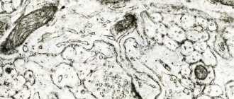
This is how the structure of the synapse looks in the figure.
Mental performance of children
A person’s performance directly determines his age, when all values increase simultaneously with the development and physical growth of children.
The accuracy and speed of mental actions vary unevenly with age, depending on other factors that determine the development and physical growth of the body. Students of any age who have health problems are characterized by a low level of performance relative to the healthy children around them.
In healthy first-graders with a reduced readiness of the body for the constant learning process, according to some indicators, the ability to act is low, which complicates the fight against problems that arise during the learning process.
The rate of onset of weakness is determined by the initial state of the children's sensory nervous system, the work tempo and the volume of load. At the same time, children are prone to overwork during prolonged immobility and when the actions performed are uninteresting to the child. After a break, performance becomes the same or becomes higher than before, and it is better to make the rest not passive, but active, switching to a different activity.
Changes in performance in children
The first part of the educational process for ordinary primary school children is accompanied by excellent performance, but by the end of the 3rd lesson they experience a decrease in concentration:
- They look out the window.
- They do not listen attentively to the teacher's words.
- Change the position of their body.
- They start talking.
- They get up from their place.
The work capacity values are particularly high among high school students studying in the 2nd shift
It is especially important to pay attention to the fact that the time for preparing for classes before the start of the educational activity in the classroom is quite short and does not guarantee complete relief from harmful changes in the central nervous system. Mental activity quickly depletes in the first hours of lessons, which is clearly reflected in negative behavior
Therefore, qualitative changes in performance are observed in students of the junior block in lessons 1 - 3, and in the middle-senior blocks in lessons 4 - 5. In turn, lesson 6 takes place in conditions of particularly reduced ability to act. At the same time, the duration of classes for grades 2–11 is 45 minutes, which weakens the condition of the children. Therefore, it is recommended to periodically change the type of work, and take an active break in the middle of the lesson.
Convolutional neural network topology
- determine the problem to be solved by a neural network (classification, prediction, modification);
- determine the limitations in the problem being solved (speed, accuracy of the answer);
- determine input (type: image, sound, size: 100×100, 30×30, format: RGB, grayscale) and output data (number of classes).
Figure 2 — Convolutional neural network topology
Convolutional layer
Figure 3 — Organization of connections between the maps of the convolutional layer and the previous one Figure 4 — Convolution operation and obtaining the values of the convolutional map (valid) Convolution operation and obtaining the values of the convolutional map. The kernel is shifted, the new map is the same size as the previous one (same) Figure 5 - Three types of convolution of the original matrix
Subsampling layer
Figure 6 - Formation of a new map of the subsample layer based on the previous map of the convolutional layer. Subsampling operation (Max Pooling)
Selecting an activation function
- continuous;
- monotonically increasing;
- differentiable.
Activation function hyperbolic tangent
- symmetric activation functions, such as hyperbolic tangent, provide faster convergence than the standard logistic function;
- the function has a continuous first derivative;
- a function has a simple derivative that can be calculated in terms of its value, resulting in computational savings.
ReLU activation function
- its derivative is equal to either one or zero, and therefore gradients cannot grow or decay, because multiplying one by the error delta we get the error delta, but if we used another function, for example, a hyperbolic tangent, then the error delta could either decrease, or increase, or remain the same, that is, the derivative of the hyperbolic tangent returns a number with a different sign and magnitude, which can greatly influence the attenuation or expansion of the gradient. Moreover, using this function leads to thinning of the weights;
- calculating sigmoid and hyperbolic tangent requires computationally intensive operations such as exponentiation, while ReLU can be implemented using a simple threshold transformation of the activation matrix at zero;
- cuts off unnecessary parts in the channel with a negative output.
Presynaptic part
Presynaptic part
contains synaptic vesicles with a neurotransmitter, cytoskeletal elements and mitochondria. Voltage-gated Ca2+ channels are built into the presynaptic membrane. When AP enters the terminal extension, the membrane is depolarized, Ca2+ channels open, Ca2+ ions enter the terminal, triggering the process of fusion of the synaptic vesicle membrane and the presynaptic membrane in the active zones, i.e. secretion (exocytosis) of the neurotransmitter (Fig. 6–6, positions 2–4).
Role of Ca2+.
Fusion of synaptic vesicles with the presynaptic membrane occurs with an increase in the concentration of Ca2+ in the cytosol of the nerve terminal. The synaptic vesicle protein synaptotagmin binds to Ca2+ and thereby takes part in the regulation of exocytosis (including through reorganization of the perimembrane cytoskeleton).
Synaptic vesicles.
Neurotransmitter molecules accumulate in the nerve terminal, located inside synaptic vesicles along with ATP and some cations. Each vesicle contains several thousand neurotransmitter molecules, which constitutes a neurotransmitter quantum.
Neurotransmitter
synthesis .
The enzymes necessary for the formation of neurotransmitters are synthesized in the perikaryon and transported to the synaptic terminal along the axons (Fig. 6–4).
The types
of bubbles
are small (diameter about 50 nm) and large (diameter 100–200 nm). Small synaptic vesicles contain “classical” transmitters (see below). Large vesicles contain neuropeptides.
Secretion.
When the AP reaches the nerve terminal, synaptic vesicles merge with the presynaptic membrane, which leads to the release of neurotransmitter quanta into the synaptic cleft. A small amount of neurotransmitter quanta is constantly (spontaneously) secreted into the synaptic cleft.
Recognition.
Preceding the fusion of synaptic vesicles and plasmalemma, the process of recognition of the presynaptic membrane by the synaptic vesicle occurs through the interaction of membrane proteins (synaptobrevin, SNAP-25, syntaxin and others).
The influence
of toxins
.
Syntaxin, SNAP-25 and synaptobrevin are targets of botulinum toxin, which irreversibly inhibits the fusion of synaptic vesicles with the presynaptic membrane.
The target of tetanus toxin is synaptobrevin. Active zones
(Figure 6-3).
Secretion of the neurotransmitter occurs in specialized areas of the presynaptic nerve ending - active zones - areas of thickening of the presynaptic membrane. The active zone consists of a “dense stripe” on the presynaptic membrane and synaptic vesicles grouped around it, voltage-dependent calcium channels, special exocytosis proteins and cytoskeletal elements. The number of active zones in the neuromuscular synapse reaches 30–40, in interneuron synapses - about a dozen. Active zones are located opposite clusters of receptors in the postsynaptic membrane, which reduces the delay in signal transmission associated with neurotransmitter diffusion in the synaptic cleft
.
Rice. 6-3. Active zones of the neuromuscular junction
located opposite the postsynaptic folds - areas of accumulation of cholinergic receptors. The presynaptic membrane on the left is split into two sheets.
Life cycle of synaptic vesicles
(Fig. 6–4). Synaptic vesicles are formed in the neuron body in the endoplasmic reticulum and Golgi complex (1) and enter nerve endings with axonal transport (2).
Rice. 6-4. Formation, transport and exocytosis of synaptic vesicles
.
At the nerve ending, small
synaptic vesicles
are filled with mediator (3) through active transport and move to the presynaptic membrane (4).
The release of the mediator (5) can be carried out through exocytosis with complete (“classical” mechanism) or incomplete (“kiss and run” mechanism) fusion. The first type of exocytosis is accompanied by the insertion of the vesicle membrane into the presynaptic membrane, emptying of the vesicle, and then through endocytosis, clathrin-coated vesicles are formed (6), which then pass through the endosome stage (7) and are again filled with mediator (3). The second type of exocytosis is characterized by the formation of a temporary pore connecting the vesicle cavity with the synaptic cleft. After the release of the transmitter, the vesicle is not integrated into the presynaptic membrane, but buds from it (8) and is refilled with the transmitter (3). Large synaptic vesicles
are filled with transmitter in the cell body (9), their exocytosis occurs in other parts of the presynaptic membrane, and endocytosis of empty vesicles is absent (10).
- Back
- Forward
Clinical significance
Genetic defects in connexins are often the cause of heart defects, since electrical synapses play a critical role in synchronizing the electrical and contractile activity of the heart. Disturbances in the functioning of connexins in Schwann cells lead to functional pathology of axons, which underlies Charcot-Marie-Tooth disease. With this disease, progressive motor and sensory movements are observed, in addition, the speed of action potential transmission along axons is reduced. The formation of gap junctions between smooth muscle cells of the uterine wall is influenced by estrogens, which stimulate their formation during pregnancy. Defects of gap junctions in the uterus and a decrease in their number often lead to premature birth.