Synonyms: hydrocephalic syndrome, hypertensive-hydrocephalic syndrome, HGS. ICD-10 code: G91
Medical editor: Druzhinkina V.Yu., neurologist. November, 2020.
Hydrocephalic syndrome is a pathological condition characterized by excess production of cerebrospinal fluid (CSF), which accumulates under the meninges and in the ventricles of the brain.
The clinical picture of hypertensive-hydrocephalic syndrome can be explained by two concepts:
- hypertension (increased intracranial pressure);
- hydrocephalus (increased amount of cerebrospinal fluid in the brain).
Important! Hydrocephalic syndrome as a term is accepted only in the former USSR (in the CIS) and in modern Russia. Western doctors attribute hydrocephalic syndrome to a manifestation of brain pathologies.
The syndrome is often diagnosed by pediatric neurologists, and, as a rule, without reason. HHS is a fairly rare pathology, and in 97% of cases the diagnosis of hydrocephalic syndrome has no right to exist.
Causes
There are congenital causes of hydrocephalic syndrome (HHS in newborns) and acquired ones.
Congenital causes of hydrocephalic syndrome
- course of pregnancy and childbirth with complications;
- hypoxic (intrauterine hypoxia and intrauterine growth retardation, their manifestation in fetal bradycardia) and ischemic (injuries during childbirth) brain damage;
- premature birth (up to 36 weeks);
- head injuries during childbirth (subarachnoid hemorrhages);
- intrauterine infections (toxoplasmosis, influenza, cytomegalovirus infection and others);
- congenital abnormalities of brain development;
- delayed birth (at 42 weeks and later);
- long water-free period (more than 12 hours);
- chronic maternal diseases (diabetes mellitus, chronic pyelonephritis, arterial hypertension and others).
Acquired causes of hydrocephalic syndrome:
- tumors, abscesses, hematomas, parasitic cysts of the brain;
- foreign bodies in the cranial cavity;
- fractures of the skull bones with the introduction of fragments into the brain;
- causeless intracranial hypertension;
- infectious diseases (malaria, tick-borne encephalitis);
- post-stroke disorders;
- metabolic disorders.
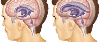
Photo: cerebrospinal fluid pressure on the brain with hydrocephalus.
Types of hydrocephalus
There are several variants of the disease depending on the cause of occurrence and localization. By origin, there is a congenital form, which develops as a result of problems suffered by the baby in the prenatal state, and an acquired form, provoked after birth by external factors (injuries, infections).
According to the type of location, they are distinguished:
- external hydrocephalus, when in a child cerebrospinal fluid accumulates mainly in the space under the membranes of the brain;
- internal form, in which cerebrospinal fluid flows into the ventricles of the brain, causing them to expand;
- mixed or general hydrocephalus, which is characterized by a combination of external and internal forms of the disease.
There are also open (communicating) and closed (obstructive) types of pathology: in the first case, the communication between the ventricles and the subarachnoid space is not broken, in the second it is absent. According to the speed of flow, acute, subacute and chronic dropsy of the brain are distinguished.
Symptoms of hydrocephalic syndrome
Signs of hydrocephalic syndrome in newborns
Parents note that the child does not latch on well, constantly cries for no apparent reason, and sometimes moans.
The child has:
- decreased muscle tone (“seal feet” and “heel feet”);
- weak innate reflexes (swallowing, grasping);
- tremor (trembling of the limbs, chin) and convulsions may occur;
- spitting up like a fountain;
- strabismus;
- when examined by a doctor, positive Graefe's sign is observed (the eyeball is directed downward - a white stripe is visible between the pupil and the upper eyelid) and the rising sun sign (the iris is almost half hidden behind the lower eyelid)
- opening of the sutures of the skull (in particular the sagittal);
- bulging, tension of the fontanelles;
- in dynamics there is an increased increase in head circumference (by 1 cm every month), which is significantly ahead of age standards;
- head thrown back, which is especially noticeable during sleep;
- cyanosis, pale skin;
- increased respiratory movements;
- When examining the fundus, swelling of the optic discs is observed.
Clinical manifestations of HGS in children
Symptoms of hypertensive-hydrocephalic syndrome in older children usually develop after an infection or brain injury.
A characteristic symptom is headache, which often occurs in the early morning hours, nausea and vomiting, which follow it and do not bring relief. The pain is dull, aching or bursting; localized in the area of the temples, forehead and brow ridges.
Children complain that it is difficult for them to raise their eyes and lower their heads. Dizziness often occurs (young children define it as “swinging on a swing” or “instability of objects”).
During an attack of pain, pale skin, weakness and lethargy are noted. Bright light and loud sound cause irritation and increase unpleasant sensations.
Walking “on tiptoes” due to increased tone of the leg muscles, squint, drowsiness and slow thinking, poor memory and attentiveness are also typical. Such children do not eat well because they feel nauseous.
Hydrocephalic syndrome in adults
HGS in adults develops as a result of traumatic brain injuries, tumors, neuroinfections and after a stroke.
Signs of hydrocephalic syndrome are similar to those in older children:
- visual impairment (double vision, strabismus);
- severe headaches;
- nausea and vomiting, especially severe in the morning;
- disturbance of consciousness up to coma and convulsions.
Hydrocephalus in children: just the facts
Hydrocephalus is a disease that occurs due to excessive production and excessive accumulation of cerebrospinal fluid in the intrathecal spaces and/or ventricles of the brain (in the subarachnoid or subdural space), leading to thinning (atrophy) of the medulla and divergence of the skull bones [1–4].
ICD-10 provides the following codes for various types of hydrocephalus: G91 (G91.0 - communicating hydrocephalus, G91.1 - obstructive hydrocephalus, G91.2 - normal pressure hydrocephalus, G91.3 - post-traumatic hydrocephalus unspecified, G91.8 - other types hydrocephalus, G91.9 - hydrocephalus unspecified).
Classification of hydrocephalus
There are several options for classifying hydrocephalus. First of all, a distinction is made between congenital and acquired hydrocephalus.
Congenital hydrocephalus is present at the time of birth of the child and is the result of inflammatory diseases of the central nervous system (CNS), malformations of the brain, as well as intracranial (birth) trauma, hemorrhages in the brain.
Acquired hydrocephalus develops after birth due to etiologically and pathogenetically diverse pathologies (neuroinfections, tumor processes, vascular diseases, traumatic brain injuries, etc.).
The disease can be active or passive. In the first case, there is an increase in intracranial pressure in the presence of dilatation of the ventricles and subarachnoid spaces. With passive hydrocephalus, it is assumed that the described changes are observed in the absence of intracranial hypertension.
There are open and closed forms of hydrocephalus. Open (communicating) hydrocephalus is diagnosed in the absence of disturbances in the connection of the ventricular system of the brain with the subarachnoid space, and closed (occlusive or non-communicating) - when this communication is disrupted.
External and internal forms of hydrocephalus are determined by the localization of the accumulation of cerebrospinal fluid (CSF); External hydrocephalus is an accumulation of CSF in the intrathecal spaces of the brain, internal hydrocephalus is an accumulation of CSF in the ventricles of the brain.
Pathogenetic classification of hydrocephalus: 1) hypersecretory forms (excessive formation of CSF); 2) aresorptive forms (impaired CSF absorption processes); 3) occlusive forms (the presence of an obstacle to the movement of CSF from the source of its formation to the areas of its resorption).
Depending on the existing level of occlusion of the cerebrospinal fluid pathways, five variants of hydrocephalus are considered: 1) occlusion of one or both foramina of Monroe; 2) blockade of the cavity of the third ventricle; 3) stenosis or occlusion of the Sylvian aqueduct; 4) occlusion or non-opening of the openings of the fourth ventricle; 5) violation of the patency of the subarachnoid spaces.
Depending on the achieved level of control over intracranial hypertension, it is customary to distinguish between compensated, subcompensated or decompensated hydrocephalus.
A separate variant of the disease is normal pressure hydrocephalus (“symptomatic”). With it, an increase in intracranial pressure is initially observed (a consequence of excessive accumulation of CSF in the ventricles of the brain) - with or without ventriculomegaly, which gradually decreases, but remains moderately elevated (150–200 mmH2O). There are two types of normal pressure hydrocephalus - idiopathic (cause unknown) and secondary (consequence of subarachnoid hemorrhage, trauma, tumor and/or infection of the central nervous system, complication of neurosurgical intervention, etc.) [1–4].
S. Oi (2010) proposed a “multi-category classification of hydrocephalus”, reflecting modern ideas about this disease (Table) [5, 6].
Etiology and pathogenesis of hydrocephalus
The most important etiological factor of hydrocephalus is intra- and perinatal pathology of the central nervous system. Pregnancy pathology and oxygen starvation of cerebral tissue are considered as etiological causes of congenital hydrocephalus; intrapartum factors leading to hypoxic-ischemic and/or traumatic brain damage; gestational immaturity of brain structures most susceptible to the described damage.
The development of hydrocephalus is caused by inflammatory diseases of the brain and its membranes, as well as intrauterine and neuroinfections, congenital malformations of the central nervous system, vascular pathology, tumors of the brain and spinal cord, traumatic injuries (including intracranial birth injuries), genetic factors, etc.
Cases of hydrocephalus associated with cerebral dysgenesis, brucellosis, mumps and other infections, diffuse villous hyperplasia of the choroid plexus, vascular anomalies, intracranial hemorrhages, etc. have been described.
Post-traumatic hydrocephalus (progressive excess accumulation of CSF in the cerebrospinal fluid spaces and brain matter due to traumatic brain injury) is caused by impaired circulation and resorption of CSF [1–4, 7].
According to C. Schrander-Stumple and JP Fryns (1998), hereditary X-linked (congenital) hydrocephalus occurs in 4% of all registered cases of the disease (according to other data in 5–15% of observations), up to 40% of cases of hydrocephalus are etiogenetically caused [8 ]. Hydrocephalus occurs in Dandy–Walker syndrome, Arnold–Chiari syndrome, etc. At least 43 mutations/loci associated with hereditary forms of hydrocephalus (in humans and laboratory animals) have been described; Under experimental conditions, 9 genes associated with this cerebral pathology were found, while in humans there is only one [1–4].
The progression of hydrocephalus is accompanied by structural and morphological changes in the brain of varying severity: 1) thinning of the cortex and white matter (up to its complete elimination); 2) atrophy of the choroid plexus; 3) atrophy/subatrophy of the basal ganglia, brainstem, cerebellum; 4) severe disorders of capillary blood flow; 5) thickening and/or fusion of the meninges; 6) excessive growth (hypertrophy) of glial tissue. In severe cases, the formation of hydroanencephaly is possible, when there is only ependyma and a thin layer of the pia mater.
The most intensively functioning and highly vascularized periventricular region suffers the most from hypoxia. The consequence of atrophy of the periventricular white matter of the brain is passive expansion of the ventricular system with the formation of ventriculomegaly.
With post-traumatic hydrocephalus, pathological processes in the brain are morphologically characterized by dilation of the ventricular system, periventricular edema and obliteration of the subarachnoid fissures. Obliteration of the CSF flow paths is determined by the following pathogenetic factors: subarachnoid hemorrhage, intracranial hematomas, focal and/or diffuse brain damage, cicatricial adhesions and atrophic processes (including after extensive craniotomy and resection trepanations), meningoencephalitis and ventriculitis. The time frame for the development of post-traumatic hydrocephalus (normotensive, hypertensive or occlusive) usually varies from 1 month to 1 year [1–4, 7].
Clinical syndromology of hydrocephalus
The main symptoms of hydrocephalus (congenital and acquired) are determined by two groups of factors: 1) the causes of the disease; 2) directly hydrocephalic syndrome.
The first group mainly includes focal manifestations (usually in the form of spastic paresis of the ascending type in the lower and/or upper extremities). The severity of group 2 symptoms depends on the form, stage and degree of progression of hydrocephalus. In the congenital form of the disease, signs of hydrocephalus may be present both at birth and appear later - by the age of 3–6 months.
More often, the first sign of the disease is a disproportionately rapid increase in head circumference. To assess it in children, special tables (centile) are used.
In children of the first year of life, there may be a predominance of the “cerebral” parts of the skull over the “facial” (as a result - a forced position with the head thrown back), increased venous pattern and congestion of the saphenous veins of the head, tension of the greater and other fontanelles, divergence of the skull bones, Graefe’s symptom . These symptoms are accompanied by a lag in psychomotor development (of varying severity), and less often in physical development. Optic nipple atrophy is a severe complication of untreated or treatment-resistant progressive hydrocephalus.
Cephalgic syndrome is more typical of acquired hydrocephalus in children over 1 year of age (when the fontanelles and cranial sutures are already closed).
Clinical manifestations of post-traumatic hydrocephalus are characterized by neurological and mental disorders caused by primary brain injury. In part, they are a reflection not so much of hydrocephalus itself, but rather a consequence of a traumatic brain injury (TBI) or premorbid pathology.
A. P. Konovalov et al. (1999) distinguish three variants of post-traumatic hydrocephalus: 1) against the background of resolved or mild residual symptoms of severe TBI, with dominance in the clinic of any specific symptom complex; 2) against the background of slowly resolving severe symptoms of severe TBI with the addition of intellectual-mnestic and ataxic syndromes; 3) against the background of a vegetative state (which prevents exit from it). Hypertensive and occlusive post-traumatic hydrocephalus (excluding normotensive hydrocephalus) is characterized by headache, vomiting, and dizziness. All patients exhibit psychopathological symptoms (intellectual-mnestic disorders, euphoria or lethargy, aspontaneity, akinetic mutism, etc.); as well as gait disturbances and ataxia (with a characteristic “sticking of the feet to the floor”), dysfunction of the pelvic organs.
With normal pressure hydrocephalus, there are usually no classic symptoms characteristic of congenital or acquired hydrocephalus, but since ventriculomegaly has a negative effect on adjacent areas of cortical tissue, the disease has its own characteristics (classic triad: gait disturbances, urinary incontinence, decreased intelligence of varying degrees of severity) [ 1–4].
Diagnostics and examination methods
Establishing a diagnosis of hydrocephalus (in addition to physical data) is based on neuroimaging data (neurosonography - with an open fontanel, computed tomography and magnetic resonance imaging of the brain - CT and MRI), which are considered in conjunction with the symptoms of the disease described above. These neuroimaging methods have replaced the previously used skull radiography. It is resorted to only in rare cases; Survey radiography of the skull allows one to indirectly judge secondary changes in the bones of the skull (in the absence of CT and MRI).
It is especially important to monitor the adequate functioning of shunts installed during neurosurgical intervention. At the slightest suspicion of shunt failure, the patient should be referred to a neurosurgeon.
Isotope cisternography with the introduction of a radioactive isotope (during a lumbar puncture) and subsequent observation of the removal of CSF from the brain makes it possible to establish the diagnosis of normal pressure hydrocephalus.
Diaphanoscopy (transillumination of the skull) has now been practically abandoned. Lumbar puncture is a traditional research method that allows you to assess blood pressure and analyze the CSF. An increase in echo pulsation up to 70–80% during an ultrasound examination of the brain is not a diagnostic sign of hydrocephalus.
The study of auditory evoked potentials often reveals their disturbances, which indicates a special sensitivity of the brain stem to intracranial hypertension.
An ophthalmological examination of the fundus allows one to identify changes characteristic of hydrocephalus (congestion in the fundus, atrophic processes, signs of inflammation, as well as hemorrhage, changes in the tone and caliber of blood vessels, etc.), which makes it possible to assess the course of the pathological process.
If a congenital infection (intrauterine infection) is suspected, serological and virological studies are justified.
Abroad, a method of non-invasive monitoring of intracranial pressure through the large fontanel in children of the first year of life, based on the principle of applanation, is used. The measurement is made using a special device (fontanometer), and the study itself is called “phonogram” or “fontanometry” [1–4, 7].
Differential diagnosis
Hydrocephalus is differentiated from the following main pathological conditions: subdural intracranial hemorrhage, megalencephaly (primary), hydroanencephaly, meningitis, brain tumors, familial (constitutional) macrocephaly, vitamin D-deficient rickets, rickets of premature infants, etc.
Less common are other types of pathology from which it is necessary to differentiate hydrocephalus: achondroplasia, Soto syndrome (cerebral gigantism), numerous so-called “neurocutaneous” syndromes, a group of leukodystrophies (Alexander’s disease, Canavan’s disease, globoid and methochromatic forms of leukodystrophies), gangliosidosis, mucopolysaccharidosis (Tay’s disease –Sachs and Sandhoff), urine disease with the smell of maple syrup, etc. [1–4, 7].
Treatment
Therapeutic measures for progressive hydrocephalus are divided into surgical and therapeutic (drug and non-drug).
If there are signs of an ongoing inflammatory process in the central nervous system, children are prescribed appropriate therapy (antibacterial agents, specific drugs, as well as glucocorticosteroids and human intravenous immunoglobulins - according to indications).
Progressive forms of hydrocephalus (occlusive), as a more serious variant of the pathology, require timely neurosurgical intervention (shunt surgery). The purpose of surgery is to create an adequate outflow of CSF using special shunts (drainage systems made of synthetic materials). There are shunts that divert CSF from the ventricles of the brain to various loci of the body (ventriculoperitoneal, lumboperitoneal, ventriculoatrial). Thus, some shunts transport excess accumulation of cerebrospinal fluid into the peritoneal cavity, others - into the right ventricle of the heart. Ventriculoperitoneal shunting is the main method of neurosurgical treatment of hydrocephalus in the Russian Federation and abroad.
There is a method of surgical treatment called “endoscopic third ventriculostomy” (perforation of the lower part of the third ventricle of the brain to eliminate the blockage of CSF flow and increase its outflow); another name for the method is “endoscopic ventriculocisternostomy of the bottom of the third ventricle.” The purpose of the described operation is to create pathways for the outflow of CSF from the third ventricle into the cisterns of the brain, through which the resorption of cerebrospinal fluid occurs.
Other types of endoscopic treatment of hydrocephalus in various clinical situations are aqueductoplasty, ventriculocystocysternostomy, septostomy, endoscopic removal of an intraventricular tumor, as well as endoscopic installation of a shunt system.
Among the neurosurgical procedures in infants, ventricular puncture (removal of CSF from the ventricles of the brain through the fontanelle) can be used. This external drainage procedure is used extremely rarely, as it can be accompanied by a large number of complications (infection, etc.).
There are isolated reports of the use of intradural spinal endoscopy in the treatment of hydrocephalus and associated conditions in children.
In the acute occurrence of intracranial hypertension, drugs with a diuretic effect are used. These include: furosemide (intramuscular) - sometimes in combination with a solution of magnesium sulfate, glycerol (per os), mannitol (intravenous drip).
If long-term treatment of hydrocephalus is necessary, acetazolamide (Diacarb) is first prescribed, a diuretic from the group of carbonic anhydrase inhibitors (an enzyme that catalyzes the reversible reaction of carbon dioxide hydration and subsequent dissociation of carbonic acid). The action of acetazolamide is associated with the suppression of carbonic anhydrase in the ventricular plexuses of the brain and with a decrease in CSF production (hypolyquor effect). The side effects of acetazolamide (acidosis and shortness of breath) are systematically corrected by the administration of sodium bicarbonate (0.5 g 3 times a day).
Patients with hydrocephalus under the age of 3 years need to be prescribed vitamin D and calcium supplements. In addition to calcium, supplementation of potassium and magnesium preparations (Asparkam, Panangin) is absolutely necessary during acetazolamide therapy.
Symptomatic treatment for hydrocephalus is determined by individual indications and usually includes: massage, physical therapy, various types of physiotherapy, biofeedback method, as well as stimulating therapy (nootropic, metabolic, vascular drugs, etc.), etc. Anticonvulsants for children with hydrocephalus prescribed if there are appropriate indications (symptomatic epilepsy, etc.).
Neurodietological measures should be aimed at maintaining nutritional status, adequate fluid intake, supplementation of vitamins and minerals, and, if necessary, nutritional support (clinical nutrition: enteral and/or parenteral) [2–4].
Prevention
Since in a significant number of cases hydrocephalus can be diagnosed during fetal development, sonographic examination of the fetus is recommended (starting from the 17th week of gestation). In some cases, MRI examination is indicated, although P. Peruzzi et al. (2010) indicate that it does not have pronounced diagnostic advantages compared to ultrasound methods [9].
In order to prevent hydrocephalus in children, timely detection and treatment of intrauterine infections in their mothers is necessary. It is necessary to fully implement the prevention of childhood injuries and neuroinfections. To prevent myelomeningocele, preventive administration of folic acid supplements is indicated [1–4, 7].
Forecast
Hydrocephalus is a cerebral pathology associated with significant neurological deficits, potential disability and decreased quality of life. There is a risk of irreversible changes in the nervous system and a decrease in intelligence (even mental retardation).
With congenital and acquired hydrocephalus, the prognosis is determined by the early onset and adequacy of treatment (pharmacological or neurosurgical).
A serious complication of hydrocephalus can be epilepsy, induced both by hydrocephalus itself (structural and morphological changes in brain structures) and by the installation of a drainage shunt [1–4, 7].
According to M. Mataro et al. (2001), the average IQ values in patients with hydrocephalus are reduced in a number of verbal and nonverbal functions. Among patients who received adequate therapy, normal levels of intellectual function are observed in 40–65% of cases [10].
Literature
- Kliegman RM, Stanton BF, St Geme III JW et al., eds. Nelson textbook of pediatrics. 20 th ed. Philadelphia. Elsevier. 2016. 3474.
- Shamansurov Sh. Sh., Studenikin V. M. Congenital and acquired hydrocephalus. Ch. 11. In the book: Neurology of early childhood. Tashkent: O'Qituvchi, 2010. 156–164.
- Studenikin V. M., Shamansurov Sh. Sh. Congenital hydrocephalus. Ch. 9. In the book: Neonatal neurology. M.: Medforum, 2014. 120–135.
- Studenikin V.M., Shelkovsky V.I., Kuzenkova L.M. Hydrocephalus and hydrocephalic syndrome in children // Doctor.ru. 2006; 5:2–5.
- Oi S. A proposal on “Multi-categorical Hydrocephalus Classification”: McHc. Critical review in 72,576 patterns of hydrocephalus // J. Hydrocephalus. 2010; 2 (2): 1–21.
- Oi S. Hydrocephalus research update: controversies in definition and classification of hydrocephalus // Neurol. Med. Chir. (Tokyo). 2010; 50:859–869.
- Kandasamy J., Jenkinson MD, Mallucci CL Contemporary management and recent advances in pediatric hydrocephalus // BMJ. 2011; 343:d4191.
- Schrander-Stumple C., Fryns JP Congenital hydrocephalus. Nosology and guidelines for clinical approach and genetic counseling // Eur. J. Pediatr. 1998; 157(5):355–362.
- Peruzzi P., Corbitt RJ, Raffel C. Magnetic resonance imaging versus ultrasonography for the in utero evaluation of central nervous system anomalies // J. Neurosurg. Pediatr. 2010; 6 (4): 340–345.
- Mataro M., Junque C., Poca MA et al. Neuropsychological findings in congenital and acquired childhood hydrocephalus // Neuropsychol. Rev. 2001; 11 (4): 169–178.
V. M. Studenikin, Doctor of Medical Sciences, Professor, Academician of the Russian Academy of Economics
LLC NPSMC "Dream Clinic", Moscow
Contact Information
Hydrocephalus in children: just the facts / V. M. Studenikin
For citation: Attending physician No. 4/2018; Issue page numbers: 66-69
Tags: cephalalgia, brain, cerebrospinal fluid, intracranial hypertension
Diagnostics
Diagnosis of hydrocephalic syndrome is difficult. Not all instrumental methods help establish a diagnosis in 100% of cases. In infants, regular measurement of head circumference, checking reflexes, dynamics of body weight gain, and neurological examinations are important.
Also in the definition of GGS is used:
- electroencephalography;
- lumbar puncture of the spinal cord to take cerebrospinal fluid to measure its pressure (the most reliable method);
- neurosonography (ultrasound examination of the anatomical structures of the brain, in particular the size of the ventricles);
- assessment of fundus vessels (swelling, congestion or vasospasm, hemorrhage);
- computed tomography () and nuclear magnetic resonance (NMR).
Treatment of hydrocephalic syndrome
Treatment of hydrocephalic syndrome is carried out by neurologists and neurosurgeons with the involvement of ophthalmologists. Patients with HGS need to be observed and treated in a specialized neurological center.
Treatment in newborns
Children require outpatient or surgical treatment, depending on the cause. Main therapeutic measures:
- prescribing a diuretic drug - diacarb (reduces the production of cerebrospinal fluid and removes excess fluid from the body, primarily from the cranial cavity);
- it is possible to take nootropics - some experts claim that these drugs improve blood supply to the brain (Encephabol, Actovegin), but there are no convincing large-scale studies on this topic;
- for hyperexcitability, sedatives are indicated;
- massage.
Treatment for infants is quite long, taking several months.
Treatment of HGS in older children and adults
In adults and older children, therapy also depends on the cause of hydrocephalic syndrome. If it is the result of a neuroinfection, then appropriate antiviral or antibacterial therapy is carried out.
In case of traumatic brain injuries and tumors, surgical intervention is indicated. Diuretics and nootropics are also used.
How to treat dropsy?
This brain pathology is practically incurable. Existing medications are designed only to slow down the process.
The most effective treatment method today is surgical, when the patient undergoes bypass surgery or endoscopic surgery.
Massage plays an important role in the treatment of hydrocephalus. Dropsy helps to increase muscle tone, and stroking and rubbing movements have a relaxing effect, helping to restore motor functions.
Another type of treatment, manual therapy, is used as an addition to the use of medications. Manual influence is aimed at activating the human body’s own reserves. The use of manual therapy for secondary dropsy is especially effective.
Recommendations for the use of treatment methods are given by a neurologist or neurosurgeon.
Complications and prognosis
Complications of hypertensive-hydrocephalic syndrome are possible at any age:
- delayed mental and physical development;
- blindness;
- deafness;
- coma;
- paralysis;
- epilepsy;
- urinary and fecal incontinence;
- death.
The prognosis is most favorable for hydrocephalic syndrome in infants. This is because they experience transient increases in blood pressure and cerebrospinal fluid, which stabilize with age.
In older children and adults, the prognosis is relatively favorable and depends on the cause of HGS, the timeliness and adequacy of treatment.
Sources:
- Bitterlich L.R. On the issue of differential diagnosis, classification and differentiated treatment of hydrocephalus and hirdocephalic syndromes in children. - International Neurological Journal, No. 1 (79), 2021.
- Order on approval of the standard of specialized medical care for children with hydrocephalus. — Ministry of Health of the Russian Federation, 2013.
- Federal clinical guidelines for the provision of medical care to children with consequences of perinatal damage to the central nervous system with hydrocephalic and hypertension syndromes. — approved Union of Pediatricians of Russia February 14, 2015
Therapeutic measures
It is important to remember that hydrocephalus syndrome in an infant can only be treated using methods approved by a doctor. Self-medication is strictly prohibited due to the possibility of causing harm to the child’s body.
Therapy is complex. Treatment measures are prescribed individually, taking into account the severity, characteristics of the clinical picture, and the patient’s age. The main method of therapy is medication.
For treatment purposes, the following groups of drugs are used:
- diuretics;
- sedatives;
- nootropic;
- antibiotics;
- venotonics;
- antitumor.
Numerous positive reviews characterize activities that allow a child to develop certain skills and abilities, develop his abilities, and adapt him to social life. A good effect is characterized by therapeutic massage and moderate physical activity. HGS can also be treated with numerous physiotherapeutic procedures.