Vegetative-vascular dystonia is one of the most frequently diagnosed diagnoses in neurological practice, especially at the outpatient stage of treatment. However, I personally am increasingly asked the question of what it is if there is information on the Internet that “there is no such diagnosis in the West.” That is why I decided to touch upon the topic of vegetative dystonia on this site and consider this issue within the framework of the symptom of dizziness. And also at the same time tell why there is no point in using information on the Internet without having the proper medical data for its full processing and understanding.
Causes of VSD: the essence of the diagnosis
In the human body, as I prefer to say during my appointment, there are three sections of the nervous system: central (brain and spinal cord), peripheral (spinal cord roots and individual nerves) and autonomic (including the vagus nerve - emerging from the structures of the brain, autonomic nerve plexuses, including the solar-celiac plexus and carotid glomerulus, as well as individual vegetative cells). So, the functions of the central nervous system are the mind, information processing, most of the sensory organs due to the cranial nerves (vision, hearing, taste, etc.), integrative tasks, as well as the development of a command to act. The peripheral nervous system provides control over the human body, facilitates the collection of information (temperature sensitivity, tactile sensitivity, etc.), and also ensures the execution of commands for action. The autonomic nervous system is practically independent of our consciousness and controls autonomic functions: vascular tone, including the brain, blood pressure, heart rate, respiratory rate, production of exocrine and internal secretion glands (including sweat glands). All this provides a double and sometimes even triple system of control of important functions of the human body. At the same time, in the autonomic nervous system there are inhibitory and excitatory influences in balance, which, by changing the strength of these influences, regulate the functions described above.
However, the autonomic nervous system is in close relationship with other departments, in particular, through the work of the vagus nerve, which is a cranial nerve. The vagus nerve nuclei, in turn, interact to some extent with the limbic structures of the brain responsible for emotions and perceptions of stress. In general, the human body is a single and very complex structure. Therefore, an imbalance in one part of the nervous system can lead to disturbances in another part. Thus, the presence of chronic or simply severe acute stress, congenital structural features of the autonomic nervous system, the influence of debilitating diseases, hormonal changes (during puberty, as well as during menopause in women), as well as some other factors lead to an imbalance of inhibitory and excitatory influences and the development of various types of disorders, including changes in cerebral vascular tone. This is exactly how, due to this imbalance and changes in vascular tone, a symptom complex of such a condition as vegetative-vascular dystonia (VSD, vegetative dystonia, vegetative dystonia syndrome, these are all synonyms) is formed.
So, VSD is not a disease in the full sense of the word, “in the West” there are no separate codings according to the ICD 10 system for various forms of VSD, this is what led to the appearance of information about the absence of such a diagnosis in European and American practice. At the same time, psychological and psychotherapeutic assistance is very well developed there, and since one of the most important factors in the development of vegetative dystonia is stress, very often this problem is dealt with there more often not by doctors, but by psychologists, and, less often, by psychotherapists (and the diagnosis implies somatoform autonomic dysfunction, which is encrypted in class F and cannot be made as the main diagnosis by a therapist or neurologist).
In domestic realities, going to a psychotherapist for a patient means the presence of a certain “stigma”, and psychological support is unsatisfactorily developed and is quite expensive. Therefore, neurologists often deal with these problems. And they are forced to make a diagnosis due to the bureaucratic aspects of paying for visits, diagnostic procedures, etc. In this case, the generally accepted ICD 10 system is used, which still contains the term other disorders of the autonomic nervous system - code G90.8. And it is the code G90.8 that is the ICD 10 code for autonomic dysfunction. At the same time, according to the data I have, in the near future, with the adoption of ICD 11, a whole list of codes for this classification system will appear, responsible for coding this diagnosis. And there is a whole section 8D** responsible for coding these diagnoses.
About vegetative-vascular dystonia
Vegetovascular dystonia (in other words, autonomic dysfunction) is a disorder of the autonomic nervous system.
The autonomic nervous system is part of the nervous system that regulates many processes in the internal organs. It is sometimes called the autonomic nervous system because... regulation occurs autonomously, i.e. without the participation of human consciousness. The autonomic nervous system conventionally has two divisions: sympathetic and parasympathetic, which are responsible for oppositely directed actions: for example, the parasympathetic division stimulates digestion, and the sympathetic one slows it down. Normally, the parasympathetic and sympathetic systems are in balance without predominance of one over the other.
Vegetovascular dystonia is an imbalance in the autonomic nervous system, the consequence of which is disruption of the functioning of internal organs. Vegetative-vascular dystonia affects up to 70% of the adult population and 15-25% of children.
Symptoms
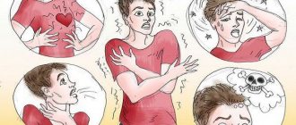
The symptoms of vegetative dystonia can be practically loved, without exaggeration. These may include changes in blood pressure, increased sweating, rapid heartbeat, sleep disturbances, bowel movements (vomiting, constipation, etc.), headaches, general weakness, and mood swings. There may be muscle pain, transient, numbness and crawling that do not have a clear localization. The person may experience discomfort in the chest and a lump in the throat. The symptoms of autonomic dysfunction are diverse and multifaceted. However, as part of the site about dizziness, I wanted to dwell on this symptom in more detail.
Dizziness with VSD may occur. However, nevertheless, it is rarely (almost never) systemic in nature, but fits more into the framework of non-systemic dizziness in the form of a fainting state or general lightheadedness, and may have signs of psychogenic dizziness. It does not have a fixed time frame and often depends on stress factors and weather conditions. Most often, it is not accompanied by tinnitus or nausea leading to vomiting, although nausea may be present.
Symptoms of VSD in the acute stage
The cardiovascular symptom complex in VSD is manifested by the following symptoms:
- changes in heart rate;
- lability of blood pressure;
- pathological vasomotor reactions (redness, cyanosis, pallor of the skin, hot flashes, chilliness of the feet and hands).
In the acute stage, burning, stabbing, pressing, throbbing pain or discomfort in the heart area is observed.
The respiratory symptom complex is manifested by increased ventilation (rapid deep breathing), psychogenic shortness of breath (sniffling, yawning, coughing, periodic deep breaths). Hyperventilation always accompanies anxiety. Shortness of breath is accompanied by dissatisfaction with inhalation, a feeling of lack of air, intermittent breathing, and a feeling of stopping breathing. In the acute stage, muscle spasms and a crawling sensation around the mouth and in the distal limbs occur. Neurologists define Chvostek's symptom - contraction of the muscle that raises the corner of the mouth when tapped in the projection of the facial nerve. Hyperventilation may cause the following symptoms:
- headache;
- fainting state;
- pain in the heart area;
- heart rhythm disturbance;
- abdominal pain, combined with increased peristalsis, nausea, and belching of air.
The gastrointestinal symptom complex is characterized by a disorder of the functions of the digestive organs. May exhibit the following symptoms:
- disturbances of appetite, motility of the esophagus, stomach or intestines;
- psychogenic dysphagia (difficulty, discomfort during the act of swallowing or the inability to take a sip);
- vomiting;
- feeling of heaviness in the epigastrium;
- transient flatulence (bloating);
- diarrhea;
- pain in the abdominal cavity.
The thermoregulatory symptom complex is manifested by an increase or decrease in temperature, chill syndrome. The vasomotor symptom complex consists of several symptoms:
- pallor, cyanosis of the skin;
- chilliness of hands, feet;
- sensations of hot or cold flashes;
- changes in dermographism (weak mechanical irritation of the skin when carried out with a blunt object causes a trace in the form of inflamed swelling);
- increased sweating of the palms and feet.
The genitourinary complex is characterized by cystalgia (frequent, painful, imperative urination in the absence of pathology of the urinary system or changes in urine) and sexual dysfunction (impaired erection or ejaculation in men, vaginismus or anorgasmia in women with preserved or reduced libido, painful menstruation). Hormonal fluctuations, including endocrine changes in the body after childbirth, and postpartum stress provoke an exacerbation of VSD symptoms.
The neurotic symptom complex is manifested by the following symptoms:
- fatigue;
- asthenia;
- low threshold of pain sensitivity;
- sleep disorders;
- irritability;
- senestopathies (pain in the heart, a feeling of dissatisfaction with inhalation, a burning sensation in different parts of the body).
VSD occurs with a wave-like increase and decrease in the manifestations of symptom complexes. This is due to changes in etiological factors and living conditions of the patient. An exacerbation of VSD may occur in the fall. Symptoms of the disease are most pronounced during a “vegetative storm” or crisis.
Diagnostics
The diagnosis of vegetative-vascular dystonia is still rather a diagnosis of exclusion. If the neurological status does not have any focal neurological symptoms, pathological reflexes, and there is vegetative stigmatization (hyperhidrosis, marbling of the skin color, increased dermographism, etc.), and there is not the slightest evidence for the presence of other diseases that could would cause this condition, we can talk about VSD. Relatively reliable studies that confirm the presence of autonomic dysfunction are REG, EEG, as well as stress autonomic tests (orthoclinoplasty and others).
Most often, “real” vegetative-vascular dystonia occurs at the age of 14-30 years, as well as in the age period of 45-55 years in women.
Vestibular vertigo (VV) is one of the most common syndromes: about 5% of people experience it annually [1]. Prevalence of V.G. increases with age. Women suffer from vestibular disorders 2-3 times more often than men. The most common causes of VH are benign paroxysmal positional vertigo (BPPV), Meniere's disease, vestibular migraine, vestibular neuronitis, stroke, and transient ischemic attack (TIA). VH is usually accompanied by severe instability, as well as nausea and vomiting. Seizures V.G. are difficult for patients to tolerate, and anxiety and anticipation of new attacks often lead to the formation of emotional disorders that complicate vestibular compensation. All this makes it especially relevant to use and search for the most effective methods for stopping CH.
The choice of modern methods for treating CH is quite wide. In this case, effective treatment is based on identifying the cause of vestibular disorders. In our country, the contribution of cerebrovascular pathology, arterial hypertension, or diseases of the cervical spine to VH is often overestimated. Meanwhile, large studies conducted in recent years show that the most common causes of dizziness may be other diseases: peripheral vestibular analyzer disorders (BPPV, Meniere's disease, vestibular neuronitis) and migraine.
Seven principles for improving the effectiveness of CH treatment
1.
In the acute period of VH, vestibular suppressants are indicated
. Classic vestibular suppressants include antihistamines and benzodiazepines. In addition to them, some other drugs can be used: calcium channel blockers and acetyl leucine.
Antihistamines.
For CH, only those H1 blockers that penetrate the blood-brain barrier are effective. Such drugs include dimenhydrinate (50-100 mg 2-3 times a day), promethazine (25 mg 2-3 times a day orally or intramuscularly), diphenhydramine (25-50 mg orally 3-4 times a day or 10-50 mg intramuscularly) and meclozine (25-100 mg/day orally) [2, 3]. Side effects of vestibular suppressants include drowsiness, dry mouth, and confusion.
Benzodiazepines.
The inhibitory transmitter of the vestibular system is GABA, and benzodiazepines enhance the inhibitory effects of GABA, which explains the effect of these drugs in V.G. Benzodiazepines, even in small doses, significantly reduce dizziness and associated nausea and vomiting. The risk of drug dependence, side effects (drowsiness, increased risk of falls, memory loss), as well as slower vestibular compensation limit their use in vestibular disorders. The most commonly prescribed drug is lorazepam, which in low doses (for example, 0.5 mg 2 times a day) rarely causes drug dependence and can be used sublingually (at a dose of 1 mg). Diazepam 2 mg twice daily can also effectively reduce VG. Clonazepam has been less studied as a vestibular suppressant but appears to be as effective as lorazepam and diazepam. It is usually prescribed at a dose of 0.5 mg 2 times a day. Long-acting benzodiazepines, such as phenazepam, are not effective for CH [4]. A recent comparative study of the effectiveness of lorazepam and promethazine demonstrated the greater effectiveness of the latter as a drug for the relief of GV [5].
Other vestibular suppressants
. Less common vestibular suppressants include acetyl leucine and calcium channel blockers. The effectiveness of acetylleucine (currently not registered in the Russian Federation) is due to its interaction with phospholipids of cell membranes and the ability to regulate the membrane potential of neurons of the vestibular nuclei and Purkinje cells [6]. Calcium channel blockers - nimodipine, verapamil, flunarizine (not registered in the Russian Federation) and cinnarizine - block voltage-gated calcium channels of neurons in the labyrinth of the inner ear. These drugs are inferior in effectiveness to antihistamines and benzodiazepines [7]. Sometimes anticholinergics (scopolamine, platyphylline) are used as vestibular suppressants. These drugs inhibit the activity of the central vestibular structures, thereby reducing dizziness [3]. However, some side effects of these drugs, in particular drowsiness and blurred vision due to accommodation disorder, superimposed on vestibular symptoms, in some cases only worsen the condition of patients, which significantly limits their use in recent times. As a result, these drugs are more often used to prevent motion sickness rather than to treat CH.
2. Antiemetics enhance and complement the effect of vestibular suppressants in the acute period of VH.
They do not affect the severity of VH, but reduce the vegetative symptoms that often accompany it: nausea and vomiting. The most common antiemetics include phenothiazines, in particular prochlorperazine (5-10 mg 3-4 times daily). Metoclopramide (10 mg intramuscularly) and domperidone (10-20 mg 3-4 times a day, orally) - peripheral D2 receptor blockers - normalize gastrointestinal motility and thereby also have an antiemetic effect [8]. Ondansetron, a serotonin 5-HT3 receptor blocker, also reduces vomiting in vestibular disorders [9]. Side effects of antiemetics include dry mouth, drowsiness, and extrapyramidal disorders.
3.
The duration of taking vestibular suppressants and antiemetics should not exceed 2-3 days.
Vestibular suppressants and antiemetics depress the central nervous system and thus slow vestibular compensation [10, 11]. Animal studies have shown that drugs such as phenobarbital, chlorpromazine, diazepam and ACTH antagonists slow vestibular compensation and delay recovery [12]. These data served as a reason to recommend limiting the use of vestibular suppressants in acute VH (2-3 days). Moreover, this period is also reduced if possible. In practice, the reason for discontinuing vestibular suppressants is to stop vomiting. Instead of vestibular suppressants, vestibular gymnastics and drugs that stimulate central vestibular compensation are prescribed.
4. Vestibular gymnastics stimulates vestibular compensation and speeds up recovery.
Among the methods of treating diseases of the vestibular system, rehabilitation occupies a special place. There are several reasons for this, first of all, its high efficiency and sometimes the lack of a serious alternative to drug therapy. As a result, today, in accordance with international recommendations, vestibular gymnastics occupies almost the main place in the complex treatment of almost any disease of the vestibular system, be it central or peripheral vestibulopathy.
Gymnastics for vestibular disorders consists of four groups of exercises: for stabilizing the gaze, for training postural stability and gait, for sensory substitution and for habituation. Vestibular gymnastics should begin as early as possible (no later than the 1st week of the disease) [13]. Delaying the start of rehabilitation to a later time will most likely slow down vestibular compensation and prolong the period of disability. In addition to early initiation, there are several rules for vestibular rehabilitation that can increase its effectiveness: do not use monotonous exercises and select them taking into account the needs of the patient’s daily activity, take into account the patient’s cognitive functions and concomitant diseases that affect balance, take measures to reduce anxiety and depression, since these conditions make vestibular compensation difficult [14].
5. The effectiveness of vestibular gymnastics can be increased with the help of drugs that stimulate vestibular compensation.
Some substances have the property of stimulating central vestibular compensation. Animal studies have shown that amphetamine, caffeine and ACTH accelerate vestibular adaptation [12]. Among drugs, the ability to stimulate vestibular compensation was found in betahistine dihydrochloride (Betaserc), piracetam and Gingko biloba extract [15-17]. The effectiveness of these drugs is due to their effect on the neuroplasticity of the central nervous system and in some cases has been confirmed not only by experimental but also by clinical studies. Thus, the results of a placebo-controlled study [17] showed that betahistine dihydrochloride accelerates the onset of the effects of vestibular rehabilitation by 3 times in patients with unilateral non-progressive peripheral vestibulopathy resulting from labyrinthectomy for Meniere’s disease.
The duration of use of these drugs is not entirely clear, but appears to be determined by the nature of the vestibular damage and, therefore, the expected timing of vestibular compensation. If, with unilateral peripheral vestibulopathy, compensation occurs after approximately 2 months, then with bilateral, as well as with central vestibular disorders, compensation occurs much more slowly: within several months or years [18].
6. Pathogenetically based treatment is based on identifying the cause of CH.
The high frequency of diagnostic errors in VH, resulting from an overestimation of the role of vascular and cervicogenic mechanisms in the development of vestibular dysfunction, significantly reduces the effectiveness of treatment of dizziness in general. The consequence of inadequate diagnosis is the widespread use of vasoactive and nootropic drugs, the value of which in the treatment of the most common causes of VH (peripheral vestibular disorders and vestibular migraine) is small. At the same time, the possibilities of pathogenetically based drug and non-drug treatment of vestibular diseases have expanded recently due to the emergence of new drugs and the improvement of vestibular gymnastics.
Treatment of BPPV primarily involves the use of therapeutic repositioning maneuvers. Maneuvers are designed to treat canalolithiasis of various semicircular canals; Their efficiency is high and approaches 100%. Drug or surgical treatment of BPPV is not required in most cases [19].
Treatment of Meniere's disease consists of several stages. At the first stage, conservative approaches are used, which boil down to a salt-free diet, the use of diuretics and betahistine dihydrochloride. If results cannot be achieved within six months and attacks of VH continue, transtympanic injections of corticosteroids or gentamicin are performed. Finally, if these measures are ineffective, they resort to surgical treatment - labyrinthectomy, or selective neurectomy of the vestibular part of the vestibular-cochlear nerve [20-23].
Treatment of vestibular neuronitis is reduced to the use of symptomatic agents (vestibular suppressants and antiemetic drugs) in the acute period of the disease, followed by the selection of vestibular rehabilitation exercises in combination with drugs that stimulate vestibular compensation [24].
For vestibular migraine, the same drugs are prescribed to prevent attacks of dizziness as for ordinary migraine: β-blockers (propranolol or metoprolol), antidepressants (tricyclics or SSRIs), antiepileptic drugs (for example, topiramate, lamotrigine or valproic acid drugs), calcium blockers channels (for example, flunarizine). To relieve vestibular migraine attacks, in addition to vestibular suppressants and antiemetics, triptans and NSAIDs are used [25, 26].
For the treatment of central vestibular disorders, which arise, for example, due to stroke, trauma or MS, symptomatic drugs are used to reduce oscillopsia and instability [27]. Among such drugs are baclofen, memantine, gabapentin, aminopyridine, acetylleucine [28].
7. According to the
VIRTUOSO , betahistine dihydrochloride can reduce the frequency of VH attacks, regardless of the cause of their occurrence.
Betahistine was first registered in Canada in 1968 and has since been widely used throughout the world for the treatment of diseases manifested by VG. Betahistine is a strong H3 receptor antagonist and a weak H1 receptor agonist [29]. By acting on the H1 receptors of the inner ear, betahistine has a pronounced vasodilator effect [30]. The reduction in the number of attacks of dizziness during treatment with betahistine is apparently due to an increase in blood flow in the inner ear, which reduces the increased endolymphatic pressure in it, restoring the balance between the production and reabsorption of endolymph [31]. In addition, various animal studies have shown that betahistine is able to improve vestibular compensation, exhibiting direct histaminergic effects; the drug accelerates the release and metabolism of histamine by blocking presynaptic histamine H3 receptors (in the vestibular and tuberomammillary nuclei) [30].
The clinical effectiveness of betahistine has been assessed in several studies. At the same time, in a number of studies the effectiveness of betahistine was confirmed [17, 32, 33], while the results of others did not differ from the effectiveness of placebo [34].
The purpose of the recently completed international observational program VIRTUOSO was to evaluate the effects of betahistine dihydrochloride (Betaserc) at a dose of 48 mg/day in tablet form in routine clinical practice in outpatients with V.G. In this case, not only the overall clinical response during the treatment period was assessed, but also the delayed effects of treatment, determined by the frequency of attacks of VH within 2 months after completion of betahistine [35].
Study design.
The study was a prospective, multicenter, noncomparative, postmarketing observational program involving 23 medical centers and 309 patients; 305 patients completed the observational program. The program included patients who were prescribed betahistine in accordance with the instructions for use of the drug and clinical recommendations for the treatment of a disease manifested by paroxysmal V.G. Betahistine was prescribed at a dose of 48 mg/day. Treatment lasted 1-2 months; The observation period after cessation of treatment was 2 months.
The study included men and women aged 18 years and older, suffering from paroxysmal CH of known or unknown etiology, who were prescribed treatment with betaserc (betahistine dihydrochloride) at a daily dose of 48 mg. All patients consented to the use and processing of personal data. Patients included in the program could not take betahistine for 5 or more days before obtaining written consent and inclusion in the study.
Treatment efficacy measures included the number and proportion of patients with a clinical response at the end of therapy during follow-up within the program, as assessed by the VH Severity and Clinical Response (SVVSLCRE) scale. In addition, changes in the frequency of VH episodes at the end of therapy were identified during program follow-up compared with the inclusion visit among patients treated with betahistine dihydrochloride. The overall clinical response was also assessed separately as assessed by the physician and the patient, determined on a 4-point scale, where 4 is marked improvement, 3 is significant improvement, 2 is slight improvement, and 1 is deterioration. We analyzed the change in the frequency of VH episodes within 2 months after the end of therapy compared with the end of the therapy period. Finally, during treatment, regression of dizziness-related symptoms (tinnitus, hearing loss, nausea, vomiting, weakness and headache) was determined according to the physician and patient.
Research results.
The clinical effectiveness of treatment according to the SVVSLCRE scale was assessed as significant improvement, marked improvement or very marked improvement in 74.1% of cases (
p
<0.001).
The number of dizziness attacks per month while taking betahistine decreased from an average of 8.0 to 3.0 ( p
<0.001).
During the 2-month follow-up period after discontinuation of the drug, the number of attacks continued to decrease until they disappeared completely at the end of the 1st month of follow-up ( p
< 0.001). This effect persisted at the end of the 2nd month of observation.
The overall clinical response to treatment with betahistine (by the end of the 2nd month of taking the drug) according to the doctor was assessed as a marked improvement or significant improvement in 94.4% of cases, and according to the patient - in 95.4% of cases.
Betahistine also had an effect on the symptoms associated with dizziness. The effectiveness of treatment of associated symptoms was assessed as a marked improvement or significant improvement in 71.4% of cases by the end of the 1st month of taking the drug and in 90.5% of cases by the end of the 2nd month, according to both the doctor and the patient.
The observational program demonstrated the high safety of betahistine dihydrochloride and a low incidence of side effects. Only one patient dropped out of the study early due to lack of treatment effectiveness and an increase in the frequency of dizziness attacks. Sereznykh N.Ya. was not registered.
Thus, the VIRTUOSO observational program confirms the results of previously conducted studies, which indicated the effectiveness of betahistine at a dose of 48 mg/day in V.G. The study demonstrated a significant reduction in the frequency of CH attacks, regardless of the etiology of the disease. An important result of the study was the discovery of a delayed effect of treatment with betahistine: the number of attacks continued to decrease for 2 months after stopping the drug. This result may indirectly indicate in favor of stimulation of central vestibular compensation under the influence of betahistine. The low incidence of side effects confirms the good tolerability and high safety of betahistine. All these data allow us to recommend the inclusion of betahistine dihydrochloride (betaserc) in the complex therapy of various diseases manifested by VH, including not only Meniere’s disease, but also other disorders of the peripheral or central parts of the vestibular analyzer.
In general, if the above principles are followed, treatment of diseases manifested by CH can be quite effective. It seems promising to search for vestibular suppressants that do not suppress vestibular compensation, improve methods of pathogenetically based therapy and conduct clinical studies aimed at confirming the effectiveness of existing treatment options, as well as finding ways to stimulate vestibular compensation in various peripheral and central vestibular disorders.
Conflict of interest: The authors declare participation in the VIRTUOSO observational study sponsored by .
Treatment
Treatment of vegetative-vascular dystonia is the subject of enormous controversy in the scientific community. Who should make this diagnosis? Neurologist, therapist or psychiatrist? How to treat this condition and how – symptomatically or try to find the mechanisms of pathogenesis of this condition?
But disputes are disputes, and on this site I share my opinion. The most important thing is non-drug therapy, namely psychological support, stabilization of lifestyle (sleep, work conditions, family situation, bad habits, etc.); in the presence of panic conditions, consultation with a psychiatrist and the prescription of anti-anxiety therapy are indicated. In all cases, restorative therapy will not be harmful (including B vitamins, their derivatives, in particular Enerion, such “soft” drugs as Mexidol, Neurox, Ethoxidol, Armadin, Fnibut, etc.). In the presence of dizziness, the prescription of vertigolytics (Betaserc, Vestibo, etc.) is controversial, because there is usually no substrate for their work. It is more important to use vestibular rehabilitation and vestibular gymnastics techniques than drug treatment.
Causes of vegetative-vascular dystonia
To get rid of vegetative-vascular dystonia, it is first necessary to identify the cause of the disease. An experienced neurologist can do this.
Autonomic dysfunction can occur for one of the following reasons or a combination of them:
- factor of hereditary predisposition (in this case, manifestations of VSD appear already in childhood)
- past acute and chronic infectious diseases or intoxication
- chronic psycho-emotional stress, depression, sleep disorders
- sedentary lifestyle, sedentary work and, as a consequence, poor posture and cervical osteochondrosis
- bad habits (smoking, drinking alcohol)
- as a consequence of chronic disease of the endocrine (hormonal changes), cardiovascular system, gastrointestinal tract and nutritional disorders
- diseases of the central and peripheral nervous system.