How does an appointment with a neurologist go: what does the patient need to know?
The most complex system in our body.
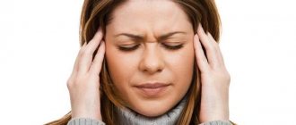
The main causes of neurological disorders are stress, sedentary lifestyle and dietary errors. Thus, today the absolute majority of the young working population is susceptible to neurological diseases, especially in large cities. Periodic headaches, dizziness, discomfort in the back, sleep problems, cramps in the limbs - perhaps these symptoms are familiar to everyone today. We are used to not paying attention to them, chalking them up to bad weather or fatigue. However, such seemingly harmless sensations are most often symptoms of a neurological disease. The nervous system, central and peripheral, are the main systems of the human body that control and perform all our vital functions. There is an opinion that the nervous system is the most important and most complex system in our body.
Fortunately, as a rule, most neurological diseases respond well to comprehensive treatment, especially in the early stages. At the BELSONO medical center in Gomel, highly qualified neurologists with many years of practice receive treatment. We will help you accurately determine the diagnosis and develop a treatment regimen that is necessary specifically in your case.
What does a neurologist treat: what diseases?
The basis of diseases of the nervous system is a disruption in the functioning of neurons, nerve cells, a weakening of the connection between them, as well as inflammatory processes in the brain and spinal cord.
The most common in practice:
· Lumbosacral and cervical radiculopathies
· Vertebrogenic thoracalgia
Vertebro-basilar insufficiency
Inflammatory processes of peripheral nerves - neuritis, neuralgia
Migraine and tension headaches
· Astheno-neurotic disorders
· Convulsive syndromes.
How does an appointment with a neurologist go: what does the patient need to know?
During an initial visit to a neurologist, the specialist will need to ask the person in detail about the complaints, that is, collect a history of the disease.
The specialist examines the appearance (appearance) of the patient for the presence of asymmetry of the face and various parts of the body.
To study the work of the oculomotor nerves, you will need to follow the movements of the hammer without turning your head.
The doctor can check your reflexes using your facial expressions. The neurologist will ask you to wrinkle your forehead, stick out your tongue, or say “Ah.”
You can check the sensitivity of your face using a needle. Don’t be scared; you will need to concentrate as much as possible and answer the neurologist’s questions about whether you experience the same sensations when receiving injections in symmetrical zones.
To determine the condition of the muscles, their tone and reflexes, the doctor will be asked to shake his hand and resist when trying to bend the elbow. Evaluation occurs by assigning points from 1 to 5.
Deep reflexes of the arms and legs are tested by striking the tendons with a hammer.
Superficial reflexes are tested by irritating the skin of the abdominal wall with a needle.
A deep examination of muscles and joints is carried out when the patient's eyes are closed, and the doctor moves his finger in different directions and asks him to name exactly in which direction he is doing it.
Palpation of paravertebral points helps determine the condition of the spinal nerves and paravertebral pain points.
Coordination of movements is tested by the Romberg pose. The patient stands with his feet together, arms extended forward, eyes closed. The neurologist will ask you to slowly move your index finger to your nose (with each hand). In this study, a person should ideally not stagger to the sides.
You may need to answer specific questions about counting or dates to assess memory.
Additional examination of patients.
Not in all cases it is possible to make a diagnosis based on clinical data. There is a need for additional studies that will provide detailed information about the patient's condition. Unfortunately, not all of the methods listed below are yet available in our medical centers. As BELSONO develops, it plans to launch MRI, angiography and radiology techniques in a phased manner.
*CT scan. Identifies areas of hemorrhage, malformations of arteries or veins. Allows you to see tissue changes, swelling, softening during a heart attack (stroke) or injury.
*Magnetic resonance imaging. A more modern diagnostic method provides more detailed and accurate data due to the greater resolution of the device and does not have such a detrimental effect on the patient. But it should be noted that in some cases CT and MRI are not mutually exclusive methods and they have their own diagnostic features.
*Angiography. Contrast X-ray examination, which reveals pathologies and changes in blood vessels, including in the brain.
*Ultrasound of BCA. Using this study, a detailed image of the large vessels of the neck is obtained and the characteristics of blood flow are determined.
*Lumbar puncture or cerebrospinal fluid analysis.
*X-ray. Contrast is used to detect intervertebral disc herniations, proliferation of vertebral bodies, and tumor processes. The study gives a clear picture of the state of the subarachnoid space of the entire spinal cord.
*Electroencephalography.
The main test prescribed for patients with suspected epilepsy.
And there are quite a lot of methods and studies that help in diagnosing diseases: biopsy of muscles, nerve tissue, genetic studies, blood tests.
Successful treatment requires finding out the cause of the pain and making the correct diagnosis.
And for this you need to contact a neurologist. You shouldn’t let the disease get started; it’s much easier to defeat it in its infancy! You can make an appointment with a neurologist in Gomel by calling our medical centers, as well as through the online appointment form on the website.
Reanimatology School of Professor Sergei Vasilievich Tsarenko
Assessment of neurological status is the leading means of neuromonitoring. This is explained by two reasons. The first is that in most intensive care units, dynamic neurological examination is the only available way to assess the state of the brain and determine the effectiveness of the therapy. The second reason for special attention to the neurological examination is that the ultimate goal of any treatment is not to achieve certain indicators of ICP and CPP, oxygen content and metabolites, but to improve the functional state of the brain and the outcome of the disease! Only neurological status can be an absolute measure of the sufficiency of brain perfusion and oxygenation for a particular patient, determination of the critical ICP threshold, and indications for surgical treatment of a hematoma detected on CT. It is the assessment of the neurological condition (restoration of the level of consciousness, relief of dislocation symptoms and resolution of focal disorders) that always puts the final point in the dispute about how to properly treat the patient.
Unlike many foreign researchers, we believe that when assessing the state of the brain, one cannot limit oneself only to determining the reaction of the pupils to light and their size, as well as the nature of motor responses to pain. With the help of ten to twelve reflexes within 5 minutes, you can evaluate in detail dislocation and neurological symptoms, as well as the effectiveness of treatment measures. Our experience allows us to propose a working scheme for assessing neurological status.
Like any diagram, it simplifies the true picture at the expense of losing details of information, but it is convenient and practical. According to this scheme, the activity of each brain structure is assessed by individual neurological symptoms. In other words, when the activity of a particular structure is disrupted, characteristic symptoms arise. The primary focus of the neurological examination is on movement disorders, cranial nerve involvement, and level of consciousness. In order to understand why exactly these disorders attract our attention, let's start with anatomy and physiology.
A bit of anatomy First, let's look at the anatomical features of the brain stem. As is known, the brain stem is the focus of vital centers, the defeat of which determines the prognosis of diseases and damage to the brain. Anatomically, the brainstem is divided into diencephalic structures (upper trunk), mesencephalic structures and pons (middle trunk), and bulbar regions (lower trunk) (Fig. 4.1). The stem formations are located in the close space of the posterior cranial fossa, surrounded by dense bone and connective tissue structures, in particular the tentorium of the cerebellum (Fig. 4.2). Because of this, with any damage to the brain that leads to its dislocation, the brain stem is pressed against these solid structures, which leads to the “loss” of its functions. In addition to dislocation, dysfunction of the trunk is caused by its primary damage due to ischemia or hemorrhage.
Damage to various parts of the brain stem is diagnosed by dysfunction of the cranial nerves (CN), the nuclei of which are located at various levels (Fig. 4.3). When the stem structures are damaged, the functions of the corresponding nuclei (the central link of nervous regulation) are disrupted. The axons of the neurons that make up these nuclei exit the brain in the form of cranial nerve roots. When these roots are pressed against the tentorium of the cerebellum or bone structures, the peripheral link of nervous regulation suffers.
The nuclei of the III nerve (oculomotor nerve) are located above all. Below them are the nuclei of the IV nerve (trochlear nerve), which is the only one whose roots emerge not on the ventral surface of the brain, but on the dorsal one. The nuclei and roots of subsequent CNs are located sequentially from top to bottom along the length of the brain stem. Nuclei III and IV of the CN belong to the midbrain (mesencephalon). The nuclei of the V (trigeminal nerve), VI (abducens nerve), VII (facial nerve) and VIII nerves (auditory and vestibular nerves) are located in the pons. Nuclei IX (glossopharyngeal nerve), X pair (vagus nerve), XI (accessory nerve) and XII pair (hypoglossal nerve) are located in the bulbar regions of the brainstem.
The next important anatomical and physiological aspect that needs to be considered is the innervation of gaze. There is a popular expression: “The eye is the window to the brain.” This is not surprising, since the III, IV, VI pairs of cranial nerves, as well as the structures of the medial longitudinal fascicle located next to the nuclei of the abducens nerve, take part in the motor innervation of the eye. The III pair provides upward, downward and inward movements of the eyeball, the IV pair - outward and downward, the VI pair - outward. Sensitive innervation of the cornea is provided by the I branch of the V pair, which, in addition, plays the role of an afferent link of the corneal reflex. Damage to the oculomotor nerves is accompanied by asymmetrical changes in the position of the eyeballs. The most striking manifestations are divergent strabismus when the III pair is affected and convergent strabismus when the VI pair is affected (Fig. 4.4.). The mechanism of strabismus is as follows: there is a dysfunction of the nucleus or root of the nerve on the side of the affected eye. Those muscles that are innervated by this nerve stop pulling the eye in their direction. The remaining eye muscles, the control of which is not affected, pull the eyeball towards themselves.
The coordinated work of the oculomotor nerves is ensured by the medial longitudinal fasciculus system. The medial longitudinal fasciculus receives afferent innervation from the muscles of the neck, vestibular nuclei, and gaze centers. There are two types of gaze centers - cortical and stem. The cortical centers are located in the middle frontal gyrus of the cerebral hemispheres. From them, impulses travel along intersecting paths and control the function of the contralateral (on the opposite side) oculomotor muscles. The stem centers of gaze are located in the midbrain, among the nuclei of the reticular formation, as well as in the pons. From them, impulses travel along axons to the ipsilateral (on the same side) oculomotor nerves. The mesencephalic gaze centers provide vertical and rotational eye movements, while the cortical and pontine centers provide horizontal movements. Coordination of the innervation of the eye with the help of the cortical and stem centers of gaze leads to the fact that, normally, when turning the head in a horizontal or vertical plane, the eyeballs synchronously shift in the same direction.
With irritation (hyperfunction) of the cortical center, excitation of the contralateral oculomotor nerves occurs. This situation often occurs in the first hours and days of ischemia (damage) to cortical structures and is combined with hemiparesis due to simultaneous damage to the pyramidal tracts. When the left hemisphere is damaged, the left cortical gaze center is affected. The left center of gaze ensures eye movement in the opposite direction, that is, to the right. In this case, right-sided hemiparesis develops. In the most acute period (approximately during the first day), the eyeballs turn to the right side due to irritation of the affected center (Fig. 4.5). Then the loss of its function leads to the predominance of the influence of the opposite center of gaze, so the eyes turn and are set in a position to the left (gaze to the left). In case of incomplete loss of functions of the cortical centers of gaze, a violation of the oculomotor innervation may not be manifested by a complete opening of the gaze, but only by the inability to turn the eyeballs in the opposite direction (under-direction of the gaze to the right). When the right hemisphere is damaged, the pathological processes are mirror in nature.
There are useful mnemonic rules: 1. “A patient with a supratentorial lesion at the beginning of the disease turns away from the lesion and looks at the affected limbs.” 2. “A patient with a supratentorial lesion on the second day of the disease and then looks at the lesion and turns away from the affected limbs, does not want to see them.”
When the pontine gaze centers are damaged, the stage of hyperfunction cannot be observed for unclear reasons.
“The patient with a subtentorial lesion always turns away from the lesion and looks at the affected limbs.” A more vivid expression: “A patient with a subtentorial lesion examines the affected limbs.”
With the development of dislocation and pressing of the brain to the tentorium of the cerebellum, the oculomotor nerves are affected and both cortical and stem centers of gaze are “switched off,” causing the characteristic symptom of “upward gaze paresis.” This symptom is diagnosed by inducing the corneal reflex. When touching the cornea with a clean piece of gauze or cotton wool, the eyeballs should normally rise above the line connecting the corners of the palpebral fissure. In the initial stages of upward gaze paresis, the eyeballs shift only to the level of this line. With advanced paresis of upward gaze, the eyeballs cannot rise even to this level and are directed downward (Fig. 4.6). Less common is irritation of these centers, which causes upward gaze and convulsive contraction of the eyelids. Resuscitators and neurologists often rejoice at this symptom completely in vain: “The patient has opened his eyes!” The patient actually continues to be in a coma. This rare form of coma is called "open-eye coma" (Fig. 4.7). In our practice, there were also several patients with upward rotation of the eyeball on only one side - they were in a coma with one eye open!
The separation of the cortical and stem centers of gaze by the brain being displaced and pressed against the tentorium leads to a loss of influence of both cortical centers of gaze and can cause disinhibition of the stem centers. This is manifested by three symmetrical symptoms. The first symptom is friendly “floating” movements of the eyeballs. The second symptom is the appearance of the oculocephalic reflex, otherwise called the “doll eye” reflex. In the presence of this symptom, turning the head to the side causes the eyeballs to move in the opposite direction, like a child's toy (Fig. 4.8). When you turn your head up, your eyes “look” down, and vice versa. The reflex is not evoked normally and appears when dislocation increases. It is interesting that the oculocephalic reflex disappears with further development of dislocation and damage to the stem centers of gaze. However, the improvement in the patient’s condition is accompanied by the disappearance of the oculocephalic reflex due to the restoration of the dominant role of the cortical gaze centers. In addition to the horizontal oculocephalic reflex, a vertical one can be evoked. When the head is bent and the chin is brought to the chest, the eyes turn upward; when the head is straightened, the eyeballs turn upward.
The third symptom behaves similarly to the oculocephalic reflex - the oculovestibular reflex (response to temperature stimulation of the eardrum). Normally, the introduction of cold water into the ear canal leads to nystagmoid movements of the eyeballs. The slow component of nystagmus is directed towards the irritated ear canal, and the fast component - in the opposite direction. An increase in dislocation symptoms leads to loss of the fast component of nystagmus, and the eyes, when inducing the oculovestibular reflex, turn towards the irritated ear. An improvement in the patient's condition is accompanied by the appearance of a fast component, while a deterioration is accompanied by the disappearance of both components. Checking for this symptom requires confidence that there is no damage to the eardrum. Bilateral irritation of the eardrums with warm water leads to a friendly deviation of the eyeballs upward, and with cold water - downward.
There are two more important anatomical and physiological aspects that need to be touched on before moving directly to assessing the neurological status. The first aspect is the features of the localization and functioning of motor pathways. The main motor pathways located in the brain stem are corticospinal (pyramidal), corticonuclear and extrapyramidal. The corticospinal and corticonuclear tracts are arranged as follows. The body of the first neuron is located in the cerebral cortex. Its axon passes to the opposite side in the region of the brain stem (the so-called decussation of motor tracts) and reaches the body of the second neuron, where the corticospinal or corticonuclear tract ends. The body of the second neuron is located in the nuclei of the CN (the end of the corticonuclear tracts) or in the motor nuclei of the spinal cord (the end of the pyramidal tracts). The axon of the second neuron conducts nerve impulses that provide motor functions on its side. The pyramidal tracts pass on the ventral half of the brain stem and are sent in transit from the cerebral cortex to the motor neurons of the spinal cord (Fig. 4.9). The extrapyramidal tracts are structured in much the same way as other motor pathways. The first neurons are located in the gray matter of the subcortical nuclei - the striatum, the red nucleus, the substantia nigra, as well as in the cerebellum and cerebral cortex. The axons of these neurons descend down to the same motor neurons of the spinal cord as the pyramidal tracts.
The pyramidal tracts control voluntary movements of skeletal muscles, the extrapyramidal tracts control the tone of striated muscles.
The function of motor pathways in traumatic brain injury is impaired in three situations:
•in supratentorial pathological processes due to damage to the first neuron; •in case of primary brain stem damage due to damage to the second neuron located in the nuclei of the brain stem; • during brain dislocation, when motor pathways are pressed against connective tissue and bone structures.
In supratentorial pathology, due to the loss of the control functions of the first neuron, the activity of the motor nuclei of the spinal cord and the nuclei of the cranial nerve on the side opposite to the source of damage is disrupted. When the pyramidal tracts are damaged, pathological foot signs appear on the side opposite to the cortical lesion - the Babinski reflex and its analogues (Fig. 4.10), as well as hemiparesis. If the focus is on the left, then hemiparesis appears on the right. Disturbances in the functions of the CN are also noted on the right, that is, on the same side. With subtentorial lesions, there is a direct impact on the CN nuclei from the side of the lesion and the pyramidal tracts passing nearby, which have already crossed to the opposite side. As a result, with a brainstem lesion on the left, the functions of the CN are lost on the left, and hemiparesis is noted on the right (the so-called alternation of symptoms).
If motor lesions are part of the dislocation syndrome, then the appearance of hemiparesis and pathological foot signs is explained not by damage to the first neuron, but by compression of the pyramidal and extrapyramidal tracts at the meso-diencephalic level. In this case, alternating syndrome also occurs. There is a combination of damage to the third pair of CN and contralateral hemiparesis, which occurs when the cerebral peduncle is pressed against the tentorium of the cerebellum due to lateral transtentorial dislocation.
With increasing compression of the brain stem, disturbances of consciousness deepen and the nature of motor reactions to painful stimuli changes (Fig. 4.11). The ability to localize pain (differentiated reaction) is replaced by undifferentiated reactions: the patient cannot determine (differentiate) the source of painful stimulation. As compression of the pyramidal tracts increases, undifferentiated reactions are replaced by postural reactions, which are first flexion in nature, then extension. When the damage to the motor tract extends to the level of the bridge and below, the application of painful stimulation causes only weak movements of the arms and legs, such as twitching of small amplitude.
The second fundamentally important anatomical and physiological aspect for a neuroresuscitator is the location of the centers of the ascending reticular formation in the upper parts of the brain stem (in the diencephalic region). When brain dislocation develops, these centers are among the first to suffer. Their activating influence on the cerebral cortex is eliminated, which is one of the reasons for the depression of consciousness to varying degrees, up to coma (Fig. 4.12). Depression of consciousness can be a consequence of two more reasons - extensive diffuse damage to cortical structures and direct damage to mesencephalic-diencephalic structures.
After brushing up on the intricacies of anatomy and physiology, you can move directly to neurological monitoring. The most important thing in neuromonitoring is a clear diagnosis of the depth of depression of consciousness. Currently, in our country there is a division into clear consciousness, mild stupor, deep stupor, stupor, moderate coma, deep coma and atonic coma (A.N. Konovalov et al., 1998). In most cases, the border between stupor and coma is the patient’s ability to open his eyes slightly to sound and other stimuli. A patient who opens his eyes slightly to pain or a cry is in stupor; if he is unable to open his eyes slightly, he is in a coma. A more detailed description of coma is as follows: “Coma is a deep degree of depression of consciousness, characterized by the absence of any manifestations of conscious behavior in response to any stimuli.” To make a detailed assessment of the depth of the comatose state, it is necessary to determine the reaction to painful stimuli. A differentiated reaction is characteristic of moderate coma. Deep coma is characterized by the appearance of undifferentiated and post-notonic reactions. In atonic coma, there are no reactions to pain and reflexes (except for spinal ones), body temperature normalizes and decreases (Table 7).
Table 7. Degrees of depression of consciousness (according to A.N. Konovalov et al., 1998)
A bit of physiology The presence of spinal reflexes often “confuses” the doctor when he is faced with the problem of diagnosing atonic coma. In a patient in an atonic coma, muscle tone and normal and pathological tendon reflexes cannot be detected. However, it may have what is called “triple flexion.” This spinal reflex is evoked by stroke stimulation of the soles and is manifested by flexion of the foot, leg and thigh (Fig. 4.13).
In addition to the domestic classification, the degree of impairment of consciousness can be assessed using the Glasgow Coma Scale (G. Teasdale, B. Jennett, 1974). This scale evaluates three indicators: speech production, reaction to pain and eye opening. Each response type is scored independently of the others. The sum of the three answers determines the depth of the disorders of consciousness. The level of coma according to the GCS can vary from 3 points (atonic coma) to 15 (clear consciousness) - table. 8.
Glasgow Coma Scale (G.Teasdale, B.Jennett, 1974)
The domestic scale of disturbances of consciousness and the GCS correlate with each other as follows. 15 points - clear consciousness, 14 points on the GCS correspond to mild stunning according to the domestic classification of disorders of consciousness, 12-13 points - deep stupor, 9-11 points - stupor, 6-8 points - moderate coma, 4-5 points - deep coma , 3 points – atonic coma.
The Glasgow Coma Scale is widely used throughout the world and is used in scientific research and practical work. However, it has a serious drawback. Very often it is impossible to check one of the most important components - the patient’s verbal reaction due to aphasic disorders and the presence of an endotracheal or tracheostomy tube in the trachea. In this regard, sometimes only one motor component of the GCS is used.
In addition to assessing the level of consciousness, a detailed diagnosis of brain stem damage at its various levels is fundamentally important (F. Plum, D.B. Posner, 1986). If a patient experiences an increase in transtentorial axial dislocation of the supratentorial brain structures of the “top-down” type, then in the clinical picture one can observe sequential damage to the diencephalic, mesencephalic, pontine and bulbar structures. If there is a direct lesion or dislocation of the subtentorial structures, then such a characteristic sequence cannot be observed. In this case, the “loss” of the functions of one or another cranial nerve can be used to assess the specific level of damage to the brain stem. Neurological symptoms with transverse displacement of the brain under the medullary falx depend on the degree of concomitant axial transtentorial dislocation.
Let's start with transtentorial dislocation of the “top-down” type. With supratentorial lesions, depression of consciousness usually increases gradually and is often accompanied by gaze in the direction opposite to the lesion and hemiparesis on the opposite side. Subsequently, the gaze turns towards the hearth. As a rule, there are pyramidal disorders that are combined with corticonuclear disorders, as a result of which the functions of the cranial nerves are lost on the same side as the hemiparesis. The most characteristic cortical-nuclear loss is central paresis of the facial muscles (damage to the nuclei of the facial nerve - VII pair) and the resulting asymmetry of the face (Fig. 4.14). For unknown reasons, there is a violation of the corneal reflex. As dislocation symptoms develop, the depth of the coma increases in parallel with the appearance of diencephalic symptoms. The interest of the diencephalic level of the trunk is determined by signs of damage to the hypothalamus. Autonomic disorders occur - tachypnea, tachycardia, arterial hypertension, hyperhidrosis, hyperthermia.
The spread of dislocation to the mesencephalic level is diagnosed based on the appearance of symptoms of damage to the nuclei and roots of the oculomotor nerve (III pair of cranial nerves). The nature of the damage to the oculomotor nerve depends on the type of dislocation - lateral or central (Fig. 4.15). With lateral dislocation, the oculomotor nerve is usually affected on the same side where the pathological process is localized (ipsilateral symptoms). Nerve damage manifests itself in suppressed pupillary response to light, mydriasis, and divergent strabismus (Fig. 4.16). Due to damage to the pyramidal tract at the mesencephalic level, along with loss of functions of the third pair of cranial nerves, hemiparesis develops on the opposite side. However, sometimes, in approximately 20% of cases, the dislocation occurs in such a way that the oculomotor nerve and the cerebral peduncle are pressed against the tentorium of the cerebellum not from the ipsilateral side, but from the opposite, contralateral side. Such cases are accompanied by damage to the third pair on the contralateral side and ipsilateral hemiparesis.
With the central type of transtentorial dislocation, a very important symptom of the mesencephalic stage of the lesion is paresis of upward gaze due to dysfunction of the quadrigeminal nuclei located in the tegmentum of the brain. Due to the loss of connections between the nuclei of the third pair of the cranial nerve and the hypothalamus, the sympathetic innervation of the muscle that constricts the pupil suffers. This leads to the predominance of parasympathetic innervation and the appearance of a characteristic symptom - narrow pupils that react poorly to light. Due to the disinhibition of the stem centers of gaze, floating movements of the eyeballs, oculocephalic and oculovestibular reflexes appear. As a rule, oculocephalic and oculovestibular reflexes are evoked symmetrically on both sides. In parallel with these processes, as a result of damage to the pyramidal tracts passing in the brain stem, differentiated reactions to pain are replaced by undifferentiated ones. Due to disruption of corticospinal connections, motor disorders such as hemiparesis develop, as well as pathological foot signs. As the dislocation increases, hemiparesis gives way to tetraparesis, flexion posture-tonic reactions appear, which are then replaced by extension ones.
With further increase in dislocation, neurological signs do not differ depending on the type of process - central or lateral. When the upper parts of the pons are damaged, signs of damage to the trochlear and abducens nerves (IV and VI pairs of cranial nerves), as well as the nearby medial longitudinal fascicle, appear. They are manifested by a difference in the distance of the eyeballs vertically and horizontally - the Hertwig-Magendie symptom (Fig. 4.17). Bilateral loss of the corneal reflex reflects damage to the adjacent nuclei of the V pair (afferent arc of the reflex) and VII pair (efferent arc).
Bilateral damage to the trigeminal nerve (V pair), or rather, its motor nuclei, leads to a decrease in the tone of the lower jaw. Bilateral damage to the facial nerves (VII pair) is manifested by a decrease in the tone of the facial muscles and drooping of the lower eyelids (lagophthalmos). The latter symptom is difficult to interpret in comatose patients, as it may be due to a general decrease in muscle tone.
With a further increase in dislocation due to the loss of functions of the III, IV and VI pairs of the cranial nerves, the eyeballs are installed in the midline. Since the lower parts of the pons provide connections between the cerebrum and the cerebellar structures, when they are damaged, the oculocephalic and oculovestibular reflexes are lost. Loss of reflexes appear, as a rule, asymmetrically, that is, first on one side while they are intact on the other, and then on both sides. It is possible to preserve vertical oculocephalic reflexes while losing horizontal ones, and vice versa. Reactions to pain manifest themselves in weak, low-amplitude movements of the arms and (or) legs.
The spread of dislocation to the structures of the lower brainstem is diagnosed based on the disappearance of almost all segmental reflexes of the brain stem:
•eyes in the midline; •the pupils are submaximally dilated and do not respond to light; •there are no corneal, oculocephalic and oculovestibular reflexes; •low tone of the muscles of the face and lower jaw; •cough reactions to irritation of the trachea with an endotracheal tube or sanitary catheter are significantly reduced, up to complete absence.
In addition, bradypnea, bradycardia and arterial hypotension are noted. Motor reactions to pain disappear.
Symptoms of brainstem damage that do not have a clear “link” to certain anatomical structures are disturbances in muscle tone and tendon reflexes. There is unevenness (dissociation) of tone and reflexes along the axis of the body. They are usually higher in the legs and lower in the arms, but it can also happen the other way around. Tone and reflexes may differ on the right and left sides of the body.
A possible explanation for reflex-tonic disorders is uneven damage to the pyramidal and extrapyramidal motor systems running nearby in the brain stem. The appearance of stereotypical acts should be assessed similarly: vomiting, yawning, stagnation of gastric contents. One should always be attentive to the appearance of the latter, since this symptom is a very sensitive and early sign of deterioration in the functional state of the brain stem, caused by both neurological causes (dislocation, direct damage) and secondary ischemia of brain stem structures.
A characteristic symptom of damage to the extrapyramidal system is also constant or periodic motor agitation of the patient. The last symptom is most often regarded by clinicians as delirium or damage to the frontal lobes, which is actually incorrect. Delirium and damage to the frontal lobes are expressed in complex behavioral disorders of the patient: hallucinations, delusions, etc. Extrapyramidal symptoms are manifested by stereotypical motor acts, often characteristic of the patient, for example, related to his professional activities. It is fundamentally important that “frontal” symptoms and delirium have nothing to do with damage to the brain stem - primary or due to dislocation.
The neurological symptoms of damage to the trunk appear somewhat differently when the subtentorial structures (posterior cranial fossa) are damaged. If these structures are damaged, primary damage to the upper trunk is possible, as well as compression of it by the cerebellum during dislocation “from bottom to top” (Fig. 4.18). This is accompanied by a sudden depression of consciousness to the point of stupor and coma, the appearance of reflex-tonic disorders and stereotypical acts. There is either hemiparesis with alternating loss of cranial functions, or tetraparesis with asymmetric dysfunction of the cranial nerves. A distinctive feature of subtentorial lesions is also the mosaic pattern of loss of cranial nerve functions without the characteristic sequence described for axial dislocation “from top to bottom.” In the clinical picture, one can observe, for example, with damage to the pons of the brain, the preservation of the functions of the III pair with damage to the VI pair and the medial longitudinal fasciculus (convergent strabismus, vertical separation of the eyeballs, pinpoint pupils) (Fig. 4.19). Primary asymmetrical loss of oculocephalic and oculovestibular reactions may be observed. Primary damage to the bulbar structures may be accompanied by a decrease in the level of consciousness only to stupor, but severe breathing disorders such as bradypnea will be observed. When the cerebellum is damaged, nystagmoid movements of the eyeballs are detected, both horizontally and vertically. Sometimes eye movements of the float or circular type are observed. The latter are called rotational nystagmus. These neurological disorders are explained by disruption of connections between the VIII pair of the cranial nerve and the cerebellum. Table 9 presents segmental reflexes reflecting dysfunction at various levels of the brain stem.
Table 9. Topic of brainstem lesions based on reflexes
In addition to focal and dislocation symptoms, the neuroreanimatologist should pay attention to meningeal symptoms. Meningeal symptoms are determined by irritation of the meninges with blood or inflammatory processes. In addition to the rigidity of the muscles of the back of the head, which is approximately quantified by the number of “transverse fingers” of the researcher, placed between the chin and the sternum of the patient, it is also necessary to identify Kernig’s sign and the upper Brudzinski’s sign. Brudzinski's symptom manifests itself in the spontaneous tightening of the patient's legs when checking the rigidity of the muscles of the back of the head. Kernig's sign is the inability to straighten the patient's lower leg after the examiner has raised the thigh vertically. Dynamic assessment of meningeal symptoms in combination with signs of purulent intoxication significantly facilitates the diagnosis of post-traumatic meningitis.
Despite modern technological capabilities, dynamic neurological assessment continues to be one of the simplest and most important ways to monitor the adequacy of intensive care. Data from instrumental methods should always be considered only in comparison with the clinical picture. An increase in the degree of depression of consciousness, the depth of motor and tonic disorders, and an increase in the number of symptoms of loss of cranial functions reflect the ineffectiveness of therapy. The opposite is also true. With the effectiveness of therapeutic measures, the level of wakefulness increases, tonic and motor disorders are leveled, and the functions of the cranial nerve are restored.
A typical example of the mandatory neurological examination is the insufficient assessment of intracranial hypertension based on instrumental monitoring of intracranial pressure. Clinical experience demonstrates the absence of a close connection between the absolute value of ICP and the development of brain dislocation. Consequently, such aggressive means of treating intracranial hypertension, such as the administration of hyperosmolal solutions, should and can be used only when dislocation symptoms appear and increase. The absolute value of the increase in ICP is important, but still auxiliary.
A neuroreanimatologist can and should assess the neurological status without the involvement of neurologists and neurosurgeons, since he is the one who is constantly near the patient and conducts intensive therapy.
The experience of our department shows that this task is not beyond the realm of possibility. ‹ Chapter 4. Neuromonitoring.Up4.2. Neuroimaging methods. ›