Organic lesions of the central nervous system are a group of various neuropsychic disorders that develop as a result of the influence of various pathological factors on the brain at different stages of ontogenetic development. This pathology has many variations and different development mechanisms.
Pathogenesis and, accordingly, clinical manifestations of cerebral-organic disorders depend on:
- from the causes of the defeat;
- the period in which the brain damage occurred;
- localization, intensity and depth of damage to brain structures;
- the time that has passed since the onset of the disease that led to brain damage.
If unfavorable factors impact the fetus at the embryonic stage of development, severe developmental defects incompatible with life may occur.
Damage to the central nervous system can occur in a child at different periods: during fetal development (antenatal), during childbirth (intranatal) and after the birth of the child (postnatal).
The classification of organic lesions of the central nervous system in children in the modern world is usually based on the causes and mechanisms that can lead to damage to the central nervous system in newborns or older children. According to this classification, there are 4 groups of perinatal central nervous system disorders:
- traumatic lesions of the central nervous system in newborns. Such a case is a leading accompanying factor in damage to the tissues of the central nervous system during childbirth or the first hours of a child’s life;
- hypoxic damage to the central nervous system: the main factor of damage is lack of oxygen;
- toxic-metabolic and dismetabolic lesions of the central nervous system in newborns: the damage factor in this condition is a metabolic disorder in the child’s body during the prenatal period;
- damage to the central nervous system in children due to infectious diseases: the main damaging effect is exerted by an infectious agent (usually a virus).
What is organic damage to the nervous system?
Organic damage to the central nervous system
- a pathological condition in which the brain does not function fully. The lesion can be congenital or appear due to trauma, stroke, infectious diseases of the brain, alcoholism and drug addiction.
Lesions are divided into grades 1-3. Stage 1 lesions are diagnosed in many people; they do not manifest themselves in any way and do not require treatment. Lesions of degrees 2 and 3 interfere with normal life and can provoke more dangerous diseases, so they need to be treated.
Reasons for the development of residual mental retardation
ZPR of residual organic origin are disorders that can be caused by:
- injury or disease affecting brain tissue;
- chemical or hormonal disorders;
- exposure to toxic materials;
- neurological disorders or abnormal changes associated with aging;
- diseases of the liver, kidneys or thyroid gland;
- vitamin deficiency.
Concussions, blood clots, or bleeding in or around the brain from injury can lead to organic brain syndrome. Low oxygen levels in the blood, high amounts of carbon dioxide in the body, strokes, brain infections and heart infections can also lead to organic mental disorder.
Degenerative disorders such as Parkinson's disease, Alzheimer's disease, Huntington's disease and multiple sclerosis may also be contributing factors.
How does organic damage to the nervous system manifest and what is dangerous?
When the central nervous system is damaged, the following symptoms are observed:
- fast fatiguability;
- impaired coordination, inability to perform simple movements and actions because of this;
- problems with hearing, vision;
- inability to concentrate on a task, the need to constantly be distracted;
- trouble sleeping, insomnia, nightmares or constant awakenings;
- urinary incontinence;
- decreased immunity, which often causes colds and other diseases.
Brain lesions are especially dangerous for children who are lagging behind in physical development and cannot study normally and communicate with peers. Also, lesions lead to oligophrenia, that is, mental retardation, and dementia - loss of skills and knowledge, and the inability to acquire new ones.
Treatment of organic mental disorders
Treatment for organic mental disorders depends on the underlying cause. Drug treatment or rehabilitation therapy may be given to help patients regain function in the parts of the brain affected by OPD.
Advice: in order to reduce the percentage of a child’s lag in mental and speech development, it is recommended to surround him with positive emotions, attention, love, as well as an active speech environment: fairy tales, cartoons, songs, reading books, commenting on his actions (to develop the child’s subject-personal connections).
Types of ZPR on a residual organic background:
- catatonic - they are characterized by problems associated with motor skills or muscle malfunction;
- mood disorder - deep emotional problems may be caused by an organic mood disorder, which can cause depression or mania;
- anxious - those who have problems with anxiety in public places;
- dissociative - characterized by problems with awareness, identity, memory, perception, or a combination of these;
- emotionally labile - wild mood swings in both directions;
- personal - associated with problems that lead to the fact that people do not fit into most of society in an extreme form;
- post-concussion syndrome - problems that can occur after a concussion due to a blow to the head;
- unspecified - the entire spectrum of syndromes that are not listed above can be classified as unspecified, but are just as serious.
Advantages of treating central nervous system lesions in our clinic
- We accept adults and children
. We enroll adult patients and preschool children. We send young patients to a doctor who specializes in brain damage in children. - We treat children in the presence of parents
. If a child experiences fear or anxiety, we conduct a consultation in the presence of the parents. If necessary, we carry out further treatment in front of the parents to reduce stress for the child. - We select therapy individually
. We assess the degree of damage, the presence of concomitant diseases and disorders. We select treatment taking into account the patient’s age, general health, and response to previous therapy. - We warn you about the duration of treatment and possible results
. We will tell you how long the treatment can last, since organic lesions require long-term therapy. We predict how effective the therapy will be in a particular case, and discuss this with the patient.
To make an appointment with a doctor, call or use the feedback form. The administrator will call you back, answer questions and help you choose a convenient appointment time.
Diagnosis of the disease
To make a correct diagnosis, a neurologist listens to the patient’s complaints and studies the medical history. According to indications, the doctor refers the patient for instrumental and laboratory examinations (diagnostic methods are selected individually):
- X-ray of the cervical spine;
- echoencephalography;
- electroencephalography;
- ultrasound examination of the brain;
- duplex scanning;
- rheovasography;
- computed or magnetic resonance imaging;
- blood and urine tests.
Types and severity of cerebral encephalopathy
This psychoorganic syndrome, as already mentioned, can be congenital, and we will talk about it below in more detail. Acquired is divided into types depending on what caused the damage and death of brain cells, and, as a consequence, the development of symptoms:
- Angioencephalopathy (dyscirculatory or vascular) is a consequence of hypertension, atherosclerosis, and venous diseases that impair the blood supply to the brain. It mainly affects older people.
- Post-traumatic encephalopathy (missed punch or boxer encephalopathy, Martland syndrome) - occurs as a result of head injury immediately or after some time. Athletes are at risk.
- Toxic encephalopathy is the result of poisoning with alcohol, a number of drugs, drugs, toxic substances, such as lead, carbon monoxide, chloroform.
- Hypoxic encephalopathy of the brain develops when there is oxygen deficiency or starvation of nerve cells in the brain and their death. In turn, it is divided into perinatal, that is, birth, residual, asphyxia (suffocation), postanoxic or resuscitation-related,
- Radiation encephalopathy is a lesion that affects all brain cells under the influence of ionizing radiation.
- Toxic-metabolic encephalopathy is provoked by a long-term presence and gradual increase in the amount of metabolic products in the body, disruption of the process of their breakdown and elimination. There are such types of encephalopathy as hepatic, bilirubin, diabetic, hypoglycemic or hyperglycemic, uremic, Reye's syndrome, etc.
Also known:
- Wernicke encephalopathy, developing against the background of vitamin deficiency, frequent vomiting, lack of nutrition, AIDS, alcoholism;
- Hashimoto encephalopathy – accompanies inflammation of the thyroid gland.
The course of encephalopathy occurs in several ways. Apathetic is characterized by severe fatigue, general weakness, asthenia, and increased irritability. This later develops into a disorder of mental activity, emotional and mental instability, and a decrease in self-criticism.
The euphoric scenario implies swagger, increased mood, and narcissism. And another option is an explosive course, with a decrease in criticality, excessive irritability, rudeness, antisocial behavior, affective lability, and loss of interests.
There are 3 degrees of severity of encephalopathy:
I degree - there are almost no symptoms, there are no external manifestations of the syndrome, but instrumental studies show diffuse changes in brain tissue.
II degree – mild disturbances of brain activity.
III degree – pronounced neurological disorders requiring assignment of a disability group to the patient.
Enuresis
Enuresis (another name is urinary incontinence) is a person’s inability to “endure” to the toilet, a constant struggle with wet sheets.
Potty training a child is one of the stages in the overall mental and physical development of a child. The age at which we can already talk about enuresis in a child should be at least 4-5 years.
The causes of enuresis are varied - mental trauma, abnormal development of the urinary tract, underdevelopment of the lumbosacral spine, improper daily routine, poor nutrition, endocrine disorders, delayed maturation of the nervous system.
Enuresis can be either an independent disease or a manifestation of some other disease. There are diurnal and nocturnal enuresis, primary and secondary. With primary enuresis, there is a lack of previous control over bladder emptying. Secondary enuresis is said to occur if a person controlled the process of urination for at least 6 months, and then again began to urinate in his pants or bed, due to the influence of urological, neurological, mental or endocrine diseases.
With enuresis, persistent sleep disturbances, problems with falling asleep and waking up, excessively deep sleep, night terrors, sleep talking and sleepwalking are noted. If such a child is forcibly awakened, then one can observe a disorientation with motor agitation and fears.
Currently, there is a widespread increase in the frequency of organic damage to the central nervous system in children, associated with both inflammatory and hypoxic-ischemic processes [1]. The most severe form of pathology in these cases is encephalitis (EF). The severity of damage to the nervous system, the rapidity of development of the disease, the frequency of disability and high mortality (4-30%) determine the relevance of their study [2]. The authors previously established [1-4] that encephalitis can have both an acute and protracted, chronic course, and in the latter case the disease can develop atypically. It should be noted that in children, the absence of general infectious, meningeal and cerebral symptoms, and a normal MRI picture at the onset of the disease do not exclude E.F. But it is also possible to develop a typical clinical picture of EF with the detection of infectious agents in the blood or CSF, when the diagnosis is beyond doubt, however, these manifestations may be a “mask” of a brain tumor, the manifestation of which is activated by infection [2, 6, 7].
We present a clinical observation that is difficult for differential diagnosis.
Sick K.
, 12 years old, was in the Clinic of Neuroinfections and Organic Pathology of the Nervous System of the Children's Research and Clinical Center for Infectious Diseases.
From the anamnesis: the boy was born from the second pregnancy, which proceeded without complications, in term labor. Body weight at birth was 3600 g, length -50 cm. The baby was put to the breast on the 1st day, and was discharged from the maternity hospital on the 5th day. The physical and mental development of the child from the moment of birth and thereafter corresponded to his age. There was no history of allergic reactions to medications or foods. There were no traumatic brain injuries. He lived with his mother and stepfather, two sisters, 17 and 5 years old, all relatives were healthy. On the paternal side, heredity is burdened with cancer of various locations, including the brain. From the age of 9, the boy was observed by an ophthalmologist at the clinic with a diagnosis of myopia.
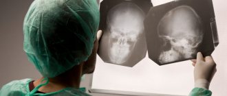
The first symptoms of the disease appeared in February, when the parents went to the ophthalmologist with the child’s complaints of pain in the eyes. A diagnosis of conjunctivitis was made, followed by abdominal pain and repeated vomiting. The child was examined by a pediatrician who diagnosed “biliary dyskinesia, chronic gastroduodenitis.” From this period, the boy vomited once a week. Over the next 2 months he attended school. At the very end of March, the condition worsened, with repeated vomiting, general weakness, ataxia, and abdominal pain. The patient was hospitalized in one of the hospitals with a diagnosis of chronic gastroduodenitis. Over the course of 24 hours, the child’s condition gradually worsened and neurological symptoms developed (ataxia, right-sided hemiparesis). During an examination by a neurologist on April 1, a suspicion of encephalitis or a space-occupying lesion in the brain arose. A computed tomography scan of the brain with the introduction of a contrast agent on 01.04: in both hemispheres of the cerebrum and the right hemisphere of the cerebellum, several areas were identified that actively accumulated a contrast agent. The largest of them, measuring 17x25 mm, was located in the region of the posterior sections of the corona radiata on the left, adjacent to the posterior sections of the body of the right lateral ventricle and extended to the splenium of the corpus callosum (Fig. 1).
Rice. 1. CT scan of the brain with intravenous contrast (ultravist-300) of patient K., 12 years old. In both hemispheres of the cerebrum and in the right hemisphere of the cerebellum, areas of accumulation of the contrast agent were identified. The largest area is adjacent to the posterior sections of the right lateral ventricle.
MRI of the brain revealed changes in both hemispheres of the cerebrum, the cerebellum, and the brainstem, involving both the white and gray matter of the brain (Fig. 2).
Rice. 2. MRI of the brain of patient K., 12 years old. Axial plane, T2-WI, T1-WI, TIRM-IP. Diffuse changes in white matter involving the gray cerebral hemispheres and cerebellum. The volumetric effect is moderate, the ventricles of the brain and the sulci of the hemispheres are narrowed. It was concluded that it was necessary to differentiate the development of the following diseases in the patient: demyelinating disease (disseminated encephalomyelitis), infectious process (encephalitis), vasculitis, focal processes of another nature.
The patient also underwent a computed tomography scan of the chest area. Single intralobular foci of tissue compaction were detected in the S9 segment of the left lung. The diameter of the bronchi in the lower lobes of both lungs is diffusely expanded. The course and patency of the trachea and bronchi are preserved. The intrathoracic lymph nodes are not enlarged. According to the radiologist, changes in the left lung represent foci of acute bronchiolitis.
On 02.04, based on clinical data and the results of radiological diagnostics, neurosurgeons excluded the diagnosis of “space-occupying brain mass”. After consulting a phthisiatrician on April 5, tuberculosis was ruled out.
After admission to the hospital, the patient received the following therapy: antiviral drug - medovir 350 mg 2 times IV (30 mg/kg/day), dexazone 8 mg 4 times a day IV (1 mg/kg/day), lendacin 1 g per day, infusion therapy, including the drug cytoflavin (1 ml/kg/day, intravenous drip, 5 infusions), diacarb, asparkam.
After a full examination, on April 6, the patient was transferred to the Children's Research and Clinical Center for Infectious Diseases with a diagnosis of encephalitis.
Upon admission to the institute's clinic, the boy was in clear consciousness. He could sit up in bed independently, but stood and moved only with support. The child was oriented in time and space and answered all questions correctly. Upon examination, a restriction in the horizontal movement of the eyeballs was revealed, as well as large-scale horizontal and vertical nystagmus, intensifying in the extreme abductions. Deviation of the tongue to the left. Phonation and swallowing were not impaired. Palatine and pharyngeal reflexes are alive. A decrease in muscle strength was detected in the right extremities, more in the leg - up to 3 points. Deep reflexes asymmetrical, D
>
S
. Pathological reflexes were determined in the lower and upper extremities: the Babinsky and Marinescu-Rodovic symptoms are bilateral, more pronounced on the right. Ataxia was noted in the trunk and in the extremities, and misses were noted when performing finger-to-toe and knee-heel tests. No superficial or deep sensitivity disorders have been identified. Abdominal reflexes were reduced and were worse evoked on the left. From the internal organs without pathology. Peripheral lymph nodes are not enlarged. The liver and spleen are not enlarged.
Repeated consultations with the ophthalmologist were carried out, which showed the presence of signs of intracranial hypertension, as well as manifestations of allergic conjunctivitis. When examining the fundus of the eye, no symptoms such as atrophy or congestion of the optic discs were detected.
Results of laboratory examination: in the clinical blood test dated 04/07, no deviations from the norm were noted. On April 10, a lumbar puncture was performed for diagnostic purposes; no changes were detected in the CSF: cytosis - 6/3 (6 mononuclear cells), protein - 0.209 g/l. Biochemical blood test dated 04/08/10: prothrombin time - 14.2 s; fibrinogen according to Rutberg - 3.22 µmol/l, prothrombin - 79.4%, ALT - 16.5 U/l; AST - 21.0 U/l, bilirubin - 9.6 µmol/l, creatinine - 67 µmol/l, urea - 3.5 mmol/l, glucose - 4.3 mmol/l (all values were within normal limits) . Laboratory diagnostic methods also included blood and CSF testing using polymerase chain reaction (PCR), immunocytochemistry, and enzyme-linked immunosorbent assay (ELISA). A negative result was obtained during a PCR study of blood and CSF for herpes viruses type 1-2, Epstein-Barr virus (EBV), cytomegalovirus (CMV), herpes type 6, enteroviruses, toxoplasma, chlamydia, and mycoplasma. Immunocytochemical examination of a smear from the conjunctiva for type 2 herpes gave a positive result; for EBV, CMV, chlamydia, toxoplasma - negative. Immunocytochemical study of CSF and blood cells for herpes type 6, herpes type 2 - positive result; for EBV, CMV, chlamydia, mycoplasma, toxoplasma - negative. Blood ELISA for herpes type 1 revealed antibodies of class M, G. IgG to CMV, herpes virus type 6 and EBV IgG (NA) were also detected in the blood. Blood ELISA for toxoplasma, chlamydia and mycoplasma IgM and IgG gave a negative result. Immunological examination: CSF CEC - 0.057 (normal up to 0.050), blood CEC from 04/08. - 134 (normal up to 135), blood immunoglobulins: IgM - 1.1 (normal), IgG - 9.0 (normal), IgA - 1.1 (normal). The CSF revealed an increased content of myelin basic protein - 5.5 ng/ml (normal up to 0.5).
Based on the presented examination results, a diagnosis of “disseminated encephalitis, herpes viral etiology, prolonged course” was made.
At the institute, the patient continued antiviral therapy (Medovir 350 mg 3 times a day intravenously, alpha-2 interferons intramuscularly), as well as neurometabolic (cytoflavin) and dehydration therapy. On April 10 and April 11, he received intravenous immunoglobulin G 7.5 g per day (0.25 g/kg per day).
By 13.04, the neurological status began to show positive dynamics in the patient’s condition: both horizontal and vertical nystagmus decreased. I started walking with support, my ataxia decreased, and my appetite improved. Positive dynamics continued until 22.04. But on April 23, the patient’s condition worsened again. In the clinical picture of the disease, the severity of focal symptoms and ataxia increased, and nystagmus intensified. To clarify the reasons for the deterioration, the patient was consulted by a neurosurgeon and a repeat MRI of the brain was prescribed. Hormone therapy was prescribed. But since the patient's condition did not improve, he was transferred to the intensive care unit. At 12:23:04 he developed tonic convulsions with loss of consciousness. Dormicum 10 mg was administered for anticonvulsant purposes, but at 3 p.m. repeated convulsions with respiratory arrest occurred and the child was transferred to mechanical ventilation.
To exclude the development of subarachnoid or intracerebral hemorrhage against the background of an ongoing infectious process, a space-occupying lesion of non-viral etiology, the patient was re-consulted by a neurosurgeon (the diagnosis of a space-occupying lesion was not made). A repeat diagnostic lumbar puncture was also performed, but the CSF parameters were not changed (cytosis - 5/3, protein - 0.209 g/l).
The deterioration of the patient's condition began to be associated with the progression of foci of demyelination against the background of the current infectious process, and therefore virological and bacteriological studies were carried out, but the next day the patient's condition sharply worsened. Signs of cerebral edema appeared. On April 25, during a clinical examination and neurophysiological studies (evoked brain potentials, Dopplerography of cerebral vessels, EEG), the patient was diagnosed with a 3rd degree coma. Despite active resuscitation measures (the patient received infusion therapy with cytoflavin, hormones - dexazone, methylprednisolone, mannitol, antiviral drugs), on April 27, at 10 a.m., cardiac arrest occurred, and at 10:30 a.m., death was pronounced.
A postmortem examination revealed a large malignant tumor in the region of the right cerebellopontine angle of the brain with invasion of pontine tissue into the right hemisphere of the cerebellum and perifocal microcirculation disorders (Fig. 3).
Rice. 3. Tumor in the area of the right cerebellopontine angle of the brain. There were also multiple foci of metastasis in the white matter of the cerebral hemispheres and areas of invasion of tumor tissue in the cortex and adjacent pia mater, growth into the walls of the lateral ventricles, tissue of the choroid plexus of the right lateral ventricle and structures of the subcortical nuclei. Histological examination revealed polymorphic cell glioblastoma (IV degree of malignancy according to WHO classification, 2007), expressing S-100, with high proliferative activity (more than 25% of tumor cells expressing Ki-67), scant vimentin-positive stroma (Fig. 4).
Rice. 4. Microscopic picture of polymorphic cell glioblastoma. Staining with hematoxylin and eosin, UV. 400. The immediate cause of death was dislocation of the brain stem with their wedging into the foramen magnum with progressive edema and swelling of the brain. Pathomorphological examination also revealed a generalized herpes viral infection (herpes virus types 2 and 6) with damage to the tissue of the brain, lungs, small and large intestines, as well as CMV infection in the form of sialadenitis (Fig. 5, 6) .
Rice. 6. Expression of herpes type 2 and 6 antigens in brain tissue. Immunohistochemical reaction, DAB, uv. 400.
Rice. 5. Expression of S-100 by tumor tissue. Immunohistochemical reaction, DAB, uv. 400.
The presented clinical observation indicates the difficulties of differential diagnosis of disseminated encephalitis and brain space-occupying lesions, when clinical symptoms of the disease manifested already with pronounced MRI changes caused by infiltrative tumor growth. Tumor growth in this case was also accompanied by ischemic and hypoxic disorders, which led to the development of widespread damage to both the white and gray matter of both hemispheres, brainstem and cerebellum. A diffuse change in the MR signal during an MRI study of the brain, as well as the absence of a pronounced mass effect, did not allow a correct diagnosis.
It is known that glioblastoma is a highly malignant glioma, which is characterized by the presence of multiple foci of necrosis, pronounced changes in the vessels in the form of arteriovenous shunts and blastomatous proliferation of adventitia and endothelial cells. Rapid growth of the tumor and hemorrhages into it usually lead to a sharp deterioration of the condition and death, which was observed in the presented case.
This observation confirms the need for wider use of brain biopsy in children, which is the standard for diagnosing not only space-occupying lesions, but also inflammatory diseases of the central nervous system. This will avoid a lengthy, sometimes erroneous, diagnostic search and ensure timely establishment of the correct diagnosis.
The authors declare no conflict of interest.
Treatment of residual encephalopathy
Neurologists at the Ardenium Clinic select individual treatment for each patient, taking into account the causes of the disease, symptoms and their severity, and the severity of the condition.
Typically, residual encephalopathy is treated with conservative methods, which may include:
- drug therapy – eliminates unpleasant symptoms, restores brain activity, normalizes metabolism and cerebral circulation;
- physiotherapy – activates the body’s defenses, mobilizes them to fight the disease;
- therapeutic massage – stimulates blood flow to the brain, providing it with nutrients and oxygen;
- physical therapy – activates blood circulation, restores motor activity, improves coordination.
If the disease is detected in a timely manner, complete recovery can be achieved. In advanced cases, it is impossible to completely restore the functioning of the brain, but it is possible to slow down the progression of the pathology and significantly improve the general condition of the patient.
What symptoms suggest perinatal brain damage?
- Perception disorders;
- Hyperactivity or lethargy;
- Nervousness of the child, frequent mood swings, hysterics;
- Speech disorders;
- Sleep disorders;
- Headache;
- Movement disorders: unnatural movements, obsessive movements, nervous tics;
- Involuntary muscle contractions (cramps);
- Sleep disorders.
And others.