1.General information
It is known that dizziness is one of the serious problems of modern medicine, and several scientific directions and medical specialties, as well as international and national associations, are working on this problem. Despite the prevalence and seeming self-evidence of dizziness as a pathological phenomenon, it is very difficult for researchers to give it a clear, comprehensive definition, and for patients to verbalize their sensations. In addition, today there are many known pathological conditions, diseases, syndromes, the clinical picture of which includes dizziness as the dominant or one of the leading symptoms; Accordingly, it is necessary to introduce various classifications of dizziness, based on etiological, clinical and other criteria.
Vestibular neuronitis, or acute peripheral vestibulopathy, is one of the most common causes of paroxysmal dizziness (according to various sources, the second or third most common). The disease was first described more than a hundred years ago, but many of its aspects remain unclear to this day and are the subject of study. It is generally accepted that the peak incidence occurs in young and middle age, although vestibular neuronitis is recorded with varying frequency in all age groups.
A must read! Help with treatment and hospitalization!
Peripheral vestibular syndrome as a cause of dizziness and balance disorders
Recently, interest in the problems of dizziness and balance disorders has increased significantly. This is due not only to the growing number of patients turning to doctors with similar complaints, but also to the emergence of new diagnostic methods, the creation of both specific pharmacological drugs and methods of rehabilitation therapy. It is still very important for the doctor to accurately establish the cause of the disease and its form in order to further prescribe adequate therapy. Based on the experience gained over five years in a specialized center for dizziness and balance disorders, we concluded that in more than 50% of patients at the time of treatment, the appearance of such symptoms is due to a whole range of reasons. This is explained by the complex structure of the balance system and the vestibular system.
For a practicing physician, it is important to familiarize yourself in detail with the complaints presented by the patient and the history of the development of the disease. The use of additional modern diagnostic methods and consultation with specialists significantly increases the efficiency of diagnosis. First of all, among additional diagnostic methods, vestibulometry and posturography (possibly stabilometry) should be noted. If the first method, which is a set of tests with computer data processing, allows you to answer questions about the functional state of the vestibular system, including changes in the vestibular receptor, central vestibular structures, brain stem, cerebellum; state of the oculomotor system; cervical receptors, etc., then posturography makes it possible to assess the state of balance (functional composition of components - vision, vestibular system, proprioceptors), including for individual selection and rehabilitation therapy. Duplex ultrasound examination of the main blood flow of the transcranial and cervical sections allows, in addition to conventional procedures, to conduct studies with functional tests and obtain information about venous blood flow. In combination with rotary tables, this method makes it possible to obtain information about increased intracranial pressure.
In practice, we are faced with vestibular pathology, which can be divided into peripheral, central and mixed vestibular syndromes. The choice of the attending physician in the existing system of specialized care most often occurs as follows. If the patient has cochleovestibular syndrome, he is referred to an audiologist; vestibular syndrome without hearing changes - see a neurologist. In our opinion, at the first stage of the examination, consultations with a neurologist and an otoneurologist (otorhinolaryngologist) are mandatory, since peripheral vestibular syndrome without audiological symptoms can be the cause of the development of diseases of the inner ear, which should be discussed in more detail.
The group of diseases of the inner ear without changes in hearing includes: benign paroxysmal positional vertigo (otolithiasis), vestibular neuronitis, labyrinth fistula, vestibular form of Meniere's disease and secondary labyrinthine hydrops.
Benign paroxysmal positional vertigo (BPPV) is one of the most common causes of vertigo associated with pathology of the inner ear. Patients with this pathology develop short-term attacks of dizziness and nystagmus (usually lasting less than 30 seconds) when changing the position of the body and head: usually when turning in bed, getting in and out of bed, bending and straightening, or stretching the neck to look up.
The basic features of BPPV and associated positional nystagmus were described by Robert Barany (Barany, 1921), but the term did not exist until 1952, when Dix and Hallpike described the provocative positional test and clearly defined the syndrome (Dix and Hallpike, 1952).
The diagnosis of BPPV continues to rely on the finding of a characteristic positional nystagmus (also called paroxysmal positional nystagmus) in a patient with a typical history of positional vertigo. Nystagmus can be observed during visual inspection in the Dix-Hallpike test, however, since it occurs due to pathology of the vestibular receptor (i.e., it is peripheral), it is susceptible to depression when fixating gaze, so diagnosis involves the use of Frenzel glasses or videonystagmography. Currently, we have accumulated experience in treating more than 100 patients with a similar pathology. Treatment allows particles to be moved from the semicircular canals into the vestibule of the labyrinth and thus relieve dizziness. The effectiveness of the procedures, according to our and foreign sources, ranges from 90-95%.
Acute vestibular neuronitis
This is a disease with an as yet unknown etiology. The most popular hypothesis is about viral etiology. It is characterized by attacks of vestibular dizziness, often accompanied by autonomic reactions (nausea and vomiting). Any movement of the head increases the feeling of dizziness. The duration of the disease ranges from several hours to several days. An important sign is the absence, as with all diseases of this group, of hearing changes. During the examination, typical vestibular peripheral nystagmus can be observed against the background of an asymmetric lesion, which will be combined with a corresponding deviation of the hands and posture when performing statokinetic tests. Treatment is usually symptomatic with the use of vestibular suppressants in the first stage. Special mention should be made of toxic neuronitis caused by exposure to aminoglycoside antibiotics, especially when it comes to gentamicin. The introduction of gentamicin, one or two times, through the eardrum, in its effectiveness, successfully replaces surgery to destroy the labyrinth (if it needs to be performed when other methods of treatment are absolutely ineffective).
A labyrinthine fistula can be no less difficult to diagnose. This disease is characterized by the appearance of a communicating opening or canal between the middle and inner ear, resulting in the leakage of fluid from the inner ear into the cavity of the middle ear. The hydrodynamic system is disrupted, and then the state of the sensory cells. Most often, a fistula is formed due to barotrauma (for example, diving, coughing, sneezing). A characteristic symptom is the appearance of dizziness during the Valsalva maneuver (or similar loads - straining or lifting heavy objects). Fistulas tend to close on their own, but due to the frequently occurring increased intralabyrinthine pressure (hydropses), they can become chronic. Electrophysiological, radiological and other research methods cannot give a reliable answer to the question of the presence of a fistula. Therefore, after an examination and with certain test results, the issue of diagnostic myringotomy (surgical separation of the eardrum for revision of the tympanic cavity) is decided. When a fistula is detected, it is repaired.
The vestibular form of Meniere's disease has been the subject of separate studies, conducted mainly abroad. Typical Meniere's disease is characterized by the following symptoms. Usually the disease begins at the age of 25-45 years, men are more often affected. The disease is accompanied by attacks of vestibular dizziness, lasting up to 6-12 hours, tinnitus, fluctuating hearing loss of the sensorineural type with a tendency to progress in the degree of hearing loss, a feeling of discomfort, fullness in the affected ear.
The existence of a large number of patients with secondary hydrops of the labyrinth is noteworthy. The term “secondary” suggests that the increase in intralabyrinthine pressure occurs due to causes that are systemic, and what occurs in the inner ear reflects the manifestation of these changes. In one of the issues of “The Treating Doctor” (No. 9, 2000) we dwelt in detail on the features of diagnosis and treatment of this pathology.
Such changes require appropriate therapy, including at least two components: normalization of the state of venous outflow and treatment of labyrinthine hydrops. At this stage, work with the patient should also be carried out with the direct participation of a neurologist-vertebrologist (chiropractor) and an otoneurologist. An individual combination of physical and physiotherapeutic methods, as well as combination therapy of hydrops (dehydration, vestibular suppression, vasoactive effect on regional blood flow in the inner ear) make it possible to achieve a cure for the patient. Electrocochleography also remains a method of stage-by-stage assessment of the state of pressure in the inner ear.
Recent research in the field of inner ear immunology suggests that labyrinthine hydrops may be a consequence of an autoimmune lesion of the inner ear. Morphological changes are characterized by: degeneration of spiral ganglion cells, atrophy of the organ of Corti, arteritis around the cochlear nerve and stria vascularis, and the development of endolymphatic hydrops. For vestibular disorders, dizziness is not a constant symptom. If it is present, it is similar to Meniere-like dizziness, combined with ataxia and attacks of sudden falling. A vestibulometric study reveals bilateral symmetrical peripheral vestibular dysfunction. The clinical picture of the disease includes various combinations of auditory and vestibular symptoms, as well as their complete absence against the background of only sudden falls. For a practicing physician, the ability to determine the cause of the development of inner ear pathology serves as an indication for prescribing corticosteroid anti-inflammatory therapy.
Treatment of peripheral vestibular syndrome can be divided into two stages - the active manifestation of the disease and the subacute state. At the first stage, it is important to carry out pathogenetic therapy in combination with the prescription of vestibular suppressants and antiemetic drugs when dizziness is combined with vegetative manifestations.
Vestibular suppressants occupy the main place in the treatment of dizziness. The term “vestibular suppressant” is a collective term: this is usually the name given to drugs that reduce nystagmus and the feeling of dizziness, vestibular instability, or stop motion sickness (motion sickness).
At least four major neurotransmitters of the vestibular system are involved in the formation of the vestibulo-ocular reflex: glutamate, acetylcholine, gamma-aminobutyric acid and glycine, histamine.
Glutamate is the main excitatory neurotransmitter. Acetylcholine is both a peripheral and central agonist acting at muscarinic receptors. These receptors are found in the brain stem and bone marrow. There are reports of their possible participation in the formation of dizziness. Gamma-aminobutyric acid and glycine are inhibitory neurotransmitters found in second-order vestibular neurons and oculomotor neurons. Histamine is found in various central vestibular structures, where it is present diffusely.
Anticholinergics act on muscarinic receptors. An important feature for this group of drugs is that they do not cross the blood-brain barrier and are therefore ineffective in treating motion sickness. Also, unlike antihistamines, anticholinergic drugs are not effective if used after dizziness has already begun.
All anticholinergics used to treat dizziness have side effects such as dilated pupils and sedation. The most commonly used drugs in this group are scopolamine and atropine.
There is evidence that centrally acting antihistamines prevent motion sickness and reduce the severity of vestibular manifestations, even when symptoms have already appeared. Most antihistamines also have a calcium channel blocking effect. However, the sedative effect that antihistamines have has an adverse effect on the processes of vestibular adaptation, so this group of drugs should not be recommended to patients for long-term use.
Benzodiazepine drugs - GABA modulators - act centrally to suppress vestibular responses. These drugs are used in small doses. Their main disadvantages are addiction, memory impairment, increased risk of falls, and negative effects on vestibular compensation.
Betaserc (betahistine dihydrochloride) is characterized by a successful combination of the properties of a pathogenetic drug with the effect of suppressing the feeling of dizziness. When exposed to two groups of H-receptors, an increase in blood flow in the labyrinthine artery and suppression of information passing through the vestibular nuclei simultaneously occur. Betaserc can be considered as the drug of choice. Depending on the pathogenesis of the disease, betaserc can be combined with other therapeutic agents (with the exception of antihistamines, the combination with which leads to a weakening of the therapeutic effect of both drugs).
Peripheral vestibular pathology against the background of persistent ataxia in the future, as a rule, is compensated independently. However, in order to speed up the patient's recovery, vestibular rehabilitation can be used. This is a set of exercises performed independently or on special installations (posturographs or stabilographs) with biofeedback. The inclusion of vestibular rehabilitation in the complex therapy of peripheral vestibular syndrome, in our opinion, is justified and should be performed under the supervision of a physician. It is recommended that such exercises be prescribed after the patient has finished using vestibular suppressants. Perhaps the betaserc is an exception, since recently works have appeared in the domestic literature indicating the effectiveness of vestibular rehabilitation when using the betaserc. With age, the balance system deteriorates due to changes in the structure of sensory systems and nervous tissue. Therefore, in older people, even in the presence of peripheral vestibular syndrome, it is recommended to use vestibular rehabilitation.
In conclusion, I would like to emphasize that the success of working with patients suffering from dizziness and balance disorders depends on the diagnostic capabilities of the medical institution, the interaction of doctors of various specialties and the use of all means effective for the treatment and rehabilitation of patients.
O. A. Melnikov, Candidate of Medical Sciences, ANO "GUTA-Clinic", Moscow
2. Reasons
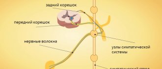
It is assumed that the source of the vestibulopathic symptom complex is inflammation of the paired vestibular nerve. Various hypotheses have been put forward as to why this particular pair of cranial nerves is involved in the inflammatory process, while the neighboring ones remain intact; In addition, the etiopathogenesis of the inflammation itself is unknown. Considering the results of numerous clinical, medical-statistical, microbiological, pathomorphological and other studies carried out in this regard, the most likely causes today seem to be neuroinfection (especially herpetic), food intoxication, immune-allergic reaction (most likely, again, this is a reaction to some kind of respiratory or other infection), or a specific, possibly genetically determined, metabolic disorder. The most well-reasoned is the viral hypothesis, which is confirmed by a large number of accumulated observations, where the chronological connection between the acute respiratory viral infection and the subsequent development of acute peripheral vestibulopathy is clearly visible; it is legitimate to assume that this connection is also cause-and-effect.
Visit our Neurology page
Description and causes of pathology
The disease is characterized by partial damage to the nerve of the vestibular apparatus, manifested by systematic attacks of dizziness with loss of balance. These attacks do not affect sound perception, however, complete restoration of coordination function can take a long period - up to several weeks. The attacks themselves are also long-lasting; dizziness can last from two to three hours to 3-4 days. The pathological condition was first described at the beginning of the 20th century, a more detailed description of the abnormal process was created in 1924, and the name and classification of the disease in scientific and practical neurology took root only by the end of the forties.
The pathology occurs quite often and is the third most common case of dizziness syndrome. The age category susceptible to the disease is adults from 30 to 60 years. The peak incidence is observed in the pre-summer months (end of April - May). The causes of the pathological condition are various etiological factors, so treatment is prescribed on an individual basis. It is important for a specialist to identify the source of the disease before planning restorative measures.
The origins of the disease have not yet been fully elucidated. Most doctors tend to assume that the source is an inflammatory process that partially affects the vestibular nerve fibers. Most often, dizziness occurs after a viral infection (ARVI). The pathology itself also provokes conditions such as developing encephalitis, which leads to the assumption that the primary source of the anomaly is the herpes virus in the body in an incubation state. The theory is also supported by numerous cases in which neuronitis of the vestibular apparatus manifested itself within the boundaries of one family living in the same house at the same time.
Some experts are inclined to think that the primary infection also affects the processes of the immune reaction, triggering subsequent allergic rejection. As a result of this reaction, inflammation of the nerve fibers occurs, and the pathogen in the form of a virus only catalyzes this reaction. Most often, the upper zone of the nerve canal of the vestibular apparatus is affected; inflammation of the lower zone is much less common. In this case, adjacent auditory nerve fibers remain untouched. There are cases when the disease starts as a result of toxic effects on the body. Typically, provocateurs are antibacterial agents (antibiotics) taken for a long period of time or in high dosages.
3.Symptoms
Vestibular neuronitis manifests itself acutely and occurs in paroxysms, usually in the form of a single episode, occasionally with distant relapses. In most cases, an attack of acute peripheral vestibulopathy resolves spontaneously within a few days, leaving residual symptoms that also gradually subside over a few weeks. It is known, however, that in elderly and senile people the period of convalescence (full recovery) often drags on for months.
Classic symptoms are intense, unbearable dizziness; nausea, vomiting; balance and coordination disorders. A neurological examination reveals nystagmoid movements of the eyeballs and ataxia in the Romberg position.
Diagnosis of vestibular neuronitis is quite complex; it requires a doctor to be highly qualified and have extensive clinical experience, since the same clinical manifestations are inherent in many other diseases. One of the most important differential diagnostic criteria is, in particular, the absence of hearing impairment. Differential diagnosis may also require additional studies (IMEP, MRI, etc.).
About our clinic Chistye Prudy metro station Medintercom page!
Signs of vestibular neuronitis
The main symptom is severe dizziness, manifested by a sensation of continued movement in space without physical effort being transmitted to the body. It seems to a person that his body is spinning, falling (failing) or swaying on invisible waves. Surrounding objects can also move illusorily. Depending on the side of the nerve fibers affected, the movement of objects will be felt. Things “move” in the direction in which the inflamed nerve is located. The severity of dizziness, as a rule, increases when trying to change the position of the body, turn the head in a different direction, or engage in physical activity. The intensity of the anomaly decreases somewhat if the patient tries to focus on one visual point or object. In parallel with the unstable condition of the patient, severe nausea and vomiting may occur. When trying to stand up or move independently, the body does not obey, staggers, and there is no balance.
Some patients note that the main long-term attack was preceded several days or hours before by episodic, momentary disturbances in coordination and balance. In an acute state, the pathology persists from a couple of hours to a couple of days, then coordination is restored, but not completely. Mild unsteadiness persists for up to several weeks. After an attack, no relapse is observed. The exception is only 2% of patients who suffer the disease a second time on the symmetrical, previously healthy side. If attacks occur several times a second time, then doctors need to reconsider the diagnosis.
If the nerve column has undergone severe inflammation, persistent vestibular dysfunction may persist. Over time, it is also compensated; this phenomenon does not significantly affect the quality of life and general nervous functions.
4.Treatment
Therapy, due to the etiological uncertainty of the disease, is palliative in nature and is aimed at eliminating the dominant symptoms. Prescribe vestibular suppressants, drugs to suppress the urge to vomit, according to indications - tranquilizers, diuretics, etc. Special vestibular gymnastics has a good effect. It is important to note that without following the instructions for the rehabilitation period, there is a very high probability of the process becoming chronic with residual clinical signs of unilateral vestibular damage.
Diagnostic methods used
At the first signs of the onset of the disease, you should seek advice from highly specialized specialists - a neurologist and an otolaryngologist. Since coordination dysfunctions persist for a long time, identifying the disease is not difficult. However, there are diseases that have similar symptoms; it is important to differentiate them from vestibular neuronitis. Initially, a thorough examination, history taking, and motor testing are performed. When the body is positioned in certain positions, it deviates towards the affected nerve. The gaze also tries to shift towards the diseased nerve. These tests are effective for two weeks after an attack.
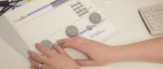
Audiometry, used by an otolaryngologist as a research method, excludes changes that have occurred in the hearing organs. If the patient does not have hearing loss, there is reason to believe that the vestibular nerve is only partially affected by the disease, which is one of the signs of the disease in question.
A mandatory step in the examination is to contact a vestibulologist, who will determine the general condition of the vestibular apparatus using instrumental methods. These methods make it possible to determine the unilateral nature of changes in nerve fibers. In clinical cases, magnetic resonance imaging of the brain comes to the aid of specialists, which will help exclude other degenerative processes in the nervous network and identify indirect signs of the disease.
If the main signs of the disease are supplemented by parallel acute otitis media, hearing impairment, a post-traumatic condition, previous exposure to sharply changing external atmospheric pressure, straining, severe cough syndrome, tinnitus, a bursting sensation inside the ear, headaches, then doctors need to revise the primary diagnosis towards other disorders. Also, if all signs of neuronitis completely disappear within the first 24 hours after the initial attack, there is a high probability that the person has suffered an ischemic attack.
Consequences and complications
A severe complication of vestibulopathy can be purulent labyrinthitis , which, in turn, sometimes develops into meningitis . Therefore, inflammation in the inner ear must be treated under the close supervision of a doctor.
Vestibulopathy, moreover, negatively affects the overall quality of life. Dizziness and migraines impair work ability. In addition, such manifestations can cause accidents and traffic accidents.
In advanced stages, the auditory nerve may be affected, leading to the development of hearing loss.
This condition can also lead to cognitive impairment.
Prevention
The following prevention methods should be practiced:
- Limit situations that provoke an attack of vestibulopathy.
- Eat right, avoiding lack of vitamins and minerals. If necessary, take vitamin-mineral complexes periodically.
- Get proper rest, sleep at least 7 hours a day.
- To refuse from bad habits.
- Exercise daily, do yoga, Pilates, swimming.
- Treat bacterial and viral diseases in a timely manner.
- If vestibulopathy occurs, consult a doctor.
Literature sources
Brandt T., Dieterich M., Strupp M. Dizziness: trans. from English // M.: Praktika, 2009.
Bronstein A., Lempert T. Dizziness; lane from English, ed. V.A. Parfenova; [preface N.N. Yakhno]. – M.: GEOTAR-Media, 2010.
Parfenov V.A., Zamergrad M.V., Melnikov O.A. Dizziness: diagnosis and treatment, common diagnostic errors: Textbook. // M.: MIA, 2009.
Kattah JC, Talkad AV, Wang DZ, Hsieh YH, Newman-Toker DE. HINTS to diagnose stroke in the acute vestibular syndrome: three-step bedside oculomotor examination more sensitive than early MRI diffusion-weighted imaging. Stroke. 2009 Nov;40(11):3504-10.
Diet
Salt-free diet
- Efficiency: 5-10 kg in 13 days
- Time frame: 13 days
- Cost of products: 4500-8000 rubles for 13 days
For some diseases that provoke vestibulopathy, it is recommended to adhere to a salt-free diet or limit the amount of salt in the menu.
In general, nutrition should be rational and varied, and the diet must include fresh fruits and vegetables, meat, fish, dairy products, nuts, and vegetable oils.
You should give up alcohol.
General information
Vestibulopathy is a disorder of the normal functioning of the vestibular apparatus , in which characteristic symptoms appear.
The ICD-10 code for vestibulopathy is H81 (Vestibular function disorders). The normal orientation of a person in space and his balance in a vertical position is ensured by the vestibular apparatus. Disturbances in its work lead to a person experiencing confusion, dizziness , and the inability to remain in a standing position. This condition is also called labyrinthopathy. It can develop due to various reasons and diseases, developing at any age in every person. Sometimes vestibulopathy is a congenital pathology.
It may manifest itself as a separate syndrome or as a sign of neuropsychiatric disorders. In the second case, it is more difficult to treat this disease. How this pathology manifests itself, and what treatment methods are advisable to practice, will be discussed in this article.