An interneuron, also known as an associative or interneuron, is present only in the tissues of the central nervous system and is interconnected exclusively with other nerve cells. This feature distinguishes it from its sensory or motor counterparts. Sensory systems interact with other systems of the body, for example, with skin receptors and sensory organs, when they convert stimuli coming from the external environment into bioelectric signals. Motor cells innervate muscle fibers and provide human motor activity.
Pyramidal neurons
Pyramidal neurons are one of the different types of neurons present in our nervous system. They are one of the most common types of multipolar neurons, accounting for about 80% of the neurons in the cortex (not for nothing, the two layers of the cortex are called the inner and outer pyramids) and are found between some of the the most relevant from the body.
They are generally considered to be projection neurons. That is, they operate by sending messages to cells that are far away and separated from the place where they were born. Discovered by Santiago Ramon y Cajal The name of this type of neuron refers to the shape of its soma, with a triangular or pyramidal appearance. These are mainly glutamatergic neurons, with glutamate being the neurotransmitter that activates them, and they typically act as excitatory type neurons. They can have different sizes, the largest of which are giant pyramidal cells or Betz cells.
Like other neurons, the structure of this type of neuron consists of a soma, which, as we have already said, has a pyramidal shape, an axon and dendrites. However, they have their own peculiarity: as for the dendrites, they are quite long compared to the rest, called the apical dendrite and numerous basal and shorter dendrites that branch.
- Processes
- Basic functions of the spinal cord
- What is it needed for
- Classification
- Neuron and its structure
- Structure of neurons
- Characteristics of neurons
- Neuron development and growth
- Conclusion
Origin of the approach
Since the mid-twentieth century, it has been known that the brain consumes a significant part of the energy resources of the whole organism: a quarter of all glucose and ⅕ of all oxygen in the case of the great ape [1–5]. This inspired William Levy and Robert Baxter from the Massachusetts Institute of Technology (USA) to conduct a theoretical analysis of the energy efficiency of information encoding in biological neural networks (Fig. 1) [6]. The study is based on the following hypothesis. Since the brain's energy consumption is high, it is beneficial for it to have neurons that work most efficiently - transmitting only useful information and expending a minimum of energy.
This assumption turned out to be true: using a simple neural network model, the authors reproduced the experimentally measured values of some parameters [6]. In particular, the optimal frequency of impulse generation they calculated varies from 6 to 43 impulses/s - almost the same as for neurons at the base of the hippocampus. They can be divided into two groups according to the pulse frequency: slow (~10 pulses/s) and fast (~40 pulses/s). Moreover, the first group significantly outnumbers the second [7]. A similar picture is observed in the cerebral cortex: there are several times more slow pyramidal neurons (~4-9 impulses/s) than fast inhibitory interneurons (>100 impulses/s) [8], [9]. Thus, apparently, the brain “prefers” to use fewer fast and energy-consuming neurons so that they do not use up all their resources [6], [9–11].
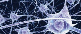
Figure 1. Two neurons are shown. In one of them, the presynaptic protein synaptophysin is colored purple. Another neuron is completely stained with green fluorescent protein. Small light specks are synaptic contacts between neurons [12]. In the inset, one “speck” is presented closer. Groups of neurons connected by synapses are called neural networks [13], [14]. For example, in the cerebral cortex, pyramidal neurons and interneurons form extensive networks. The coordinated “concert” work of these cells determines our higher cognitive and other abilities. Similar networks, only made up of different types of neurons, are distributed throughout the brain, connected in a certain way and organize the work of the entire organ.
website embryologie.uni-goettingen.de
What are interneurons?
Neurons of the central nervous system are divided into activating (forming activating synapses) and inhibitory (forming inhibitory synapses). The latter are largely represented by interneurons , or intermediate neurons. In the cerebral cortex and hippocampus, they are responsible for the formation of gamma rhythms in the brain [15], which ensure the coordinated, synchronous work of other neurons. This is extremely important for motor functions, perception of sensory information, and memory formation [9], [11].
Interneurons are distinguished by their ability to generate significantly higher frequency signals than other neurons. They also contain more mitochondria, the main organelles of energy metabolism and ATP production factories. The latter also contain a large amount of proteins cytochrome c oxidase and cytochrome c, which are key for metabolism. Thus, interneurons are extremely important and, at the same time, energy-consuming cells [8], [9], [11], [16].
The work of Levy and Baxter [6] develops the concept of “economy of impulses” by Horace Barlow from the University of California (USA), who, by the way, is a descendant of Charles Darwin [17]. According to it, during the development of the body, neurons tend to work only with the most useful information, filtering out “extra” impulses, unnecessary and redundant information. However, this concept does not provide satisfactory results, since it does not take into account the metabolic costs associated with neuronal activity [6]. Levy and Baxter's extended approach, which focuses on both factors, has been more fruitful [6], [18–20]. Both the energy expenditure of neurons and the need to encode only useful information are important factors guiding brain evolution [6], [21–24]. Therefore, in order to better understand how the brain works, it is worth considering both of these characteristics: how much useful information a neuron transmits and how much energy it spends.
Recently, this approach has found many confirmations [10], [22], [24–26]. It allowed us to take a new look at the structure of the brain at various levels of organization - from molecular biophysical [20], [26] to organ [23]. It helps to understand what the trade-offs are between the function of a neuron and its energy cost and to what extent they are expressed.
How does this approach work?
Suppose we have a model of a neuron that describes its electrophysiological properties: action potential ( AP ) and postsynaptic potentials ( PSP ) (more on these terms below). We want to understand whether it works efficiently and whether it wastes an unreasonable amount of energy. To do this, it is necessary to calculate the values of the model parameters (for example, the density of channels in the membrane, the speed of their opening and closing), at which: (a) the maximum ratio of useful information to energy consumption is achieved and at the same time (b) realistic characteristics of the transmitted signals are preserved [6 ], [19].
Search for the optimum
In fact, we are talking about an optimization problem: finding the maximum of a function and determining the parameters under which it is achieved. In our case, the function is the ratio of the amount of useful information to energy costs. The amount of useful information can be approximately calculated using Shannon's formula, widely used in information theory [6], [18], [19]. There are two methods for calculating energy costs, and both give plausible results [10], [27]. One of them - the “ion counting method” - is based on counting the number of Na+ ions that entered the neuron during a particular signaling event (AP or PSP, see sidebar “What is an action potential”) with subsequent conversion to the number of adenosine triphosphate (ATP) molecules ), the main energy “currency” of cells [10]. The second is based on the description of ionic currents through the membrane according to the laws of electronics and allows one to calculate the power of the equivalent electrical circuit of a neuron, which is then converted into ATP costs [17].
These “optimal” parameter values must then be compared with those measured experimentally to determine how different they are. The overall picture of the differences will indicate the degree of optimization of a given neuron as a whole: how real, experimentally measured, parameter values coincide with the calculated ones. The less pronounced the differences, the closer the neuron is to the optimum and the more energetically it works optimally. On the other hand, a comparison of specific parameters will show in what specific quality this neuron is close to the “ideal”.
Next, in the context of the energetic efficiency of neurons, two processes on which the encoding and transmission of information in the brain are based are considered. This is a nerve impulse, or action potential, thanks to which information can be sent to the “addressee” over a certain distance (from micrometers to one and a half meters) and synaptic transmission, which underlies the actual transmission of a signal from one neuron to another.
Processes
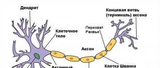
Neurons have axons (one in number) and dendrites (one or more).
Axon
It is a long outgrowth of the cytoplasm. It carries signals from the body to organs and other neurons. Its diameter is several microns, and its length in humans is several tens of centimeters. Growth depends on the soma: if damaged, its peripheral parts can die, but the main one continues to function.
The structure of axoplasm (axonal protoplasm) suggests the presence of neurofibrils (carrying out the supporting and drainage functions of neurons), microtubules (protein structures), mitochondria and the endoplasmic reticulum. In humans, the axons are covered and form pulpy nerve fibers. In such a shell there are oligodendrocytes, between which there are small parts freed from it. An action potential arises on them. The impulse is able to propagate through the pulp fibers in steps - due to this, the speed of information propagation increases.
Dendrites
Short and branched shoots. These parts of the neuron are fundamental for the formation of synapses, which influence the neuron and transmit excitation to the soma. Dendrites, unlike axons, do not have a myelin sheath.
How many input signals a nerve cell receives depends on the branching of the dendritic network and its complex structure. The main functions of dendrites are to increase the surface area for synapses, which makes it possible to integrate a large amount of information entering the nerve cell. In addition, they are able to generate action potentials and influence the occurrence of such potentials in axons.
The impulse is transmitted from the dendrite or soma to the axon. Once an action potential is generated, it is transmitted from the initial axonal portion back to the dendrites. When an axon articulates with the soma of a subsequent neuron, the contact is called axo-somatic. If with dendrites - axo-dendritic, and with the axon of another neuron - axo-axonal.
The structure of axons implies the presence of terminals - the so-called terminal sections. They branch and come into contact with other cells in the body (muscle, glandular, etc.). The axon has a synaptic terminal - the part that contacts the target cell. The postsynaptic membrane of such a cell, together with the synaptic ending, forms a synapse through which excitation is transmitted and through which cell interaction occurs.
How many connections can one neuron make? One nerve cell with the ability to communicate can make 20,000 connections.
Basic functions of the spinal cord
The work of this most important organ of the human body cannot be overestimated. The functions of the spinal cord are reflex and conductive. The first is responsible for such reflexes as:
- Flexion or flexion.
It is aimed at moving away from a dangerous stimulus, for example, withdrawing your palm from a hot object. - Stretch reflex.
It prevents excessive stretching of muscle fibers. - Other reflexes
, for example, tonic, rhythmic.
Each such reaction passes through a certain part of the nervous system - the nerve center. Here, information coming from different departments and bodies is analyzed, and in response, impulses are generated and sent to the executive bodies. For example, motor neurons are responsible for muscle contraction, and the sacrum is the center for urination - all these are functions of the spinal cord.
The conduction function is carried out through ascending and descending pathways. Through the first, the impulse is transmitted to the brain, and through the second, it returns back to the executing organs. Impulse channels with a long tract provide communication between the spinal cord and various parts of the brain; with the help of short ones, communication is carried out between neighboring elements of the spinal cord.
Conducting function of the spinal cord
You can learn more about its work if you understand the meaning of the pathways:
- Ascending paths.
The conductive function of the spinal cord is carried out thanks to centripetal nerve impulses, which transmit information from the spinal cord to the brain, notifying about changes occurring in the body. First, the signal, moving along the nerves, enters the dorsal roots, is processed by sensory neurons and sent either to the dorsal horns or to the cerebral hemispheres. - Descending paths.
The functions of the spinal cord ensure the transmission of impulses from the main organ of the central nervous system to motor neurons. From here, excitation travels through the spinal nerves to the executive organs.
Reflex function of the spinal cord
Those who ask what functions the spinal cord performs should answer that reflex work is carried out through the activation of a simple reflex arc. We are talking, for example, about flexion and extension movements of the limbs. In this case, the brain may be involved in the process. The spinal cord performs autonomic functions. It controls the functioning of the human internal environment, is responsible for digestion, urination, blood flow and more. Autonomic and motor reflexes are controlled by proprioceptors located deep in the spinal tissue.
Autonomic functions of the spinal cord
Those who are interested in what the spinal cord is needed for should answer that it is also responsible for autonomic reflexes. This is a reaction of internal organs, through which they respond to stimulation of somatic and visceral receptors. Autonomic centers are located in the lateral horns of the spinal cord. They regulate blood pressure, are responsible for heart rate and other operating parameters of the main “motor” of the body, motility and secretion of the digestive tract.
Order of interaction
The reflex regulation of body functions in an interpreted, simplified form is described in a biology textbook for the 8th grade. Interneurons, sensory and motor neurons are interconnected. The nature of the interaction depends on the type of functions of the nervous system. An approximate order of interaction in the case of the functions of sensory neurons that are localized in the skin area:
- Perception of an external stimulus by a nerve receptor located in the skin.
- Transmission of stimulus by sensory cells to areas of the brain. Typically, the signal passes through 2 synapses (in the spinal cord and thalamus), then enters the sensory zone of the cerebral cortex.
- Converting an impulse into a universal form.
- Transmission of the converted impulse to all cortical parts of the hemispheres with the help of interneurons, which are located only in the central nervous system.
Voluntary muscle movements are carried out due to the activity of motor neurons located in the cortical motor zone. Motor neurons initiate movement - the signal enters the skeletal muscles via efferent fibers. While the main signals sent by motor neurons travel to muscle tissue, the excitation spreads to other areas of the brain, for example, to the olive and cerebellum, where the planned action is fine-tuned.
Intercalary cells play the role of intermediaries, providing communication between efferent and afferent nerve cells.
What is it needed for
The spinal cord is the center that collects all information coming from the periphery. It then sends commands to the muscles and tissues, toning them. This is how all movements are born. This is complex and painstaking work, because a person makes hundreds of thousands of tiny movements per day. Its physiology is characterized by the complex organization and interaction of all parts of the central nervous system.
Important: Insomnia in children. causes and how to deal with it
The spinal cord is reliably protected by three membranes at once:
- hard;
- soft;
- arachnoid.
Inside there is cerebrospinal fluid. The center of the brain is filled with gray matter. In cross-section, this area looks like a butterfly with its wings spread. Gray matter is a concentrate of neurons; they are the ones capable of transmitting a bioelectric signal.
Each segment consists of tens and even hundreds of thousands of neurons. They ensure full functioning of the musculoskeletal system.
There are three types of projections (horns) in the gray matter:
- front;
- rear;
- side.
Different types of neurons are distributed between the zones. This is a complex and well-organized system that has its own characteristics. There are a huge number of large motor neurons in the anterior horn area. Small intercalary neurons are located in the dorsal horns, and visceral (sensory and motor) neurons are located in the lateral horns.
In total, scientists have counted more than thirteen million nerve fibers in the human spinal cord. The protective function for them is performed by the external vertebrae that form the spine. It is in them that the inner delicate and vulnerable spinal cord is located.
The gray matter is surrounded on all sides by many nerve fibers. The transmission of bioelectric signals occurs through the thinnest processes of neurons. Each person may have from one to many such processes. Neurons themselves are extremely small in size. Their diameter is no more than 0.1 mm, but the processes are striking in their length - it can reach one and a half meters.
There are different types of cells in gray matter. The anterior sections consist of motor cells and are very large. As the name itself suggests, they are responsible for motor functions. These are thin but very long fibers that go from the spinal cord directly to the muscles and set them in motion. Such fibers form large bundles and exit the spinal cord. These are the anterior roots. One of them goes out to the right, and the other goes out to the left.
In each section there are such sensitive fibers, from which a pair of roots are formed. Some sensory fibers connect to the brain. The second part is directed directly to the gray matter. The fibers end there. The end for them are different types of cells - motor, intermediate, intercalary. Through them, continuous regulation of movements and organs is carried out.
Classification
Structural classification
Based on the number and arrangement of dendrites and axons, neurons are divided into axonless neurons, unipolar neurons, pseudounipolar neurons, bipolar neurons, and multipolar (many dendritic arbors, usually efferent) neurons.
Axonless neurons
- small cells, grouped near the spinal cord in the intervertebral ganglia, which do not have anatomical signs of division of processes into dendrites and axons. All processes of the cell are very similar. The functional purpose of axonless neurons is poorly understood.
Unipolar neurons
- neurons with a single process, present, for example, in the sensory nucleus of the trigeminal nerve in the midbrain. Many morphologists believe that unipolar neurons do not occur in the body of humans and higher vertebrates.
Bipolar neurons
- neurons with one axon and one dendrite, located in specialized sensory organs - the retina, olfactory epithelium and bulb, auditory and vestibular ganglia.
Multipolar neurons
- neurons with one axon and several dendrites. This type of nerve cells predominates in the central nervous system.
Pseudounipolar neurons
- are unique in their kind. One process extends from the body, which immediately divides in a T-shape. This entire single tract is covered with a myelin sheath and is structurally an axon, although along one of the branches the excitation goes not from, but to the body of the neuron. Structurally, dendrites are branches at the end of this (peripheral) process. The trigger zone is the beginning of this branching (that is, it is located outside the cell body). Such neurons are found in the spinal ganglia.
Functional classification
Based on their position in the reflex arc, afferent neurons (sensitive neurons), efferent neurons (some of them are called motor neurons, sometimes this not very accurate name applies to the entire group of efferents) and interneurons (interneurons) are distinguished.
Afferent neurons
(sensitive, sensory, receptor or centripetal). Neurons of this type include primary cells of the sensory organs and pseudounipolar cells, whose dendrites have free endings.
Efferent neurons
(effector, motor, motor or centrifugal). Neurons of this type include the final neurons - ultimatum and penultimate - non-ultimatum.
Association neurons
(interneurons or interneurons) - a group of neurons communicates between efferent and afferent ones.
Secretory neurons
- neurons that secrete highly active substances (neurohormones). They have a well-developed Golgi complex, the axon ends at axovasal synapses.
Morphological classification
The morphological structure of neurons is diverse. Several principles are used to classify neurons:
- take into account the size and shape of the neuron body;
- number and nature of branching of processes;
- axon length and the presence of specialized sheaths.
According to the shape of the cell, neurons can be spherical, granular, stellate, pyramidal, pear-shaped, fusiform, irregular, etc. The size of the neuron body varies from 5 μm in small granular cells to 120-150 μm in giant pyramidal neurons.
Based on the number of processes, the following morphological types of neurons are distinguished:
- unipolar (with one process) neurocytes, present, for example, in the sensory nucleus of the trigeminal nerve in the midbrain;
- pseudounipolar cells grouped near the spinal cord in the intervertebral ganglia;
- bipolar neurons (have one axon and one dendrite), located in specialized sensory organs - the retina, olfactory epithelium and bulb, auditory and vestibular ganglia;
- multipolar neurons (have one axon and several dendrites), predominant in the central nervous system.
Excitatory type of interneurons
Interneurons are divided into two types: excitatory and inhibitory. When the former are activated, the transfer of data from one neural group to another is facilitated. This task is performed by “slow” neurons, which have the ability to activate for a long time. They transmit signals for quite a long time. In parallel with these actions, intermediate neurons activate their “fast” “colleagues”. When the activity of “slow” neurons increases, the reaction time of “fast” ones decreases. At the same time, the latter somewhat slow down the work of the “slow” ones.
Neuron and its structure
You can often hear that a person’s mental abilities are guaranteed by the presence of gray matter. What is this substance and why is it gray? This is the color of the cerebral cortex, which consists of microscopic cells. These are neurons or nerve cells that ensure the functioning of our brain and control of the entire human body.
How does a nerve cell work?
A neuron, like any living cell, consists of a nucleus and a cell body called the soma. The size of the cell itself is microscopic - from 3 to 100 microns. However, this does not prevent the neuron from being a real repository of various information. Each nerve cell contains a complete set of genes - instructions for producing proteins. Some of the proteins are involved in the transmission of information, others create a protective shell around the cell itself, others are involved in memory processes, others provide mood changes, etc.
Even a small malfunction in one of the programs for the production of a certain protein can lead to serious consequences, illness, mental impairment, dementia, etc.
Each neuron is surrounded by a protective sheath of glial cells; they literally fill the entire intercellular space and make up 40% of the brain substance. Glia or a collection of glial cells performs very important functions: it protects neurons from unfavorable external influences, supplies nerve cells with nutrients and removes their waste products.
Glial cells guard the health and integrity of neurons, and therefore prevent many foreign chemicals from entering nerve cells. Including medications. Therefore, the effectiveness of various drugs designed to enhance brain activity is completely unpredictable, and they affect each person differently.
Important Trimethine (trimethadione): instructions for use, reviews, analogues
Dendrites and axons
Despite the complexity of the neuron, it itself does not play a significant role in the functioning of the brain. Our nervous activity, including mental activity, is the result of the interaction of many neurons exchanging signals. The reception and transmission of these signals, more precisely, weak electrical impulses, occurs with the help of nerve fibers.
The neuron has several short (about 1 mm) branched nerve fibers - dendrites, so named because of their resemblance to a tree. Dendrites are responsible for receiving signals from other nerve cells. And the axon acts as a signal transmitter. A neuron has only one fiber, but it can reach a length of up to 1.5 meters. Connecting with the help of axons and dendrites, nerve cells form entire neural networks. And the more complex the system of relationships, the more complex our mental activity.
Neuron operation
The most complex activity of our nervous system is based on the exchange of weak electrical impulses between neurons. But the problem is that initially the axon of one nerve cell and the dendrites of another are not connected; between them there is a space filled with intercellular substance. This is the so-called synaptic cleft, and the signal cannot cross it. Imagine that two people reach out to each other and just barely reach each other.
This problem is easily solved by a neuron. Under the influence of a weak electric current, an electrochemical reaction occurs and a protein molecule, a neurotransmitter, is formed. This molecule blocks the synaptic cleft, becoming a kind of bridge for the passage of the signal. Neurotransmitters also perform another function - they connect neurons, and the more often a signal passes along this nerve chain, the stronger this connection. Imagine a ford across a river. Walking along it, a person throws a stone into the water, and then each subsequent traveler does the same. The result is a strong, reliable transition.
This connection between neurons is called a synapse, and it plays an important role in brain activity. It is believed that even our memory is the result of the work of synapses. These connections provide a high speed of passage of nerve impulses - the signal along the chain of neurons moves at a speed of 360 km/h or 100 m/sec. You can calculate how long it takes for a signal from a finger that you accidentally pricked with a needle to reach your brain. There is an old riddle: “What is the fastest thing in the world?” Answer: "Thought." And this was very accurately noted.
Action potential
Action potential ( AP ) is a signal that neurons send to each other. APs can be different: fast and slow, small and large [28]. They are often organized in long sequences (like letters in words) or in short, high-frequency “packs” (Fig. 2).
Figure 2. Different types of neurons generate different signals. In the center is a longitudinal section of the mammal's brain. The boxes show different types of signals recorded by electrophysiological methods [15], [38]. a — Cortical (Cerebral cortex) pyramidal neurons can transmit both low-frequency signals (Regular firing) and short explosive, or burst, signals (Burst firing). b - Purkinje cells of the cerebellum (Cerebellum) are characterized only by burst activity at a very high frequency. c — Relay neurons of the thalamus (Thalamus) have two modes of activity: burst and tonic (Tonic firing). d — Neurons in the middle part of the leash (MHb, Medial habenula) of the epithalamus generate low-frequency tonic signals.
[14], figure adapted
The wide variety of signals is due to the huge number of combinations of different types of ion channels, synaptic contacts, as well as the morphology of neurons [28], [29]. Since neuronal signaling processes are based on ionic currents, it is expected that different APs require different energy inputs [20], [27], [30].
What is an action potential?
- Membrane and ions. The plasma membrane of a neuron maintains an uneven distribution of substances between the cell and the extracellular environment (Fig. 3b) [31–33]. Among these substances there are also small ions, of which K+ and Na+ are important for describing PD. There are few Na+ ions inside the cell, but many outside. Because of this, they constantly strive to get into the cage. On the contrary, there are a lot of K+ ions inside the cell, and they strive to leave it. The ions cannot do this on their own, because the membrane is impermeable to them. For ions to pass through the membrane, it is necessary to open special proteins—membrane ion channels.
- Ion channels. The variety of channels is enormous [14], [36], [38], [39]. Some open in response to a change in membrane potential, others - upon binding of a ligand (a neurotransmitter in a synapse, for example), others - as a result of mechanical changes in the membrane, etc. Opening a channel involves changing its structure, as a result of which ions can pass through it. Some channels allow only a certain type of ion to pass through, while others are characterized by mixed conductivity. In the generation of AP, a key role is played by channels that “sense” the membrane potential—voltage-dependent ion channels. They open in response to changes in membrane potential. Among them, we are interested in voltage-gated sodium channels (Na channels), which allow only Na+ ions to pass through, and voltage-gated potassium channels (K-channels), which allow only K+ ions to pass through.
- Ion current and PD. The basis of AP is the ion current—the movement of ions through the ion channels of the membrane [38]. Since the ions are charged, their current leads to a change in the net charge inside and outside the neuron, which immediately entails a change in the membrane potential. Generation of APs, as a rule, occurs in the initial segment of the axon—in that part that is adjacent to the neuron body [40], [14]. Many Na channels are concentrated here. If they open, a powerful current of Na+ ions will flow into the axon, and membrane depolarization will occur—a decrease in membrane potential in absolute value (Fig. 3c). Next, it is necessary to return to its original meaning - repolarization. K+ ions are responsible for this. When the K channels open (shortly before the AP maximum), K+ ions will begin to leave the cell and repolarize the membrane. Depolarization and repolarization are the two main phases of AP. In addition to them, there are several more, which, due to lack of necessity, are not considered here. A detailed description of PD generation can be found in [14], [29], [38], [41]. A brief description of PD is also available in articles on Biomolecule [15], [42].
- Initial axon segment and AP initiation. What causes Na channels to open at the axon initial segment? Again, a change in membrane potential “coming” along the dendrites of the neuron (Fig. 3a). These are postsynaptic potentials (PSPs) that arise as a result of synaptic transmission. This process is explained in more detail in the main text.
- Conducting PD. Na-channels located nearby will be indifferent to the AP in the initial segment of the axon. They too will open in response to this change in membrane potential, which will also cause AP. The latter, in turn, will cause a similar “reaction” on the next section of the axon, further and further from the neuron body, and so on. In this way, AP conduction occurs along the axon [14], [15], [38]. Eventually it will reach its presynaptic terminals (magenta arrows in Fig. 3a), where it can cause synaptic transmission.
- Energy consumption for the generation of APs is less than for the operation of synapses. How many molecules of adenosine triphosphate (ATP), the main energy “currency”, does PD cost? According to one estimate, for pyramidal neurons of the rat cerebral cortex, the energy consumption for generating 4 APs per second is about ⅕ of the total energy consumption of the neuron. If we take into account other signaling processes, in particular synaptic transmission, the share will be ⅘. For the cerebellar cortex, which is responsible for motor functions, the situation is similar: the energy consumption for generating the output signal is 15% of the total, and about half is for processing input information [25]. Thus, PD is far from the most energy-intensive process. The operation of a synapse requires many times more energy [5], [19], [25]. However, this does not mean that the PD generation process does not exhibit energy efficiency features.
Figure 3. Neuron, ion channels and action potential. a — Reconstruction of a candelabra cell in the rat cerebral cortex. The dendrites and body of the neuron are colored blue (blue spot in the center), the axon is colored red (in many types of neurons the axon is branched much more than the dendrites [8], [11], [35]). Green and crimson arrows indicate the direction of information flow: the dendrites and body of the neuron receive it, the axon sends it to other neurons. b - The membrane of a neuron, like any other cell, contains ion channels. Green circles are Na+ ions, blue circles are K+ ions. c — Change in membrane potential during the generation of an action potential (AP) by a Purkinje neuron. Green area: Na channels are open, Na+ ions enter the neuron, depolarization occurs. Blue area: K channels are open, K+ comes out, repolarization occurs. The overlap of the green and blue regions corresponds to the period when simultaneous entry of Na+ and exit of K+ occurs.
[34], [36], [37], figures adapted
AP is a relatively strong in amplitude stepwise change in membrane potential.
Analysis of different types of neurons (Fig. 4) showed that invertebrate neurons are not very energy efficient, while some vertebrate neurons are almost perfect [20]. According to the results of this study, the most energy efficient were the interneurons of the hippocampus, which is involved in the formation of memory and emotions, as well as thalamocortical relay neurons, which carry the main flow of sensory information from the thalamus to the cerebral cortex.
Figure 4. Different neurons are efficient in different ways. The figure shows a comparison of the energy consumption of different types of neurons. Energy consumption is calculated in models with both initial (real) parameter values (black columns) and optimal ones, in which, on the one hand, the neuron performs its assigned function, and on the other, it spends a minimum of energy (gray columns). The most effective of the presented ones turned out to be two types of vertebrate neurons: hippocampal interneurons (rat hippocampal interneuron, RHI) and thalamocortical neurons (mouse thalamocortical relay cell, MTCR), since for them the energy consumption in the original model is closest to the energy consumption of the optimized one. In contrast, invertebrate neurons are less efficient. Legend: SA (squid axon) - squid giant axon; CA (crab axon) - crab axon; MFS (mouse fast spiking cortical interneuron) - fast cortical interneuron of the mouse; BK (honeybee mushroom body Kenyon cell) - mushroom-shaped Kenyon cell of a bee.
[20], figure adapted
Why are they more effective? Because they have little overlap of Na- and K-currents. During the generation of APs, there is always a period of time when these currents are present simultaneously (Fig. 3c). In this case, practically no charge transfer occurs, and the change in membrane potential is minimal. But in any case, you have to “pay” for these currents, despite their “uselessness” during this period. Therefore, its duration determines how much energy resources are wasted. The shorter it is, the more efficient the energy use [20], [26], [30], [43]. The longer, the less effective. In just the two above-mentioned types of neurons, thanks to fast ion channels, this period is very short, and APs are the most effective [20].
By the way, interneurons are much more active than most other neurons in the brain. At the same time, they are extremely important for the coordinated, synchronous operation of neurons, with which they form small local networks [9], [16]. Probably, the high energy efficiency of AP interneurons is some kind of adaptation to their high activity and role in coordinating the work of other neurons [20].
Structure of neurons
Neuron diagram
Cell body
The body of a nerve cell consists of protoplasm (cytoplasm and nucleus), bounded on the outside by a membrane of lipid bilayer. Lipids consist of hydrophilic heads and hydrophobic tails. The lipids are arranged with hydrophobic tails facing each other, forming a hydrophobic layer. This layer allows only fat-soluble substances (eg oxygen and carbon dioxide) to pass through. There are proteins on the membrane: in the form of globules on the surface, on which growths of polysaccharides (glycocalyx) can be observed, thanks to which the cell perceives external irritation, and integral proteins that penetrate the membrane through, in which ion channels are located.
A neuron consists of a body with a diameter ranging from 3 to 130 microns. The body contains a nucleus (with a large number of nuclear pores) and organelles (including a highly developed rough ER with active ribosomes, the Golgi apparatus), as well as processes. There are two types of processes: dendrites and axons. The neuron has a developed cytoskeleton that penetrates its processes. The cytoskeleton maintains the shape of the cell; its threads serve as “rails” for the transport of organelles and substances packaged in membrane vesicles (for example, neurotransmitters). The cytoskeleton of a neuron consists of fibrils of different diameters: Microtubules (D = 20-30 nm) - consist of the protein tubulin and stretch from the neuron along the axon, right up to the nerve endings. Neurofilaments (D = 10 nm) - together with microtubules, provide intracellular transport of substances. Microfilaments (D = 5 nm) - consist of actin proteins and, unlike other cells, do not contain myosin, which makes contraction impossible in these cells; microfilaments themselves are especially pronounced in growing nerve processes and in neuroglia. ( Neuroglia
, or simply glia (from ancient Greek νεῦρον - fiber, nerve + γλία - glue), is a collection of auxiliary cells of the nervous tissue. Makes up about 40% of the volume of the central nervous system. The number of glial cells in the brain is approximately equal to the number of neurons).
A developed synthetic apparatus is revealed in the body of the neuron; the granular endoplasmic reticulum of the neuron is stained basophilically and is known as the “tigroid”. The tigroid penetrates the initial sections of the dendrites, but is located at a noticeable distance from the beginning of the axon, which serves as a histological sign of the axon. Neurons vary in shape, number of processes, and functions. Depending on the function, sensitive, effector (motor, secretory) and intercalary are distinguished. Sensory neurons perceive stimuli, convert them into nerve impulses and transmit them to the brain. Effector (from Latin effectus - action) - generate and send commands to the working organs. Intercalary neurons - communicate between sensory and motor neurons, participate in information processing and the generation of commands.
There is a distinction between anterograde (away from the body) and retrograde (toward the body) axon transport.
Dendrites and axon
Main articles: Dendrite
and
Axon
Scheme of neuron structure
An axon is a long extension of a neuron. Adapted for carrying excitation and information from the body of a neuron to a neuron or from a neuron to an executive organ. Dendrites are short and highly branched processes of a neuron, which serve as the main site for the formation of excitatory and inhibitory synapses affecting the neuron (different neurons have different ratios of axon and dendrite lengths), and which transmit excitation to the body of the neuron. A neuron may have several dendrites and usually only one axon. One neuron can have connections with many (up to 20 thousand) other neurons.
Dendrites divide dichotomously, while axons give off collaterals. Mitochondria are usually concentrated at branching nodes.
Dendrites do not have a myelin sheath, but axons may have one. The place of generation of excitation in most neurons is the axon hillock - a formation at the point where the axon departs from the body. In all neurons, this zone is called the trigger zone.
Synapse
Main article: Synapse
Synapse
(Greek σύναψις, from συνάπτειν - hug, clasp, shake hands) - the place of contact between two neurons or between a neuron and the effector cell receiving the signal. It serves to transmit a nerve impulse between two cells, and during synaptic transmission the amplitude and frequency of the signal can be adjusted. Some synapses cause depolarization of the neuron and are excitatory, while others cause hyperpolarization and are inhibitory. Typically, stimulation from several excitatory synapses is necessary to excite a neuron.
The term was introduced by the English physiologist Charles Sherrington in 1897.
What else?
The energy efficiency of brain cells has also been studied in relation to their morphology [35], [52–54]. Studies show that the branching of dendrites and axons is not chaotic and also saves energy [52], [54]. For example, an axon branches so that the total length of the path that passes through the AP is minimal. In this case, the energy consumption for conducting AP along the axon is minimal.
A reduction in neuron energy consumption is also achieved at a certain ratio of inhibitory and excitatory synapses [55]. This is directly related, for example, to ischemia (a pathological condition caused by impaired blood flow in the vessels) of the brain. In this pathology, the most metabolically active neurons are most likely to fail first [9], [16]. In the cortex, they are represented by inhibitory interneurons that form inhibitory synapses on many other pyramidal neurons [9], [16], [49]. As a result of the death of interneurons, the inhibition of pyramidal neurons decreases. As a consequence, the overall level of activity of the latter increases (activating synapses fire more often, APs are generated more often). This is immediately followed by an increase in their energy consumption, which under ischemic conditions can lead to the death of neurons.
When studying pathologies, attention is paid to synaptic transmission as the most energy-consuming process [19]. For example, in Parkinson's [56], Huntington's [57], and Alzheimer's diseases [58–61], there is a disruption in the functioning or transport to the synapses of mitochondria, which play a major role in the synthesis of ATP [62], [63]. In the case of Parkinson's disease, this may be due to disruption and death of highly energy-consuming neurons of the substantia nigra, which is important for the regulation of motor functions and muscle tone. In Huntington's disease, the mutant protein huntingtin disrupts the delivery mechanisms of new mitochondria to synapses, which leads to “energy starvation” of the latter, increased vulnerability of neurons and excessive activation. All this can cause further disruption of neuronal function with subsequent atrophy of the striatum and cerebral cortex. In Alzheimer's disease, mitochondrial dysfunction (in parallel with a decrease in the number of synapses) occurs due to the deposition of amyloid plaques. The effect of the latter on mitochondria leads to oxidative stress, as well as apoptosis - cell death of neurons.
Characteristics of neurons
The structural and functional elements of the central system are glial cells and neurons. The former predominate quantitatively, although they are entrusted with solving auxiliary, secondary tasks. Neurons are capable of performing many operations. They interact with each other, form connections, receive, process, encode and transmit nerve impulses, and store information.
Important Basic reflexes of newborn children: unconditioned and conditioned reflexes
Neuroglia performs a supporting, delimiting and protective (immunological) function in relation to neurons and is responsible for their nutrition. In case of damage to a section of nervous tissue, glial cells replenish the lost elements to recreate the integrity of the brain structure. The number of neurons in the central nervous system is about 65-100 billion. Brain cells form neural networks that cover all parts of the human body.
Data transmission within the network is carried out using impulses - electrical discharges that are generated by cells of the nervous tissue. It is believed that the number of neurons that are located in the human brain does not change throughout life, unless we take into account situations when, due to certain reasons (neurodegenerative processes, mechanical damage to brain structures), their death and decrease in number occur.
Irreversible damage to a section of nervous tissue is accompanied by neurological disorders - seizures, epileptic seizures, disorders of tactile perception, hearing and vision. A person loses the ability to feel, talk, think, and move. The development of human intellectual abilities is identified with an increase in the number of neural connections in the brain while the number of neurons remains unchanged.
A neuron looks like an ordinary cell, consisting of a nucleus and cytoplasm. It is equipped with processes - axon and dendrites. With the help of a single axon, information is transmitted to other cells. Dendrites serve to receive information from other cells. In the axoplasm (part of the cytoplasm of a nerve cell, which is located in the axon), substances that transmit information - neurotransmitters (acetylcholine, catecholamine and others) are synthesized.
Neurotransmitters interact with receptors, provoking processes of excitation or inhibition. Neurons form groups, ensembles, columns, taking into account their location in a certain part of the brain, depending on how many and what functions they perform during human life. For example, an ensemble may consist of hundreds of nerve cells, which include:
- Cells that receive signals from the subcortical regions (for example, from the thalamic nuclei - sensory or motor).
- Cells that receive signals from other parts of the cortex.
- Cells of local networks forming vertical columns.
- Cells that send signals back to the thalamus, other areas of the cortex, and elements of the limbic system.
A synapse is a place where bioelectrical contact occurs between two cells and information is transferred due to the conversion of an electrical impulse into a chemical signal and then again into an electrical one. Similar transformations occur in the synapse during the passage of a nerve impulse through the presynaptic membrane, the synaptic cleft and the postsynaptic membrane.
Impulse transmission is possible between individual neurons or a neuron and an effector cell (a cell of an organ that performs the task encoded in the signal). Classification of synapses involves division according to the following criteria:
- Location (central, peripheral systems).
- Type of action (excitation, inhibition).
- The type of neurotransmitter involved in the signal transmission process (cholinergic, adrenergic, serotonergic).
The number of synapses in one neuron located in the brain can reach 10 thousand. The transmission speed of the bioelectric signal is about 3-120 m/s. In addition to synaptic transmission, there is another way of transmitting the signal - through the blood. The movement of encoded data occurs due to the fact that the nerve processes contact the blood vessel and release neurohormone into the blood.
Nerve cells responsible for motor activity can create thousands of synaptic contacts. Synapses formed on dendrites predominate quantitatively. Fewer synaptic connections are formed on axons. In the process of activation of some cells, inhibition of others occurs. As a result, a person can focus on a specific thought or perform a voluntary movement.
Synapse
The transmission of a signal from one neuron to another occurs in a special contact between neurons, in the synapse [12]. We will consider only chemical synapses (there are also electrical ones), since they are very common in the nervous system and are important for the regulation of cellular metabolism and nutrient delivery [5].
Most often, a chemical synapse is formed between the axon terminal of one neuron and the dendrite of another. His work resembles... “relaying” a relay baton, the role of which is played by a neurotransmitter - a chemical mediator of signal transmission [12], [42], [44–48].
At the presynaptic end of the axon, the AP causes the release of a neurotransmitter into the extracellular environment - to the receiving neuron. The latter is looking forward to just this: in the membrane of the dendrites, receptors - ion channels of a certain type - bind the neurotransmitter, open and allow different ions to pass through them. This leads to the generation of a small postsynaptic potential (PSP) on the dendrite membrane. It resembles AP, but is much smaller in amplitude and occurs due to the opening of other channels. Many of these small PSPs, each from its own synapse, “run” along the dendrite membrane to the neuron body (green arrows in Fig. 3a) and reach the initial segment of the axon, where they cause the opening of Na channels and “provoke” it to generate APs.
Such synapses are called excitatory : they contribute to the activation of the neuron and the generation of AP. There are also inhibitory synapses. They, on the contrary, promote inhibition and prevent the generation of AP. Often one neuron has both synapses. A certain ratio between inhibition and excitation is important for normal brain function and the formation of brain rhythms that accompany higher cognitive functions [49].
Oddly enough, the release of a neurotransmitter at the synapse may not occur at all - this is a probabilistic process [18], [19]. Neurons save energy in this way: synaptic transmission already accounts for about half of all energy expenditure of neurons [25]. If synapses always fired, all the energy would go into keeping them functioning, and there would be no resources left for other processes. Moreover, it is the low probability (20–40%) of neurotransmitter release that corresponds to the highest energetic efficiency of synapses. The ratio of the amount of useful information to the energy expended in this case is maximum [18], [19]. So, it turns out that “failures” play an important role in the functioning of synapses and, accordingly, the entire brain. And you don’t have to worry about signal transmission when synapses sometimes don’t work, since there are usually many synapses between neurons, and at least one of them will work.
Another feature of synaptic transmission is the division of the general flow of information into individual components according to the modulation frequency of the incoming signal (roughly speaking, the frequency of incoming APs) [50]. This occurs due to the combination of different receptors on the postsynaptic membrane [38], [50]. Some receptors are activated very quickly: for example, AMPA receptors (AMPA comes from α- a mino-3-hydroxy-5- m ethyl-4-isoxazole p ropionic acid ). If only such receptors are present on the postsynaptic neuron, it can clearly perceive a high-frequency signal (such as, for example, in Fig. 2c). The most striking example is the neurons of the auditory system, which are involved in determining the location of a sound source and accurately recognizing short sounds such as clicks, which are widely represented in speech [12], [38], [51]. NMDA receptors (NMDA - from N - m ethyl- D - a spartate) are slower. They allow neurons to select signals of lower frequency (Fig. 2d), as well as to perceive a high-frequency series of APs as something unified—the so-called integration of synaptic signals [14]. There are even slower metabotropic receptors, which, when binding a neurotransmitter, transmit a signal to a chain of intracellular “second messengers” to adjust a wide variety of cellular processes. For example, G protein-associated receptors are widespread. Depending on the type, they, for example, regulate the number of channels in the membrane or directly modulate their operation [14].
Various combinations of fast AMPA, slower NMDA, and metabotropic receptors allow neurons to select and use the most useful information that is important for their functioning [50]. And “useless” information is eliminated; it is not “perceived” by the neuron. In this case, you don’t have to waste energy processing unnecessary information. This is another aspect of optimizing synaptic transmission between neurons.
Neuron development and growth
Growth cone
The issue of neuronal division currently remains controversial. According to one version, a neuron develops from a small precursor cell, which stops dividing even before it releases its processes. The axon begins to grow first, and dendrites form later. At the end of the developing process of the nerve cell, a thickening appears, which makes a path through the surrounding tissue. This thickening is called the growth cone of the nerve cell. It consists of a flattened part of the nerve cell process with many thin spines. The microspinuses are 0.1 to 0.2 µm thick and can reach 50 µm in length; the wide and flat region of the growth cone is about 5 µm in width and length, although its shape can vary. The spaces between the microspines of the growth cone are covered with a folded membrane. Microspikes are in constant motion - some are retracted into the growth cone, others elongate, deviate in different directions, touch the substrate and can stick to it.
The growth cone is filled with small, sometimes connected to each other, membrane vesicles of irregular shape. Under the folded areas of the membrane and in the spines there is a dense mass of entangled actin filaments. The growth cone also contains mitochondria, microtubules and neurofilaments, similar to those found in the body of the neuron.
Microtubules and neurofilaments elongate mainly due to the addition of newly synthesized subunits at the base of the neuron process. They move at a speed of about a millimeter per day, which corresponds to the speed of slow axonal transport in a mature neuron. Since the average speed of advancement of the growth cone is approximately the same, it is possible that during the growth of the neuron process, neither the assembly nor destruction of microtubules and neurofilaments occurs at its far end. New membrane material is added at the end. The growth cone is an area of rapid exocytosis and endocytosis, as evidenced by the many vesicles present there. Small membrane vesicles are transported along the neuron process from the cell body to the growth cone with a stream of fast axonal transport. Membrane material is synthesized in the body of the neuron, transported to the growth cone in the form of vesicles and incorporated here into the plasma membrane by exocytosis, thus lengthening the process of the nerve cell.
The growth of axons and dendrites is usually preceded by a phase of neuronal migration, when immature neurons disperse and find a permanent home.
Sphere of influence
What determines the area of influence of an interneuron? First of all, his own structure. Basically, cells of this type have axons whose synapses end on neurons of the same center, which ensures their unification. Some interneurons are activated by others, from other centers, and then deliver information to their neural center. Such actions increase the impact of the signal, which is repeated in parallel paths, thereby extending the storage period of information data in the center. As a result, the location where the signal was delivered increases the reliability of the influence on the executive structure. Other interneurons can receive activation from connections of motor “brothers” from their center. Then they become transmitters of information back to their center, thereby creating feedback connections. Thus, the interneuron plays an important role in the formation of special closed networks that extend the storage period of information in the nerve center.
Conclusion
Human physiology is striking in its coherence. The brain has become the greatest creation of evolution. If we imagine the body in the form of a coherent system, then neurons are wires through which signals pass from the brain and back. Their number is huge, they create a unique network in our body. Thousands of signals pass through it every second. This is an amazing system that allows not only the body to function, but also contact with the outside world.
Without neurons, the body simply cannot exist, so you should constantly take care of the state of your nervous system
It is important to eat right, avoid overwork, stress, and treat diseases in a timely manner.