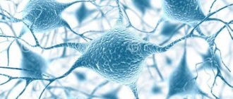
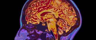
The human body is a rather complex and balanced system that functions in accordance with clear rules. Moreover, outwardly it seems that everything is quite simple, but in fact our body is an amazing interaction of every cell and organ. This whole “orchestra” is conducted by the nervous system, consisting of neurons. Today we will tell you what neurons are and how important a role they play in the human body. After all, they are the ones responsible for our mental and physical health.
What are neurons?
Every schoolchild knows that we are controlled by the brain and nervous system. These two blocks of our body are represented by cells, each of which is called a nerve neuron. These cells are responsible for receiving and transmitting impulses from neuron to neuron and other cells of human organs.
To better understand what neurons are, they can be represented as the most important element of the nervous system, which plays not only a conducting role, but also a functional one. Surprisingly, neuroscientists still continue to study neurons and their work in transmitting information. Of course, they have achieved great success in their scientific research and managed to uncover many secrets of our body, but they still cannot once and for all answer the question of what neurons are.
Nerve cells: features
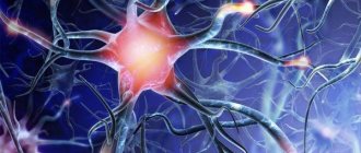
Neurons are cells and in many ways similar to their other “brethren” that make up our body. But they have a number of features. Due to their structure, such cells in the human body, when connected, create a nerve center.
A neuron has a nucleus and is surrounded by a protective membrane. This makes it similar to all other cells, but that’s where the similarity ends. Other characteristics of a nerve cell make it truly unique:
- Neurons don't divide
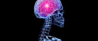
Neurons of the brain (brain and spinal cord) do not divide. This is surprising, but they stop developing almost immediately after their appearance. Scientists believe that a certain precursor cell completes division even before the neuron is fully developed. In the future, it increases only connections, but not its quantity in the body. Many diseases of the brain and central nervous system are associated with this fact. With age, some neurons die, and the remaining cells, due to the low activity of the person himself, cannot build up connections and replace their “brothers”. All this leads to imbalance in the body and, in some cases, to death.
- Nerve cells transmit information
Neurons can transmit and receive information using processes called dendrites and axons. They are able to perceive certain data using chemical reactions and convert it into an electrical impulse, which, in turn, passes through synapses (connections) to the necessary cells of the body.
Scientists have proven the uniqueness of nerve cells, but in fact they now know about neurons only 20% of what they actually hide. The potential of neurons has not yet been revealed; in the scientific world there is an opinion that the revelation of one secret of the functioning of nerve cells becomes the beginning of another secret. And this process currently seems endless.
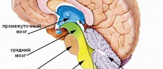
Anatomy and physiology[edit]
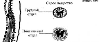
Spinal cord tracts
Location of lower motor neurons in the spinal cord
Upper motor neurons[edit]
Upper motor neurons originate in the motor cortex located in the precentral gyrus. The cells that make up the primary motor cortex are Betz cells, which are a type of pyramidal cell. The axons of these cells descend from the cortex to form the corticospinal tract. [10] Corticomotor neurons project from the primary cortex directly to motor neurons in the ventral horn of the spinal cord. [11] [12] Their axons synapse on spinal motor neurons of many muscles, as well as on spinal interneurons. [11] [12] They are unique to primates, and their function has been proposed to be adaptive control of the hands, including relatively independent control of individual fingers. [12] [13] Corticomotor neurons have so far only been found in the primary motor cortex, but not in the secondary motor areas. [12]
Neural pathways[edit]
Nerve tracts are bundles of white matter axons that carry action potentials to their effectors. In the spinal cord, these descending pathways carry impulses from different regions. These tracts also serve as the origin of lower motor neurons. Seven major descending motor tracts can be found in the spinal cord:[14]
- Lateral corticospinal tract
- Rubrospinal tract
- Lateral reticulospinal tract
- Vestibulospinal tract
- Medial reticulospinal tract
- Tectospinal tract
- Anterior corticospinal tract
Lower motor neurons[edit]
Lower motor neurons are those that originate in the spinal cord and directly or indirectly innervate effector targets. The targets of these neurons are different, but in the somatic nervous system the target will be some muscle fiber. There are three main categories of lower motor neurons, which can be divided into subcategories. [15]
According to their purposes, motor neurons are divided into three broad categories: [16]
- Somatic motor neurons
- Special visceral motor neurons
- General visceral motor neurons
Somatic motor neurons[edit]
Somatic motor neurons originate in the central nervous system and project their axons to skeletal muscles [17] (such as the limb, abdominal, and intercostal muscles) that are involved in locomotion. The three types of these neurons are alpha efferent neurons
,
beta efferent neurons
and
gamma efferent neurons
. They are called efferents to indicate the flow of information from the central nervous system (CNS) to the periphery.
- Alpha motor neurons innervate extrafusal muscle fibers, which are the main force-generating component of the muscle. Their cell bodies are found in the ventral horn of the spinal cord and are sometimes called ventral horn cells
.
One motor neuron can synapse on average with 150 muscle fibers. [18] A motor neuron and all the muscle fibers to which it connects are a motor unit. Motor units are divided into 3 categories: [19] Main article: Motor units.
Slow (S) motor units stimulate small muscle fibers that contract very slowly and produce little energy but are very resistant to fatigue, so they are used to maintain muscle contraction, for example to keep the body upright. They obtain energy through oxidative means and therefore require oxygen. They are also called red fibers. [19] - Fast-fatiguing (FF) motor units stimulate large muscle groups that exert large amounts of force but fatigue very quickly. They are used for tasks that require large, short bursts of energy, such as jumping or running. They obtain energy through glycolytic means and therefore do not require oxygen. They are called white fibers. [19]
- Fast fatigue-resistant motor units stimulate medium-sized muscle groups that do not respond as quickly as FF motor units, but can be held on much longer (as the name suggests) and provide greater force than S motor units. They use both oxidative and and glycolytic agents for energy. [19]
In addition to the voluntary contraction of skeletal muscles, alpha motor neurons also contribute to muscle tone, the sustained force produced by non-contracting muscles to resist stretch. When a muscle is stretched, sensory neurons in the muscle spindle detect the degree of stretch and send a signal to the central nervous system. The CNS activates alpha motor neurons in the spinal cord, which cause extrafusion muscle fibers to contract and thereby resist further stretch. This process is also called the stretch reflex.
- Beta motor neurons innervate intrafusal muscle fibers from the muscle spindles, with connections to extrafusal fibers. There are two types of beta motor neurons: Slow twitch - these innervate extrafusal fibers. Fast contraction - they innervate intrafusal fibers. [20]
- Gamma motor neurons innervate intrafusal muscle fibers located in the muscle spindle. They regulate the sensitivity of the spindle to muscle stretch. When gamma neurons are activated, intrafusal muscle fibers contract so that only a small stretch is required to activate spindle sensory neurons and the stretch reflex. There are two types of gamma motor neurons: dynamic - these focus on Bag1 fibers and increase dynamic sensitivity. Static - Focuses on Bag2 fibers and increases stretch sensitivity. [20]
- Regulatory Factors of Lower Motor Neurons Size Principle
- This refers to the soma of the motor neuron. This limits larger neurons to receive more excitatory signal to stimulate the muscle fibers they innervate. By reducing unnecessary muscle fiber recruitment, the body can optimize energy consumption. [20] - Constant inward current (PIC)
- A recent animal study has shown that the constant flow of ions such as calcium and sodium through channels in the soma and dendrites influences synaptic input. Another way to think about it is that the postsynaptic neuron is activated before receiving an impulse. [20] - After hyperpolarization (AHP)
- A trend has been identified that slow motor neurons have more intense AHPs for longer periods of time. One way to remember this is that slow-twitch muscle fibers can take longer to contract, so it makes sense that their corresponding motor neurons fire at a slower rate. [20]
Special visceral motor neurons[edit]
They are also known as branchial motor neurons
, which are involved in facial expressions, chewing, phonation and swallowing. The associated cranial nerves are the oculomotor, abducens, trochlear, and hypoglossal nerves. [16]
| NS branch | Position | Neurotransmitter |
| Somatic | n/a | Acetylcholine |
| Parasympathetic | Preganglionic | Acetylcholine |
| Parasympathetic | Ganglionic | Acetylcholine |
| Sympathetic | Preganglionic | Acetylcholine |
| Sympathetic | Ganglionic | Norepinephrine* |
| *Excluding fibers of sweat glands and some blood vessels, neurotransmitters of motor neurons. | ||
General visceral motor neurons[edit]
These motor neurons indirectly innervate the cardiac muscle and smooth muscles of the splanchnic muscles (muscles of the arteries): they synapse on neurons located in the ganglia of the autonomic nervous system (sympathetic and parasympathetic), located in the peripheral nervous system (PNS), which themselves directly innervate the visceral muscles (as well as some gland cells).
As a consequence, the motor command of the skeletal and branchial muscles is monosynaptic and
includes only one motor neuron,
somatic
or
branchial
, which synapses with muscles.
By comparison
, visceral muscle control is
disynaptic
, involving two neurons:
a common visceral motor neuron
, located in the CNS, synapses with a ganglion neuron located in the PNS, which synapses with the muscle.
All vertebrate motor neurons are cholinergic, meaning they release the neurotransmitter acetylcholine. Parasympathetic ganglion neurons are also cholinergic, whereas most sympathetic ganglion neurons are noradrenergic, meaning they release the neurotransmitter norepinephrine. (see table)
Neuromuscular junctions[edit]
A single motor neuron can innervate many muscle fibers, and a muscle fiber can be subject to many action potentials in the time required for a single muscle twitch. As a result, if the action potential is reached before the spasm has been completed, the twitches may overlap each other, either through summation or tetanic contraction. To summarize, the muscle is repeatedly stimulated so that additional action potentials originating from the somatic nervous system arrive before the end of the twitch. In this way, the twitches overlap each other, resulting in greater force than a single twitch. Tetanic contraction is caused by constant, very high-frequency stimulation—action potentials occur at such a high rate that individual twitches are indistinguishable and tension gradually increases, eventually reaching a plateau. [4]
The interface between a motor neuron and a muscle fiber is a specialized synapse called the neuromuscular junction. When adequately stimulated, the motor neuron releases a flood of acetylcholine (Ach) neurotransmitters from axon terminals from synaptic vesicles that bind to the plasma membrane. Acetylcholine molecules bind to postsynaptic receptors located in the motor end plate. Once the two acetylcholine receptors bind, an ion channel opens and sodium ions can enter the cell. The influx of sodium into the cell causes depolarization and triggers a muscle action potential. T-tubules of the sarcolemma are then stimulated to cause the release of calcium ions from the sarcoplasmic reticulum. It is this chemical release that causes the targeted muscle fiber to contract. [18]
In invertebrates, depending on the neurotransmitter released and the type of receptor it binds, the muscle fiber response can be either excitatory or inhibitory. However, in vertebrates, the response of a muscle fiber to a neurotransmitter can only be excitatory, in other words, contractile. Relaxation of muscles and suppression of muscle contractions in vertebrates is achieved only by suppression of the motor neuron itself. This is exactly how muscle relaxants work, acting on motor neurons that innervate muscles (reducing their electrophysiological activity) or on cholinergic neuromuscular junctions, rather than on the muscles themselves.
Structure of neurons
Each nerve cell consists of three parts:
- neuron body (soma);
- dendrites;
- axons.
It is still unknown which of the processes develop first in the cell body, but the distribution of responsibilities between them is quite obvious. The axon process of a neuron is usually formed in a single copy, but there can be a lot of dendrites. Their number sometimes reaches several hundred; the more dendrites a nerve cell has, the more cells it can be connected to. In addition, an extensive network of processes allows you to transmit a lot of information in the shortest possible time.
Scientists believe that before the formation of processes, the neuron spreads throughout the body, and from the moment they appear, it is already in one place without changing.
Transmission of information by nerve cells
To understand how important neurons are, it is necessary to understand how they perform their function of transmitting information. Neuron impulses can travel in chemical and electrical forms. The dendrite extension of a neuron receives information as a stimulus and transmits it to the body of the neuron; the axon transmits it as an electronic impulse to other cells. The dendrites of another neuron receive the electronic impulse immediately or with the help of neurotransmitters (chemical messengers). Neurotransmitters are captured by neurons and are subsequently used as their own.
How does a motor neuron work?
In order for a bioelectric impulse to occur, a potential difference is required on the membrane of the nerve cell. This occurs as a result of changes in the concentration of potassium and sodium ions from the outer and inner surfaces of the membrane.
Subsequently, the impulse travels to the end of the long process, the axon, and reaches the junction with another cell. The place of such contact is called a synapse.
On the other side of the synapse, a short branching process, a dendrite, is adjacent to the point of contact. Signal transmission across a synapse is driven by active chemicals called transmitters.
Having arisen on the dendrite, the signal spreads along its membrane and passes further to the axon. To contract a skeletal muscle, the signal originates in the motor neuron of the cortex, passes along the pyramidal tract, passes to the interneuron and then to the region of the anterior horns of the spinal cord. This chain ends in muscle tissue.
The result of excitation of the motor center of the cortex will be a contraction of a group of muscle fibers.
Types of neurons by number of processes
Scientists, observing the work of nerve cells, have developed several types of their classification. One of them divides neurons by the number of processes:
- unipolar;
- pseudounipolar;
- bipolar;
- multipolar;
- axonless.
A multipolar neuron is considered classic; it has one short axon and a network of dendrites. The most poorly studied are axonless nerve cells; scientists only know their location - the spinal cord.
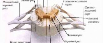
Reflex arc: definition and brief description
In neurophysics, there is such a term as “reflex arc neurons.” Without it, it is quite difficult to get a complete understanding of the work and significance of nerve cells. Stimuli that affect the nervous system are called reflexes. This is the main activity of our central nervous system, it is carried out with the help of a reflex arc. It can be thought of as a kind of road along which an impulse passes from a neuron to the implementation of an action (reflex).
This path can be divided into several stages:
- perception of irritation by dendrites;
- transmission of impulse to the cell body;
- transformation of information into an electrical impulse;
- transmission of impulse to the organ;
- change in organ activity (physical response to a stimulus).
Reflex arcs can be different and consist of several neurons. For example, a simple reflex arc is formed from two nerve cells. One of them receives information, and the other forces human organs to perform certain actions. Usually such actions are called an unconditioned reflex. It occurs when a person is hit, for example on the kneecap, and when touching a hot surface.
Basically, a simple reflex arc conducts impulses through the processes of the spinal cord; a complex reflex arc conducts an impulse directly to the brain, which, in turn, processes it and can store it. Subsequently, when receiving a similar impulse, the brain sends the necessary command to the organs to perform a certain set of actions.
Links[edit]
- Tortora, Gerard; Derrickson, Brian (2014). Principles of Anatomy and Physiology (14th ed.). New Jersey: John Wiley & Sons, Inc., pp. 406, 502, 541. ISBN 978-1-118-34500-9.
- Pocock, Gillian; Richards, Christopher D. (2006). Human physiology: fundamentals of medicine
(3rd ed.). Oxford: Oxford University Press. pp. 151–153. ISBN 978-0-19-856878-0. - Schacter D.L., Gilbert D.T. and Wegner D.M. (2011) Psychology Second Edition. New York, New York: worth it
- ^ ab Russell, Peter (2013). Biology - the study of the diversity of life
. Toronto: Nelson Education. item 946. ISBN 978-0-17-665133-6. - Tortora, Gerard; Derrickson, Brian (2011). Principles of Anatomical Physiology (14th ed.). New Jersey: John Wiley & Sons, Inc., pp. 1090–1099. ISBN 978-1-118-34500-9.
- Jump up
↑ Sadler, T. (2010).
Langmann's Medical Embryology
(11th ed.). Philadelphia: Lippincott William and Wilkins. pp. 299–301. ISBN 978-0-7817-9069-7. - ^ a b Davis-Dusenbury, B.N. Williams, Louisiana; Klim, junior; Eggan, K. (February 2014). "How to Make Spinal Motor Neurons". Development
.
141
(3):491–501. DOI: 10.1242/dev.097410. PMID 24449832. - Edgar R, Mazor Y, Rinon A, Blumenthal J, Golan Y, Bujor E, Livnat I, Ben-Ari S, Leader I, Shitrit A, Gilboa Y, Ben-Yehuda A, Edry O, Shraga N, Bogoch Y, Leshansky L., Aharoni S., Western M.D., Varshavsky D., Shtrikhman R. (2013). "LifeMap Discovery™: A portal to embryonic development, stem cell and regenerative medicine research". PLoS ONE
.
8
(7):e66629. Bibcode: 2013PLoSO...866629E. DOI: 10.1371/journal.pone.0066629. ISSN 1932-6203. PMC 3714290. PMID 23874394. - Filippidou, Polyxeni; Walsh, Caroline; Aubin, Jose; Jeannotte, Lucy; Dasen, Jeremy S. (2012). "Sustained Hox5 gene activity is required for respiratory motor neuron development". The Nature of Neuroscience
.
15
(12): 1636–1644. DOI: 10.1038/nn.3242. ISSN 1097-6256. PMC 3676175. PMID 23103965. - Fitzpatrick, D. (2001) Primary motor cortex: Upper motor neurons that initiate complex voluntary movements. In D. Purves, G. J. Augustine, D. Fitzpatrick, et al. (Ed.), Neurology. Retrieved from Archived Copy. Archived from the original on June 5, 2021. Retrieved November 30, 2021 .CS1 maint: archived copy as title (link)
- ^ ab Mack, Sarah; Kandel, Eric R.; Jessell, Thomas M.; Schwartz, James H.; Siegelbaum, Steven A.; Hudspeth, A. J. (2013). Principles of Neuroscience
. Kandel, Eric R. (5th ed.). NY. ISBN 9780071390118. OCLC 795553723. - ^ abcd Limon, Roger N. (April 4, 2008). "Descending pathways in motor control". Annual Review of Neuroscience
.
31
(1):195–218. DOI: 10.1146/annurev.neuro.31.060407.125547. ISSN 0147-006X. PMID 18558853. - Jump up
↑ Isa, T (April 2007).
"Direct and indirect corticomotor neuronal pathways and the control of hand/arm movements." Physiology
.
22
(2): 145–152. DOI: 10.1152/physiol.00045.2006. PMID 17420305. - Tortora, G. J., Derrickson, B. (2011). Spinal cord and spinal nerves. In B. Roesch, L. Elfers, K. Trost, et al. (Ed.), Principles of Anatomy and Physiology
(pp. 443-468). New Jersey: John Wiley & Sons, Inc. - Fitzpatrick, D. (2001) Lower motor neural circuits and motor control: A review. In D. Purves, G. J. Augustine, D. Fitzpatrick, et al. (Ed.), Neurology. Retrieved from Archived Copy. Archived from the original on June 5, 2021. Retrieved November 30, 2017.CS1 maint: archived copy as title (link)
- ^ a b "CHAPTER NINE". www.unc.edu
. Archived from the original on November 5, 2021. Retrieved December 8, 2017. - Silverthorn Dee Unglaub (2010). Human Physiology: An Integrated Approach
. Pearson. p. 398. ISBN 978-0-321-55980-7. - ^ ab Tortora, G. J., Derrickson, B. (2011). Muscle. In B. Roesch, L. Elfers, K. Trost, et al. (Ed.), Principles of Anatomy and Physiology
(pp. 305-307, 311). New Jersey: John Wiley & Sons, Inc. - ^ abcd Purves D, Augustine GJ, Fitzpatrick D, et al., editors: Neuroscience. 2nd edition, 2001. "Archival copy". Archived from the original on June 5, 2021. Retrieved September 5, 2021 .CS1 maint: archived copy as title (link)
- ^ abcde Manuel, Marin; Zhitnitsky, Daniel (2011). "Alpha, beta, and gamma motor neurons: functional diversity in the final motor system pathway". Journal of Integrative Neuroscience
.
10
(3): 243–276. DOI: 10.1142/S0219635211002786. ISSN 0219-6352. PMID 21960303.
Classification of neurons by functionality
Neurons can be classified according to their direct purpose, because each group of nerve cells is intended for specific actions. The types of neurons are presented as follows:
- Sensitive
These nerve cells are designed to perceive irritation and transform it into an impulse that is redirected to the brain.
2. Motor neurons
They perceive information and transmit impulses to the muscles that move parts of the body and human organs.
3. Insert
These neurons carry out complex work; they are in the center of the chain between sensory and motor nerve cells. Such neurons receive information, carry out preliminary processing and transmit a command impulse.
4. Secretory
Secretory nerve cells synthesize neurohormones and have a special structure with a large number of membrane sacs.
Symptoms of central motor neuron damage
Damage to central motor nerve cells most often occurs during stroke. When ischemia or hemorrhage occurs in the substance of the cerebral hemispheres, a section of tissue dies. Such lesions are almost always unilateral.
As a result, when central motor neurons are damaged, muscle dysfunction on one side is observed. The most noticeable symptom is unilateral paralysis, leading to the inability to actively move the arm and leg.
On the same side, muscle tone in the torso and facial muscles decreases. Damage to the central motor areas is accompanied by a number of changes in reflex activity.
Clinically, this is expressed in the appearance of various pathological reflexes. Their combination, decreased muscle tone and sensory disturbances allow the doctor to make a diagnosis.
Motor neurons: characteristics
Efferent neurons (motor) have a structure identical to other nerve cells. Their network of dendrites is the most branched, and axons extend to the muscle fibers. They cause the muscle to contract and straighten. The longest axon in the human body is the motor neuron axon, which runs to the big toe from the lumbar region. On average, its length is about one meter.
Almost all efferent neurons are located in the spinal cord, because it is responsible for most of our unconscious movements. This applies not only to unconditioned reflexes (for example, blinking), but also to any actions that we do not think about. When we peer at an object, the brain sends impulses to the optic nerve. But the movement of the eyeball to the left and right is carried out through commands from the spinal cord; these are unconscious movements. Therefore, as we age and the accumulation of unconscious habitual actions increases, the importance of motor neurons appears in a new light.
Motor neuron functions
Central and peripheral motor nerve cells work in concert. Together they provide contraction of certain muscle groups and allow a person to perform any action.
For coordinated movements of the limbs, simultaneous contraction of the flexors and extensors is necessary. When the flexors work, the initial excitation signal arises in the area of the precentral gyrus of the corresponding hemisphere.
Cells called pyramidal cells are responsible for this action. Their processes collected together form the so-called pyramidal motor pathway. Next, the signal goes to the anterior horns of the spinal cord, from where it is transmitted directly to the myofibrils.
Special centers of the posterior sections of the cerebral hemispheres have an activating effect on the motor neurons of the extensor muscles. They form the dorsal and ventral pathways. Thus, two areas of the brain are involved in the formation of coordinated movement.
Based on the nature of their function, the nerve cells involved in the process of muscle contraction are divided into motor neurons and interneurons. The former are responsible for the executive function, while the intercalary ones serve to coordinate nerve impulses. This particular variety is smaller in size and more numerous.
For comparison, in the area of the anterior horns there are 30 times more of them than motor horns. When excitation is carried out along the axon of the motor nerve, it initially passes to the interneuron. Depending on the nature of the signal, it can be amplified or weakened, after which it is transmitted further.
Intercalated cells have more processes and are more sensitive. They have a large number of processes and are also called multipolar.
To optimize the signals emanating from the axons and going to the muscle fibers, special Renshaw cells are used, which transmit excitation from one process to another. This mechanism serves to equalize the intensity of the nerve signal.
Along the process of the motor neuron, the impulse reaches the muscle fiber, which contracts. Each group of motor neurons and the muscle fibers they innervate are responsible for specific movements.
Nerve cells providing motor function:
| Types of neurons | Localization | Function |
| central innervating flexors | precentral gyrus area | contraction of skeletal flexor muscles by transmitting impulse to the anterior horns |
| central innervating extensors | hindbrain region | contraction of skeletal extensor muscles by transmitting impulses to the anterior horns |
| peripheral alpha | anterior horn of the spinal cord | direct contraction of skeletal muscles |
| peripheral gamma | anterior horn of the spinal cord | regulation of tone |
| insertion | all parts of the central nervous system | communication of signals within the central nervous system |
Large alpha neurons conducting a strong impulse cause myofibril contraction. Small ones conduct weak signals and serve to maintain muscle tone.
In addition to the fibers responsible for contraction, muscle tissue also contains special spiral fibrils that regulate the force of muscle tension.
These extrafusal muscle fibers are innervated by gamma neurons.
Excitation of the gamma motor neuron leads to an increase in the stretch of myofibrils and facilitates the passage of the tendon reflex impulse. An example would be the passage of a nerve signal along the arc of the knee reflex.
The coordinated work of peripheral motor neurons achieves fine tuning of muscle tone, which allows for precise coordinated movements. When peripheral motor neurons are damaged, muscle tone disappears and movement is impossible.
Types of motor neurons
In turn, efferent cells have a certain classification. They are divided into the following two types:
- a-motoneurons;
- y-motoneurons.
The first type of neurons has a denser fiber structure and attaches to various muscle fibers. One such neuron can involve a different number of muscles.
Y-motoneurons are slightly weaker than their “brothers”; they cannot engage several muscle fibers at the same time and are responsible for muscle tension. We can say that both types of neurons are the controlling organ of motor activity.
What muscles do motor neurons connect to?
Neuron axons are connected to several types of muscles (they are working muscles), which are classified as:
- animal;
- vegetative.
The first group of muscles is represented by skeletal muscles, and the second belongs to the category of smooth muscles. The methods of attachment to the muscle fiber are also different. Skeletal muscles form peculiar plaques at the point of contact with neurons. Autonomic neurons communicate with smooth muscle through small swellings or vesicles.