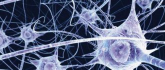
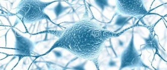
The functions of neurons are diverse. A neuron is a nerve cell, although the word is sometimes also used to mean a nerve cell. Our nervous system is primarily made up of two types of cells—nerve cells (technically called “neurons”) and supporting cells (technically called “glia” or “glial cells”). Neurons form the functional unit of the nervous system, which means it performs all activities such as learning, memory, emotions, calculations and so on. For our brains to function, millions of neurons communicate with each other using electrical and chemical signals. Glia are thought to provide physical support to neurons by encasing them and providing nutrition. The functions of neurons and types of neuronal diseases are the topic of the next article.
Neuron structure
A typical neuron consists of three important parts:
- central body of the cell,
- short branched projections known as dendrites,
- a long projection known as an axon.
The last two carry information to and from the cell body of the neuron. The cell body contains a nucleus and other organelles, just like any other cell in the body. The axon is covered by thick layers of myelin sheath, which is formed by supporting glial cells. This myelin sheath insulates each neuron and increases the speed at which electrical signals travel along the axon.
Neuron structure
Content
- Neurons Types of neurons
- Nerve fibers and nerves
- List of cranial nerves with dominant fiber designation
To get started, I recommend watching a short video that talks about different human tissues. But it is the nervous tissue that will interest us. In a more colorful and visual form, it will be easier for you to grasp the basics, and then you can expand your knowledge.
The main tissue from which the nervous system is formed is nervous tissue, which consists of cells and intercellular substance. Tissue is a collection of cells and intercellular substance that are similar in structure and functions.
Nervous tissue is of ectodermal origin. Nervous tissue differs from other types of tissue in that it lacks intercellular substance. The intercellular substance is a derivative of the glial cell and consists of fibers and amorphous substance.
The function of nervous tissue is to ensure the receipt, processing and storage of information from the external and internal environment, as well as the regulation and coordination of the activities of all parts of the body.
Nerve tissue consists of two types of cells: neurons and glial cells . Neurons play a major role in providing all functions of the central nervous system. Glial cells have an auxiliary role, performing supporting, protective, trophic functions, etc. On average, the number of glial cells exceeds the number of neurons in a ratio of 10:1, respectively.
Each neuron has an expanded central part: a body - soma and processes - dendrites and axons . Impulses travel through dendrites to the nerve cell body, and through axons from the nerve cell body to other neurons or organs.
The shoots can be long or short. The long processes of neurons are called nerve fibers . Most dendrites (dendron - tree) are short, highly branched processes. The axon (axis - process) is often a long, slightly branched process.
Types of neurons
Neurons are usually classified in one of the following ways:
- depending on the number of processes from the cell body,
- depending on the direction of transmission of information regarding the brain and spinal cord,
- depending on the type of neurotransmitter (chemical messengers that neurons use to communicate with each other) the neuron uses.
Bipolar and multipolar
Depending on the number of processes extending from the cell body, a neuron can be classified as bipolar, pseudounipolar, or multipolar.
- A bipolar neuron has an axon and dendrites.
- A pseudounipolar neuron has two axons.
- A multipolar neuron has one axon but several dendritic processes extending from its cell body.
Sensory and motor
Depending on the direction of information flow, neurons are divided into three types - sensory neurons, motor neurons and interneurons.
- Sensory neurons carry signals from sensory receptors such as the skin, ears, eyes, tongue, nose, etc. to the brain or spinal cord. It sends information to the brain about various sensations such as temperature, pain, sound, smell and vision.
- Motor neurons work in the opposite direction: they transmit signals from the brain or spinal cord to the glands or muscles of the body. In this way, the brain can control the various muscles of the body and the activities of various glands.
- Interneurons provide information transfer between sensory neurons and motor neurons in the brain and spinal cord.
Neurotransmitters
Although neurons transmit information through electrical impulses, they also communicate with each other through chemical messengers known as neurotransmitters. Each neuron predominantly uses one type of neurotransmitter, and this makes it possible to classify these neurons.
Although neurons transmit information through electrical impulses, they also communicate with each other through chemical messengers known as neurotransmitters.
Some of the commonly used neurotransmitters are:
- dopamine
- GABA
- serotonin
- acetylcholine
Neurons
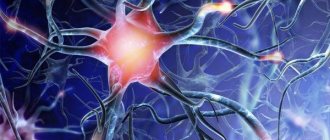
A neuron is a complex, highly specialized cell with processes capable of generating, perceiving, transforming and transmitting electrical signals, and also capable of forming functional contacts and exchanging information with other cells.
Each neuron has only 1 axon, the length of which can reach several tens of centimeters. Sometimes lateral processes extend from the axon - collaterals . Axon endings typically branch and are called terminals . The place where the axon extends from the cell soma is called axonal hillock .
In relation to the processes, the soma of the neuron performs a trophic function, regulating metabolism. A neuron has characteristics common to all cells: it has a membrane, a nucleus and a cytoplasm in which organelles are located (endoplasmic reticulum, Golgi apparatus, mitochondria, lysosomes, ribosomes, etc.).
In addition, the neuroplasm contains special-purpose organelles: microtubules and microfilaments , which differ in size and structure. Microfilaments represent the internal skeleton of the neuroplasm and are located in the soma. Microtubules stretch along the axon along the internal cavities from the soma to the end of the axon. Biologically active substances spread through them.
In addition, a distinctive feature of neurons is the presence of mitochondria in the axon as an additional source of energy. Adult neurons are not capable of division.
Types of neurons
There are several classifications of neurons based on different characteristics: the shape of the soma, the number of processes, the functions and effects that the neuron has on other cells.
Depending on the shape of the soma, they are distinguished: 1. Granular (ganglionic) neurons , in which the soma has a round shape; 2. Pyramidal neurons of different sizes - large and small pyramids; 3. Stellate neurons ; 4. Fusiform neurons .
Based on the number of processes (by structure) there are: 1. Unipolar neurons (single-processed) , having one process extending from the cell soma, are practically never found in the human nervous system; 2. Pseudounipolar neurons (false unipolar), such neurons have a T-shaped branching process, these are cells of general sensitivity (pain, temperature changes and touch); 3. Bipolar neurons (bipolar) having one dendrite and one axon (i.e. 2 processes), these are cells of special sensitivity (vision, smell, taste, hearing and vestibular stimulation); 4. Multipolar neurons (multi-process) , which have many dendrites and one axon (i.e. many processes); small multipolar neurons are associative; medium and large multipolar, pyramidal neurons - motor, effector.
Unipolar cells (without dendrites) are not typical for adults and are observed only during embryogenesis. Instead, in the human body there are pseudounipolar cells, in which a single axon divides into 2 branches immediately after leaving the cell body. Bipolar neurons are present in the retina and transmit excitation from photoreceptors to ganglion cells that form the optic nerve. Multipolar neurons make up the majority of cells in the nervous system.
According to the functions they perform, neurons are: 1. Afferent (receptor, sensory) neurons - sensory (pseudo-unipolar), their somas are located outside the central nervous system in ganglia (spinal or cranial). Along sensory neurons, nerve impulses move from the periphery to the center.
The shape of the soma is granular. Afferent neurons have one dendrite that connects to receptors (skin, muscle, tendon, etc.). Through dendrites, information about the properties of stimuli is transmitted to the soma of the neuron and along the axon to the central nervous system.
Example of sensory neurons: a neuron that responds to skin stimulation.
2. Efferent (effector, secretory, motor) neurons regulate the work of effectors (muscles, glands, etc.). Those. they can send orders to the muscles and glands. These are multipolar neurons, their somas have a stellate or pyramidal shape. They lie in the spinal cord or brain or in the ganglia of the autonomic nervous system.
Short, abundantly branching dendrites receive impulses from other neurons, and long axons extend beyond the central nervous system and, as part of the nerve, go to effectors (working organs), for example, to skeletal muscle.
Example of motor neurons: spinal cord motor neuron.
The cell bodies of sensory neurons lie outside the spinal cord, while motor neurons lie in the anterior horn of the spinal cord.
3. The betting neurons (contact, interneurons, associative, closing) make up the bulk of the brain. They communicate between afferent and efferent neurons and process information coming from receptors to the central nervous system.
These are mainly multipolar stellate-shaped neurons. Among interneurons, neurons with long and short axons are distinguished.
Example of interneurons: neuron of the olfactory bulb, pyramidal cell of the cerebral cortex.
The chain of neurons consisting of sensory, intercalary and efferent neurons is called the reflex arc. All activities of the nervous system, as defined by I.M. Sechenov, is of a reflex nature (“reflex” means reflection).
According to the effect that neurons have on other cells: 1. Excitatory neurons have an activating effect, increasing the excitability of the cells with which they are connected. 2. Inhibitory neurons reduce cell excitability, causing an inhibitory effect.
Nerve fibers and nerves
Nerve fibers are processes of nerve cells covered with a glial sheath that carry out nerve impulses. Through them, nerve impulses can be transmitted over long distances (up to a meter).
The classification of nerve fibers is based on morphological and functional characteristics.
Based on morphological characteristics, they are distinguished: 1. M myelinated (meat) nerve fibers are nerve fibers that have a myelin sheath; 2. Non -myelinated (non-myelinated) nerve fibers are fibers that do not have a myelin sheath.
According to functional characteristics, they are distinguished: 1. Afferent (sensitive) nerve fibers; 2. Efferent (motor) nerve fibers.
Nerve fibers extending beyond the nervous system form nerves. A nerve is a collection of nerve fibers. Each nerve has a sheath and a blood supply.
There are spinal nerves connected to the spinal cord (31 pairs) and cranial nerves (12 pairs) connected to the brain. Depending on the quantitative ratio of afferent and efferent fibers within one nerve, sensory, motor and mixed nerves are distinguished (see table below).
In sensory nerves, afferent fibers predominate, in motor nerves, efferent fibers predominate, in mixed nerves, the quantitative ratio of afferent and efferent fibers is approximately equal. All spinal nerves are mixed nerves. Among the cranial nerves, there are three types of nerves listed above.
List of cranial nerves with dominant fiber designation
I pair - olfactory nerves (sensitive); II pair - optic nerves (sensitive); III pair - oculomotor (motor); IV pair - trochlear nerves (motor); V pair - trigeminal nerves (mixed); VI pair - abducens nerves (motor); VII pair - facial nerves (mixed); VIII pair - vestibulo-cochlear nerves (sensitive); IX pair - glossopharyngeal nerves (mixed); X pair - vagus nerves (sensitive); XI pair - accessory nerves (motor); XII pair - hypoglossal nerves (motor).
How does a neuron work?
Neurons exchange information with each other at axonal and dendritic terminals. These terminals form a special structure known as a synapse (Greek "syn" = together + "haptein" = "to interlock"). Communication between two neurons begins with a stream of electrical impulse, known as an action potential, in one of the neurons. This action potential travels down the axon and reaches the synaptic terminal. Here it triggers the release of neurotransmitters at the synaptic cleft, the tiny space between the terminals of two interacting neurons. The released neurotransmitter then binds to receptors on the synaptic terminal of another neuron and also induces an action potential in that neuron. Now the electrical impulse will travel through the neuron.
Types of Neuronal Diseases
Neurons differ from other cells in the body in one important way - they generally do NOT regenerate. This makes neurological disorders debilitating and difficult to treat. It is known that different nervous diseases have different causes.
Some defects are characterized by the death of neurons, while others cause disruption of the normal functioning of neurons. Regardless of the mechanism, a decrease in the number or function of nerve cells will have characteristic symptoms depending on the area of the brain affected.
Dopamine
There are neurodegenerative diseases, which include conditions in which neurons progressively degenerate. Examples include Parkinson's disease, Alzheimer's disease, and Huntington's disease.
Dopamine is an important neurotransmitter produced by neurons present in an area of the midbrain known as the substantia nigra. For unknown reasons, these neurons die, causing the motor symptoms of Parkinson's disease. Altered dopamine levels are also associated with schizophrenia, one of the most common psychotic conditions.
Neuron loss
Massive loss of neurons and synapses characterizes Alzheimer's disease, which is a disorder of the cognitive and functional abilities of the brain. A condition that affects muscle coordination is Huntington's disease, in which motor neurons are damaged due to the accumulation of unwanted protein in them. Degeneration of motor neurons in an area of the brain known as the cerebellum can lead to cerebellar ataxia, which is characterized by uncoordinated movements and an unstable gait.
Massive loss of neurons and synapses characterizes Alzheimer's disease, which is a disorder of the cognitive and functional abilities of the brain.
Selective degeneration of motor neurons is a characteristic feature of motor neuron diseases such as amyotrophic and primary lateral sclerosis, progressive muscular atrophy, and progressive and pseudobulbar palsies.
Myelin sheath
Some neurodegenerative diseases are associated with degeneration of the myelin sheath, which negatively affects the functioning of neurons. Myelin degradation results in loss of axonal conduction and the neuron eventually dies. Diseases affecting the myelin sheath include multiple sclerosis, Guillain-Barré syndrome, acute disseminated encephalomyelitis , transverse myelitis, central pontine myelinosis and demyelinating diseases such as Charcot-Marie-Tooth and leukodystrophy, etc. and myelinated brain axons do not regenerate. Abnormal functioning associated with action potentials can also lead to conditions such as epilepsy and seizures.
Nerve inflammation
Inflammation is known to be a mechanism of progressive neurodegenerative conditions such as inflammatory demyelinating polyneuropathy, amyotrophic lateral sclerosis, and so on. Encephalitis can also develop in the brain due to inflammation resulting from a viral infection. Inflammation is the body's response to injury, although sometimes it occurs even when the body is not injured or injured.
Often in these cases the immune system attacks the body, which is not normal.
The nerves can become inflamed, and this is called neuritis. When any part of the body becomes inflamed, its normal functioning is impaired. These functions will likely return to normal when the inflammation subsides. However, severe inflammation and severe trauma can render the nerve cell useless. Post Views: 585
Neuron, its physiological properties and functions
Before you start training, you need to know not only the techniques, methods and methods of developing the skills you need, but also understand the laws by which the dog lives. The theoretical basis for dog training is the teaching of Academician I. P. Pavlov on higher nervous activity.
Neuron (nerve cell) is a structural and functional unit of the nervous system.
The ability to form skills depends on the functions and properties of neurons.
Basic properties of neurons:
Irritability is the ability of a nerve cell to perceive and respond to various irritations. Irritability is inherent in all cells, and especially nerve cells associated with the sensitive perception of smell, sound, light and other stimuli. Irritability is a trigger for the manifestation of another property - excitability.
Excitability is the ability of individual parts of a nerve cell to generate electrochemical impulses, that is, to respond to stimulation with excitation. For a nerve cell to transition into a state of excitation, it is necessary that the strength of the current stimulus reaches a critical limit - a threshold value. The amount of excitation of a neuron depends on the strength of the stimulus.
Conductivity is the ability of a neuron to conduct excitation impulses at a certain speed, in a constant rhythm and strength. Excitation along the nerve fiber can spread in both directions from the irritated area.
Lability (mobility) is the ability of a nerve cell to receive and transmit the maximum number of impulses per unit of time without distortion. Lability ensures the directed distribution and conduction of excitation impulses of the desired frequency along certain nerve pathways. In the process of growth and development of the body, as well as with systematic training, lability increases and ensures the dynamism of the nervous system. In other words: lability is the speed of a neuron.
Inhibition is the opposite process to excitation. Consists of weakening, stopping or preventing the occurrence of excitation. Inhibition is an active process, spreading through nerve cells, it ensures the coordinated functioning of individual organs and the entire organism as a whole.
Main functions of neurons:
The receptor function ensures the perception of certain stimuli from the external and internal environment of the body. Receptor cells are modified neurons that perceive a certain type of energy coming from the external or internal environment.
The sensory function of sensory neurons provides analysis of perceived stimuli, the formation of certain sensations and clear differentiation of numerous stimuli acting from the external and internal environment.
The information function of intermediate neurons ensures the accumulation, storage and output of information received from the external and internal environment. Information in neurons is encoded as memory and, when necessary, is issued in the form of weak excitation impulses.
The motor function of motor neurons ensures the formation and transmission of excitation impulses of a certain strength and frequency to the corresponding organs of movement or other executive organs and tissues.
Neurons consist of dendrites and axons. Dendrites are responsible for the perception of excitation, and axons for its transmission. Dendrites have many processes through which neurons connect to each other, creating synaptic connections (that is, connections between neurons). Synapses produce a mediator (a substance that is a catalyst, that is, an accelerator, of processes occurring in the cell).
Thus, a neuron is a structural and functional unit of the nervous system. The main properties of a neuron include excitation and inhibition. Excitation is characterized by the ability to generate nerve impulses and conduct them to other neurons or to executive organs. The function of inhibition is to prevent the conduction of a nerve impulse. Excitation is sensed by dendrites and transmitted by axon. Various associations of neurons in circuits and networks ensure the processing of incoming information.