“But he’s brainless, he doesn’t understand anything!”, “She’s stupid, she has no brains at all!”, “But those guys are smart, their brains work as they should!” As a rule, in everyday life, people remember the brain exclusively in the context of someone’s ability to think. Although from the school curriculum you should vaguely remember that the tasks of the brain are much broader than thinking and remembering.
You can improve your thinking skills in our Cognitive Science program, and today we will talk about the variety of functions of the human brain and how the human brain works. In the course of the presentation, we will move from simple to more complex, so as not to get confused in the intricacies of structure and functions.
General brain structure
To simplify the task, we will take as a basis material from a school biology textbook for grade 8, where the structure of the human body is explained [V. Pasechnik, 2010]. And to make it more interesting, we will supplement the explanations with interesting facts from other sources.
So, the brain is located in the cranium and occupies up to 80% of its volume. The weight of the brain on average is 2% of the total weight of a person. This explains the difference between the average brain weight of men and women: men weigh more on average than women. Interestingly, no linear relationship was found between the weight and size characteristics of the brain and human intelligence, and bigger does not mean better.
History shows that a record-breaking brain weighing 2850 grams was discovered in a young man who suffered from epilepsy and idiocy [G. Elliot, 1925]. At the same time, according to the head of the embryology department of the Research Institute of Human Morphology, Professor Sergei Savelyev, 72% of gifted people have a brain that exceeds the average weight [S. Kuzina, 2010].
One way or another, despite all the differences in size and abilities, all people have the same brain structure. The structure of the brain is considered, firstly, in the context of the functions of the hemispheres, and secondly, in the context of the functions of various parts or lobes of the brain. From the school biology course we remember (or don’t remember) that the brain consists of the following parts:
- Frontal lobe.
- Parietal lobe.
- Temporal lobe.
- Occipital lobe.
- Cerebellum.
- Diencephalon (thalamus, hypothalamus, pituitary gland).
- Midbrain.
- Bridge.
- Medulla.
In addition, the spinal cord originates from the brain. Since the spinal cord is not the topic of today’s material, we will not consider it, but we suggest studying the general diagram of the structure of the brain
The most attentive of you have noticed that the diagram does not show some parts of the brain from the list above. Everything is correct, because the diagram shows only the external structure of this organ. Everything inside is hidden by the outer lobes and parts of the brain.
In order not to clutter the diagram and not to confuse you in the details and nuances of how the human brain is structured, we will give an idea of the location of the internal components of the brain relative to the external ones in separate pictures as we look at the parts of the brain. Now let's move on to studying the functions of each of them.
Medulla
It is a continuation of the spinal cord, and its main function is conduction. Through the medulla oblongata, information is transmitted to other parts and there is a reverse transfer of information from the brain to the spinal cord.
In addition, this part of the brain is responsible for many different protective reflexes, in particular sneezing and coughing. The centers of respiratory and digestive reflexes, such as swallowing and salivation, are also located here.
Bridge
The pons is adjacent to the medulla oblongata and its main function is directly related to it. It is through this “way station” that the medulla oblongata transmits signals to the rest of the brain and through it receives signals coming from different parts of the brain.
In the medical literature you can find the second name “Varoliev's bridge”, named after the Italian physician and anatomist Constanzo Varolius (1543-1575), who made a huge contribution to the study of the structure of the brain.
Midbrain
This part of the brain is responsible for the primary processing of visual and auditory information, including the so-called “hidden” or “lateral vision.” Information about everything that comes into our field of vision enters the brain, but remembering what we saw with hidden vision is somewhat more difficult than what we consciously focused our gaze on.
In addition, the midbrain is responsible for some vital reflexes, such as the orientation reflex. When we startle from an unexpected loud sound (thunder, falling object, squeaking brakes) or a bright flash (lightning, explosion), and then try to figure out the origin of the sound or flash, this is an example of the work of the orienting reflex and the midbrain.
As we just found out, the midbrain conducts the primary processing of visual and auditory information, and the orientation reflex is directly related to these functions.
Diencephalon (thalamus, hypothalamus, pituitary gland)
Here we will talk about the main components of the diencephalon: the thalamus, hypothalamus and pituitary gland. The hypothalamus is responsible for reflexes such as thirst and hunger, regulates sleep, maintains the stability of the internal environment of the whole body and is involved in the formation of emotions. For example, such as love and aggression.
The thalamus is sometimes called the “central relay station of the brain,” where almost all sensory and motor information is collected, with the exception of signals from the olfactory organs. In addition, the thalamus is involved in controlling motor functions, speech and memory.
The pituitary gland, in principle, is an organ of the endocrine system in which the synthesis of hormones that affect metabolism, growth and reproductive function is carried out. The pituitary gland is closely interconnected with the hypothalamus and forms the hypothalamic-pituitary system, which regulates many body functions related to metabolism, reproductive function, and growth.
Cerebellum
The cerebellum is anatomically located behind the medulla oblongata and the pons and is responsible for coordination of movements, balance, maintaining the desired body position and muscle tone. The traditional way to test cerebellar function is to stretch your arms out in front of you with your eyes closed and then touch the tip of your nose without opening your eyes. A healthy person can easily do this, despite the fact that about 30 muscles are involved in this simple movement.
In addition to coordinating movements, this part of the brain performs an adaptive-trophic function, which ensures the body’s adaptation to changing conditions.
Occipital lobe
The occipital lobe borders the parietal and temporal lobes. Visual analyzers are concentrated in this part of the brain. They include the so-called “primary visual cortex” and visual association areas.
To make it clearer what we are talking about, let's say that disturbances in the primary visual cortex lead to a specific visual disorder, Anton Babinski syndrome. This is when people do not distinguish objects by appearance, but at the same time they do not even suspect their disorder and are sure that they see exactly what they really are. You can learn more about this syndrome from the video lecture dedicated to this problem:
Temporal lobe
The temporal lobe is adjacent to the frontal, occipital and parietal lobes. Auditory and taste analyzers are concentrated in this part of the brain. Thanks to auditory analyzers, we recognize speech and can perceive music. If there are disturbances in the right lobe, a person loses the ability to perceive music; if in the left, he will have a speech disorder.
Here, in the temporal lobes, is the hippocampus. This is a paired structure that, together with the cortex (outer covering) of the temporal lobe, is involved in the formation of emotions, long-term and spatial memory.
In addition, the so-called “ amygdala ” is located in the temporal lobes. This is also a pair structure, one “body” on each side. The amygdala is responsible for the formation of many emotional reactions, such as fear, and decision making.
If it is destroyed, say, due to illness, a person will not experience a feeling of fear and will not be able to make a decision adequate to the threatening danger.
Parietal lobe
The parietal lobe is adjacent to the frontal, temporal and occipital parts of the brain. It is responsible for processing and integrating sensory information. For example, for the perception of the relationship between tactile sensations and pain. Thus, a person learns that if you touch something hot, it will hurt and you can get burned.
In addition, this part of the brain allows you to orient yourself in space and understand which part of the body is affected by sensations. For example, you touched a hot object with your finger or palm, or accidentally caught it with your elbow.
Frontal lobe
The frontal lobe covers the anterior parts of the cerebral hemispheres. It is adjacent to the parietal and temporal lobes. The frontal lobe is responsible for learning, perception of information, memory and thinking. Thus, when someone is called “brainless”, it essentially means that the frontal lobes are not functioning very well, not the entire brain.
In general, the frontal lobes can be thought of as the “command post” of the entire brain. The health and safety of these lobes of the brain determines a person’s ability to analyze the situation, take initiative, make independent decisions, and control their behavior.
When this part of the brain is damaged, a person often experiences symptoms similar to those of laziness: lack of interest in what is happening, lack of initiative, inadequate carelessness. In addition, a person may lose social control over his behavior and, for example, begin to use profanity in public places.
Another difficulty that accompanies frontal lobe disorders is the loss of previously acquired skills. For example, a person forgets how to cook a particular dish that he prepared before. In this case, learning new skills is also difficult and sometimes impossible.
A sign of a disorder in this part of the brain is also perseveration in speech (for example, repetition of words unreasonable by the situation) or in actions (meaningless shifting of things from place to place). And finally, the frontal lobes are responsible for upright posture and maintaining an upright body position. When the lobes are affected, a specific “mincing” gait and constant stoop may be observed.
In addition to all of the above, in the medical literature you can find the term “telebrum”. This refers to the cerebral hemispheres covered with cortex, the corpus callosum, the striatum and the olfactory brain. We will tell you more about the cerebral cortex a little later, when we consider the structure of brain tissue. Let’s say a few words about the remaining components of the telencephalon right now.
The corpus callosum is responsible for coordinating the hemispheres and transmitting information from one hemisphere to the other:
The striatum regulates muscle tone, takes part in the formation of conditioned reflexes and regulation of the functioning of internal organs:
The olfactory brain , as the name implies, is responsible for the sense of smell and is part of the so-called “ limbic system ” of the brain, which refers to the totality of brain components and their connections involved in controlling instinctive behavior and autonomic functions:
This is, let’s say, “knowledge for the advanced,” so we will not dwell in detail on the structure of this part of the brain. Here we have provided this information so that you understand what we are talking about if you suddenly come across these terms in the specialized literature.
By the way, the limbic system is sometimes called the “animal part” of the brain because it is more about reflexes than consciousness. In this regard, the book “Will and Self-Control” is very interesting. How genes and the brain prevent us from fighting temptations” [I. Yakutenko, 2020].
So, we have found out how the human brain is structured, and now we know that the brain is symmetrical. From a school course in biology and general development, we know that the brain is divided by a fissure into two hemispheres, right and left, and that the functions of these hemispheres are different.
A natural question arises: if we have already studied in general terms the functions of all parts of the brain, how exactly should the functions of the right and left hemispheres differ? To understand this nuance, you should know that the left hemisphere processes information that comes in gradually, and the right hemisphere instantly creates a complete image. The left hemisphere stores information that needs to be thought through and coordinated, while the right hemisphere stores previously created and imprinted images.
Thus, when you are preparing for a report or exam, the left hemisphere is working, and when a spontaneous idea comes to your mind, even regarding the topic of the report or exam paper, this means that the right hemisphere is “involved” in the process. If you are right-brain dominant, you can visualize the difference between the hemispheres by simply looking at the picture with examples of the work of the right and left hemispheres:
Even a cursory glance is enough to understand: how the human brain is structured and human psychology are closely interconnected. The foundations of the processes that predetermine a person’s behavior, his success or lack of achievement in life, the presence or absence of talents for music, painting or poetry, stem from the depths of the brain.
This is why scientists are so interested in the structure of the brain, its mysteries and secrets. Moreover, it is believed that the human brain has not yet been fully studied, and new discoveries are yet to come.
If it’s easier for you to perceive information by ear than from text, we can recommend a video on the topic “How the human brain works” - a biology lesson for grade 8:
It tells how the human brain works for children, so the presentation is simple, understandable and gives a general idea of the topic, quite sufficient for a person who, due to his profession, is not related to medicine. From this video you can learn about both the structure and structure of brain tissue. We will dwell on the structure of tissues in a little more detail.
The Last Fireworks: What Happens to the Body When We Die
As a patient approaches death, changes occur in his body. They concern breathing, blood circulation, consciousness. We explain in detail what happens: if we understand death, perhaps we will be less afraid of it.
Author: Jacob Zimmank
The author of the article is a former employee of the palliative care service, which supervises dying people at home. He was amazed at how calmly many people faced their own death.
When does dying begin? At what point does a person begin his path to death?
Dying begins long before we are born. It is laid in the womb, in a cluster of cells from which the unborn child is formed. Some cells are redundant and need to make room for new ones. This is the only way the child’s organs can appear. It is due to this that there are only two kidneys, and only ten fingers on the hand. The genome of each cell already contains programs that act as a kind of “catapult”. It turns on at the moment when the cell becomes unnecessary or dangerous for the body. In this case, the cell voluntarily destroys itself.
Becoming a person is a process that involves both life and death. According to palliative medicine doctor Jean-Domenico Borasio, death is “a necessary condition so that we can, in principle, be born as viable organisms.”
“What we know for certain is that a person does not die suddenly, overnight” - Jean-Domenico Borasio, palliative care doctor
Death is omnipresent - but once we are born, we forget about it. And, if all is well, then death will appear in our lives again only after a few decades. Often this will be a condition that cannot be cured: cancer, heart disease, or kidney disease that means they can no longer cleanse the blood. And then the process of dying begins.
“What we know for certain is that a person does not die suddenly, overnight. The organs of the human body stop functioning gradually, not all at the same time, and then at some point their work stops,” says Dr. Borasio. As a result of the chain reaction, the liver, kidneys, lungs and heart stop working.
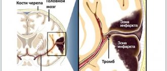
There are a variety of diseases, but at the end of life everything always follows the same pattern: the heart stops beating, breathing stops and, last of all, consciousness fades away. At the same time, it is very difficult to separate the work of the heart and brain. If the heart stops pumping oxygenated blood, brain cells begin to die within seconds. And after a few minutes, brain death occurs: at this moment the EEG will show a straight line instead of the usual curves and teeth. The reflexes that control the deep parts of the brain responsible for breathing, swallowing and consciousness also stop working. Thus, when the heart stops, the brain also stops working.
It also happens the other way around. The brain contains centers that regulate all vital functions: blood pressure, heart function, breathing. If these centers are damaged, breathing will stop and the heart rhythm will be disrupted. Often this damage occurs when intracranial pressure rises sharply as a result of a car accident or accident. The bones of the skull are hard, so with a strong blow, the soft tissue of the brain is squeezed into the only natural opening in the skull - the foramen magnum. Through it, the cranial cavity communicates with the spinal canal (the spinal cord passes into the brain stem). If the brain stem is pinched or damaged, the person dies.
There are no clear signs, but there are general patterns
Dying people live the last years, months and days of their lives in different ways. “The dying process is very individual,” says Lukas Radbruch, president of the German Society for Palliative Medicine and professor at the University Hospital Bonn. It often lasts for months or even years. Doctors divide dying into three stages: at the beginning comes the “terminal” stage, which lasts one to two years. At this time, the functioning of individual internal organs gradually decreases, and the dying person increasingly experiences fatigue. This is followed by the “pre-final” stage, which can last several weeks or months. At this time, symptoms such as shortness of breath and pain appear. And finally, the “final” stage - the last days. The patient stops eating and drinking and slowly fades away. “It is important to remember that this division of dying into stages is nothing more than a convention,” notes Radbruch. It is important for providing treatment and care, but does not provide an accurate picture and “does not help determine when the patient will die.”
“It used to be believed that the approach of death was indicated by pale skin in the nasolabial triangle,” says Radbruch. But this is also an unreliable sign. Currently, research that allows us to accurately determine the time of death is at an early stage. Some scientists are testing blood samples for markers that can be used to calculate remaining life expectancy. Radbruch says the best thing to do is ask the attending physician: Would she be surprised if she learned that the patient died that night or next weekend? If the doctor says that she is not surprised, then it is time to prepare for death.
Part 2. Death rattle
Although dying is a highly individual process, there are certain physical changes that are common to most patients (Palliative Care Review: Plonk & Arnold, 2005). As we approach death, there are more and more similar features, although the cause of death may be different. The dying are exhausted, have difficulty breathing and experience pain. Breathing changes: at first it becomes shallow, then intermittent, but after a while the dying person can again take a deep breath.
Doctors suggest that by this time the centers of the brain responsible for breathing have already been affected. They react late to the presence of carbon dioxide in the blood, allowing it to accumulate in large quantities. In some cases, breathing noises occur, one of which used to be called the “death rattle.” Due to the inability to expectorate and clear the throat, secretions accumulate in the pharynx and bronchi, which affects the passage of air flow during breathing.
The death rattle is often a terrible sound, but there is no evidence that the dying person is suffering at this moment. The same applies to an increase in the level of carbon dioxide in the blood - it is assumed that this has a rather calming and soporific effect. The death rattle (as the name suggests) indicates that death is imminent (American Journal of Hospice and Palliative Medicine: Morita et al, 1998). With very little time left, wheezing may become uncontrollable.
In addition, in the last days and hours before death, changes in blood circulation are observed. The body tries to deliver the small amount of oxygen that the heart is still able to pump through the blood to vital organs. “The pulse weakens and is often barely palpable, the hands become cold and the lips may turn blue,” explains Lukas Radbruch. “It’s a stress reaction.” In some cases, the heart begins to beat faster, the blood pressure drops, and a slight fever may even appear.
When consciousness fades
Radbruch says: “Someone remains conscious until his last breath. Another may experience anxiety and even hallucinations. And the third one just quietly fades away.”
There are enough reasons for such changes. On the one hand, due to uneven flow of blood and oxygen, brain metabolism is disrupted. On the other hand, some organs stop working, and because of this, toxic substances accumulate in the blood. For example, urea, which is excreted through the kidneys, can damage nerve cells in high concentrations. (But the dying person does not suffer from this either. Oblivion caused by a high concentration of urea in the blood is similar to anesthesia - it does not hurt the person, it is even pleasant.)
In addition to urea, ketone bodies accumulate in the blood of a dying person in large quantities, which also affect a person’s consciousness. They are produced from fat and, in conditions of energy deficiency, are used by the body as a substitute for glucose, which is necessary to nourish the brain and muscles. This is exactly what happens when dying: a person stops eating, but does not experience hunger (JAMA: McCann et al., 1994).
When dying, many patients quietly fade away or, on the contrary, become excited and whisper something excitedly. It may seem that they have already left this world, but despite this, “we must treat the dying as if they understood everything,” emphasizes Lukas Radbruch. “We don’t know exactly how much they can take in.”
Many patients continued to perceive what was happening even after cardiac arrest. This is evidenced by the results of a large study. Scientists interviewed 140 people from the UK, Austria and the US who had experienced cardiac arrest. Nine percent of those surveyed reported having had a near-death experience such as feeling fear, seeing a light or seeing family members.
Two of the respondents remembered the process of their own resuscitation. One of them talked about watching the doctors’ actions from above, from the corner of the room. He remembers that the doctors turned on the defibrillator and tried to “start” the heart, make it beat at the right rhythm again. And this coincided with what was happening in reality. It is interesting that the patient regained consciousness only a few minutes after cardiac arrest.
Last fireworks
When the heart stops and stops supplying the brain with oxygen, nerve cells do not die immediately. On the contrary, their activity increases sharply. This was reported by scientists who studied the electrical activity of the brain in laboratory rats (the rat’s brain is in many ways similar to the human brain). A few minutes after the rodents' hearts stopped beating, a burst of electrical activity could be observed—exceptionally strong. “This may explain why the memories of patients who have experienced clinical death are so real and accurate,” writes one of the study authors, anesthesiologist George Mashour of the University of Michigan Medical School.
When the heart stops, a real fireworks display occurs in the brain. Nerve cells release enormous amounts of norepinephrine, which affects the frontal lobes of the brain and sharpens attention. There is also a release of serotonin, so hallucinations and mystical insights are possible. And at the very last moment, dopamine arrives from the midbrain. It is responsible for the feeling of satisfaction, gives a feeling of warmth and joy. Perhaps even happiness.
The original interview is on ZEIT ONLINE.
We thank Vera Foundation volunteer Veniamin Sapozhnikov for translating this article.
How to support hospice patients?
It is very easy to support hospice patients. You can sign up for monthly donations (by checking the box next to “I want to donate monthly”) or make a one-time donation :
Thank you always.
Share
Tweet
Share
Share
SIMILAR
Structure of brain tissue
The brain is a very complex and very fragile structure, so nature has created a triple system of brain protection. As a result, the brain is covered in three protective layers.
This is, firstly, a soft choroid that fuses with the brain and fills its entire space.
Secondly, this is the arachnoid membrane, which fuses with the soft one and connects with the hard one.
And thirdly, it is a hard shell that fuses with the periosteum of the skull and connects with the arachnoid.
The structure of the shells can be schematically represented as follows:
The terms “epidural space”, “subdural space”,
The “subarachnoid space,” which you can read on the diagram, refers to the space between the layers of the brain. CSF channels are channels through which cerebrospinal fluid circulates. Sometimes it is also called cerebral fluid.
Liquor is generated from blood plasma, penetrates into the space under the membranes and “goes” to the lymph nodes. Cerebrospinal fluid nourishes nerve cells, maintains stability of intracranial pressure, protects the brain from concussions and removes harmful metabolic products.
The brain itself consists of gray and white matter. Gray matter is on the outside, forming the cerebral cortex, white matter is on the inside. The gray matter layer can range from 1.3 to 4.5 millimeters, with the layer being thicker at the front of the brain.
The gray matter contains neurons and glia, the supporting cells of the central nervous system. It is the number of neural connections that determines the effectiveness of a person’s thinking, the ability to compare new information with existing information, etc.
The number of neurons in the brain is in the billions. Thus, there is evidence that the adult male brain contains on average 86.1 billion neurons +/- 8.1 billion [F. Azevedo et al., 2009]. And some scientists believe that the number of neurons in the human brain can reach 100 billion [R. Hodson, 2019].
Neurons have processes called axons and dendrites. The function of axons is the propagation of nerve impulses, the function of dendrites is the receipt of nerve impulses. The relationship between axons and dendrites can be schematically represented as follows:
Glial cells (glia) are responsible for the coordinated functioning of neurons, providing neurons with nutrients. The cerebral cortex plays a leading role in higher nervous activity, ensures communication between brain cells, and corrects deviations in the functioning of human systems and organs.
The white matter of the brain consists primarily of axons, which are covered with myelin. Hence the name “white matter” because myelin is white in color. Please note that the so-called unmyelinated axons, which are not covered with myelin, are concentrated in the gray matter.
White matter connects different areas of gray matter, where nerve cells are located, to each other, and provides the transfer of impulses between neurons. The myelin coating in this case functions as a signal accelerator.
The brain contains 12 pairs of nerves, each of which provides a specific function:
- 1 pair – olfactory.
- 2nd pair – visual.
- 3 pairs – oculomotor.
- 4th pair – trochlear (innervate the muscles that rotate the eyeballs outward and downward).
- 5 pair - trigeminal (responsible for the sensitivity of the facial muscles).
- 6th pair – abductors (innervate the muscles responsible for abduction of the eyeball).
- 7th pair – facial (innervates facial muscles).
- 8th pair – vestibulocochlear (responsible for transmitting auditory signals and impulses from the inner ear).
- 9th pair – glossopharyngeal.
- 10th pair – vagus (innervates internal organs).
- 11th pair – accessory (innervate the muscles responsible for turning the head).
- 12 pair - sublingual.
In general, the functions of the nerves are clear from their names, so explanations in parentheses are provided only where the names are not very informative. You can see the location of the nerves in the brain in the following picture:
The cerebral cortex is divided into lobes, the location and functions of which we discussed above, and is dotted with convolutions, due to which the surface area of the cortex can reach 2-2.5 square meters. The more convolutions, the more gray matter is “fitted” in the brain and the more neural connections can potentially be formed, and the higher the brain's performance. The appearance of a large number of convolutions is a product of evolution, as a result of which complex cognitive structures can develop without increasing the volume of the cranium.
So, we have examined in the most general simplified form how the human brain works, its structure and the structure of the tissues of which it consists. This, in general, is enough to understand the general patterns of brain function and what thought processes actually depend on. You can develop thinking techniques more deeply by studying our Cognitive Science program.
We wish that your brain always functions flawlessly and never fails you. And in conclusion, we suggest you take a short test on the topic of the article :
We also recommend reading:
- Storytelling
- Books about development and for development
- Brain digest
- “Are you out of your mind?” or 32 exercises for brain development
- Creativity and logic: the myth of functional asymmetry of the cerebral hemispheres
- 10 simple ways to keep your brain sharp
- Brain structure
- Neuroplasticity
- 3 tasks for quick brain training
- 5 neurolifehacks: how to improve brain productivity
- Intelligence and its development: several recommendations
Key words:1Cognitive science
Vision as it is
In its simplest sense, vision is primarily two eyes that receive and process information about the world around us. In fact, human vision, of course, is much more complex, and information from the senses (that is, the eyes) goes through several stages of processing: both by the eye itself and then by the brain. Together with the 3Z ophthalmology clinic, we explain how the human visual system forms an image of reality, and explain why we do not see the world upside down, small, shaking and divided into two parts.
From your school physics course, you may remember about lenses - devices made of a transparent material with a refractive surface, capable, depending on their shape, of collecting or scattering light falling on them. It is to lenses that we owe the fact that there are cameras, video cameras, telescopes, binoculars and, of course, contact lenses and glasses that people wear in the world. The human eye is exactly the same lens, or rather, a complex optical system consisting of several biological lenses.
Projection of an object through a biconvex lens
Share
The first of these is the cornea, the outer shell of the eye, its most convex part. The cornea is a concave-convex lens that receives rays emanating from each point of the object and transmits them further through the anterior chamber filled with moisture and the pupil to the lens. The lens, in turn, is a biconvex lens, shaped like an almond or a flattened sphere.
A biconvex lens is a converging lens: rays passing through its surface are collected behind it into one point, after which a copy of the observed object is formed. An interesting point is that the image of an object formed at the back focus of such a lens is real (that is, corresponds to the very object being observed), inverted and reduced. The image that is formed behind the lens is therefore exactly the same.
The fact that the image is reduced allows the eye to see objects that are several tens, hundreds and thousands of times larger in size. In other words, the lens compactly folds the image and in the same form gives it to the retina, which lines most of the inner surface of the eye - the rear focal point of the lens. Together, the cornea and lens are thus a component of the visual system that collects scattered rays emanating from an object into a single point and forms their projection on the retina. Strictly speaking, there is actually no “picture” on the retina: these are just traces of photons, which are then converted by the receptors and neurons of the retina into an electrical signal.
Internal structure of the eye
Share
This electrical signal then travels to the brain, where it is processed by parts of the visual cortex. Together, these regions are responsible for converting signals about the location of photons—the only information the eye itself receives—into meaningful images. At the same time, the brain is an interconnected system, and not only our eyes and visual system, but also other sensory organs capable of receiving information are responsible for how we perceive what is happening in reality. We do not see the world upside down due to the fact that our vestibular apparatus has information that we are standing straight, with two feet on the ground, and a tree growing from the ground, accordingly, should not be upside down.
This is confirmed by an experiment that American psychologist George Stratton performed on himself in 1896: the scientist invented a special device - an invertoscope, whose lenses can also invert the image that the person wearing them is looking at. Stratton spent a week in his device and did not go crazy from the need to move in an inverted space. His visual system quickly adapted to the changed circumstances, and within a couple of days the scientist saw the world as he was accustomed to seeing it since childhood.
In other words, there is no special section in the brain that reverses the image received by the retina: the entire visual system of the brain is responsible for this, which, taking into account information from other senses, allows us to accurately determine the orientation of objects in space.
3Z Clinics is the largest network of ophthalmology clinics in Russia, which has 36 diagnostic centers and clinics in eight regions of Russia. Over 15 years of work, 3Z ophthalmic surgeons have performed more than 210 thousand operations, of which about 65 thousand were performed using advanced vision correction technologies.
Share
As for the retina itself, in order to understand how vision works, we also need to take a closer look at its functioning and structure. The retina is a thin, multi-layered structure that contains neurons that receive and process light signals from the eye's optical system and send them to each other and to the brain for further processing. In total, the retina contains three layers of neurons and two more layers of synapses that receive and transmit signals from these neurons.
The first and main neurons involved in processing a light stimulus are photoreceptors (light-sensitive sensory neurons). The two main types of photoreceptors in the retina are rods and cones, named for their rod- and cone-shaped shapes, respectively. Rods and cones are filled with light-sensitive pigments - rhodopsin and iodopsin, respectively. Rhodopsin is many times more sensitive to light than iodopsin, but only to light with one wavelength (about 500 nanometers in the visible region) - this is why rods containing rhodopsin are responsible for human vision in the dark: they capture even the smallest rays, helping us to distinguish outlines of objects, without allowing you to accurately determine their color. But the “daytime” photoreceptors, the cones, are responsible for color perception.
The light-sensitive iodopsin, which is part of the cones, comes in three types, depending on the wavelength of light it is sensitive to. Normally, the cones of the human eye respond to light with long, medium and short wavelengths, which roughly correspond to the colors red-yellow, yellow-green and blue-violet (or, more simply, red, green and blue). There are different numbers of cones that contain one or another type of iodopsin in the retina, and their balance is what helps to distinguish all the colors of the surrounding world. In the case when there are not enough cones with one or another type of iodopsin, or simply not there, they speak of the presence of color blindness - a visual feature in which recognition of all or some colors is not possible. The type of color blindness directly depends on which cones “do not work”, but the most common in humans is considered to be deuteranopia - in it there are no cones, whose iodopsin is sensitive to light with a medium wavelength (that is, they perceive green color poorly or do not perceive it at all) .
Red apple with normal vision and apple with deuteranopia
Share
At the same time, rods and cones do not cover the entire corresponding layer of the retinal surface: it contains a so-called blind spot, which does not contain photosensitive receptors at all. Since they are not there, there is nothing to process the light within the boundaries of the spot - that is why those objects that fall into the “field of view” of the blind spot are invisible to humans. The vision of any person (fortunately or unfortunately) does not allow seeing these blind spots, but some diseases lead to the appearance of a scotoma (that is, a blind spot in the visual field) outside the corresponding place on the retina.
Image of an apple with a central scotoma
Share
The signal received and processed by photoreceptors then passes to another layer of neurons - bipolar cells. Such cells are a kind of messengers that connect cones and rods with ganglion cells - retinal neurons that generate nerve impulses and then transmit them along the optic nerve to the visual cortex of the brain through the lateral geniculate body (a small tubercle on the surface of the thalamus).
The lateral geniculate body, which receives signals from the retinal ganglion cells, first transmits them to the primary visual cortex, the most evolutionarily ancient part of the visual system of the brain (for convenience and brevity, it is also called V1). This is where the actual image of what is happening around us begins to form - the photons received by the eye begin to take shape, and the color, shape, presence of movement and other aspects of the image turn into electrical activity. Depending on what these signals convey (the movement of an object in space or its shape), they are then sent for processing along the ventral and dorsal pathway to other parts of the visual cortex. For example, the middle temporal visual area (its serial number is five, that is, briefly called V5) is considered part of the dorsal pathway, since it is responsible for processing motion, and the fourth area (V4) is responsible for processing color, and therefore belongs to the ventral pathway.
Modern technologies help solve vision problems. To correct myopia, farsightedness and astigmatism, 3Z clinics have collected 6 of the world's best vision correction practices: ReLEx SMILE, ReLEx FLEx, Femto Super LASIK, Super LASIK, PRK and implantation of phakic intraocular lenses. The technology is selected individually for each patient to ensure the best result. Therefore, visual acuity after surgery is often 120% or even 150%.
Share
The sections responsible for processing information from the senses and, as we have already found out, helping the visual system to recreate a picture of the real world, are not the only areas of the brain that are involved in the process of vision. The motor cortex, which is responsible for processing movements, also plays an important role. The motor cortex is important because the eyes move all the time: moving your gaze helps you follow a moving image or see something that is not entirely in the field of view.
In a calm state (when we look at a static object or even at the background), the eyes still move, making very fast synchronous movements (up to 80 milliseconds) - saccades. Information that the eye needs to change position is sent to it from the motor cortex. A little earlier, exactly the same (or at least similar) signal is sent to the visual cortex as a so-called “efferent copy”. This allows the visual cortex to receive information that the eye will move before the movement begins - this helps the visual cortex to ignore possible small movements.
Approximate image of a static object without stabilization using an efferent copy
Share
Finally, there is one more point left to figure out - why the picture of reality that we see is not divided into two parts. Humans, like other vertebrates, have one pair of eyes. They are located quite close to each other: the holes in the eye sockets of the skull provide the location of the eyes in such a way that each eye, on the one hand, has its own field of view (about 90 degrees for each eye - that is, a little more than 180 in total), and on the other - 60 degrees of central field of vision, which intersect from each eye. Thanks to this intersection, the images received by one and the other eye are combined into one image in the center of the common field of view. The same intersection of visual fields gives us stereoscopic (or binocular) vision and the ability to perceive depth. Binocular vision is lost in some forms of strabismus - and in them the normal ability to perceive depth is also lost.
Therefore, the mechanism of how the image of reality is formed in our brain is not only optics and chemical reactions occurring on the retina. The most important role in creating this picture is played by our brain - not only the visual cortex, which makes figures three-dimensional, separates them from the background and paints them in the right colors, but also other sections that are responsible for vital functions.
At the 3Z Clinic we work with all types of visual impairment that arise due to irregular eye shape (myopia and farsightedness) or excessive curvature of the cornea (astigmatism). Until July 15, vision correction at 3Z can be done in installments without an advance payment or overpayments. The promotion applies to all types of laser vision correction, as well as to the implantation of phakic intraocular lenses (PIOL).
Share
Elizaveta Ivtushok