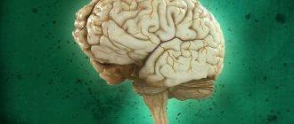
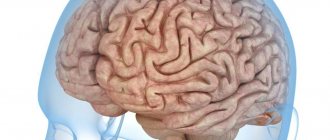
A tumor in the brain is a serious and dangerous pathology in which brain cells degenerate into malignant ones. It can lead to irreversible disorders of brain activity and a significant deterioration in quality of life. Treatment of a brain tumor should begin as early as possible to avoid serious abnormalities. It must be modern, accurate and efficient. This requires innovative equipment and drugs, as well as an experienced and properly qualified specialist. Our center is provided with all this.
Signs of developing a brain tumor
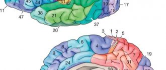
The primary clinical picture is unremarkable, so it is almost impossible to detect cancer. The first signs appear when the tumor has already reached a significant size and compresses healthy brain tissue. Symptoms of malignancy depend on its location:
- Occipital lobe: Loss or deterioration of vision in one eye, and vision may become blurred.
- Frontal region: behavioral disorders, character changes.
- Cerebellum: loss of coordination, nausea and vomiting, weakened muscle fibers at the back of the head, involuntary movements of the eyeballs.
- Parietal lobe: loss of coordination, difficulty speaking and understanding speech.
- Brain stem: bifurcation of the visible image, poor orientation in space, difficulty swallowing, slurred speech, unsteady gait.
- Temporal region: memory impairment, unexplained feelings of fear or anxiety, frequent loss of consciousness, difficulty pronouncing words, hallucinations.
In addition, oncology may be accompanied by:
- partial or complete loss of sensation in the lower or upper extremities;
- general weakness;
- ringing in the ears;
- violation of writing;
- severe headache that is not relieved by medications, often worsening with sudden movements or changes in body position, as well as in the morning;
- problems maintaining balance;
- increased fatigue.
Find out the price
Find out the price
Error! Please fill in all required fields
Thank you! We will contact you shortly
✕
Can't fly to Turkey right now? Sign up for an online consultation with an Anadolu specialist.
Diagnosis of brain tumors
With the help of an initial examination, an experienced oncologist or neurologist determines the most obvious symptoms of brain damage. To do this, the patient's coordination, behavior, sensitivity to pain, and various reflexes are tested. To determine the presence of a malignant formation, laboratory and instrumental diagnostic techniques are also used:
- radiography;
- checking blood for tumor markers;
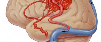
- angiography;
- vision check by an ophthalmologist;
- magnetic resonance, positron emission or computed tomography;
- spinal tap;
- Ultrasound;
- electroencephalography.
A histological analysis of tumor tissue provides a 100% accurate examination result. If the brain is affected by a malignant process, performing a biopsy becomes a complex neurosurgical intervention. It can be performed using a craniotomy or stereotactically, where a needle is inserted into a small hole and monitored by a computer.
Algorithm
Neurologists at the Yusupov Hospital perform a lumbar puncture according to the procedure algorithm. The nurse prepares a lumbar puncture kit:
- sterile gloves;
- sterile forceps;
- alcohol (70%) or 0.5% alcohol solution of chlorhexidine for skin treatment;
- adhesive plaster and sterile beads, adhesive plaster;
- 5 syringes with a capacity of 5 ml and needles for them;
- 0.25% or 0.5% solution of novocaine for anesthesia of the puncture site;
- 1-2% trimecaine solution for administration into the subdural and epidural space;
- sterile needles with a mandrel 10-12 cm long (Bier needle for lumbar puncture);
- sterile tubes for collecting cerebrospinal fluid.
The patient is psychologically prepared for lumbar puncture according to the algorithm. Successful spinal puncture largely depends on the correct position of the patient. Immediately before the puncture, the patient is given a fetal position - laid on his side, head tilted as much as possible, legs bent at the hip and knee joints.
At the level of the line that connects the superior posterior iliac spines, the doctor determines the gap between the spinous processes of the third and fourth lumbar vertebrae. Before puncture, the skin is treated with iodine. After this, the iodine is carefully removed with alcohol so that it does not enter the subarachnoid space. The spinal puncture site is surrounded with a sterile drape.
The site of the intended puncture is anesthetized with a 0.5% novocaine solution. A mandrel is inserted into the needle for lumbar puncture, the skin is pierced, and the direction of the needle is specified. As the needle is inserted, the doctor sequentially overcomes the resistance of the ligamentum flavum and dura mater. After puncture of the dura mater, the spinal puncture needle is inserted very slowly. From time to time, the mandrel is removed to check whether cerebrospinal fluid is leaking. When the needle enters the subarachnoid space, a sinking sensation occurs. When cerebrospinal fluid appears, the needle is advanced another 1–2 mm.
The patient is then asked to relax and carefully straighten his legs and head. The doctor removes the mandrin, preventing the cerebrospinal fluid from leaking. A pressure gauge is attached to the needle and the cerebrospinal fluid pressure is measured. It is normally 100–150 mm of water column. The flow of cerebrospinal fluid, if necessary, is increased by coughing, pressing the jugular veins or abdomen, or the jugular veins.
Cerebrospinal fluid is collected in at least 3 sterile tubes:
- firstly, to determine the concentration of glucose and protein;
- in the second - for serological research and determination of the cellular composition of the cerebrospinal fluid;
- in the third - for bacteriological examination of cerebrospinal fluid.
If doctors suspect tuberculous meningitis in a patient, collect cerebrospinal fluid in a fourth tube to identify the fibrin film. After collecting the cerebrospinal fluid, the mandrin is removed and the needle is removed. When a lumbar puncture is performed, the technique used in children has its own peculiarities. In children, the needle is placed perpendicular to the line of the spine, and in adults - slightly at an angle, open in the direction of the horse's tail.
Treatment of cancer in Anadolu
The main treatment method is surgery to remove the tumor. Small tumors are eliminated using microsurgical methods under the control of computer navigation. Large formations require craniotomy with opening of the skull. Malignant tumors in the pituitary gland can be removed through the nasal passages. One of the popular modern methods is stereotactic radiosurgery without harm to healthy cells, which is performed using the most precise equipment: Varian Edge (new generation TureBeam) or CyberKnife. Our clinic is equipped with a unique hybrid operating room.
Chemotherapy for brain cancer or radiation irradiation of the tumor can be used as auxiliary techniques.
Medical provides patients with a level of service that meets all international standards. Unlike domestic institutions, our clinic helps patients with any type of cancer, including complex and advanced cases of its development. Doctors with professionalism and an individual approach to each patient achieve impressive results. At the same time, the cost of a treatment course is significantly lower than in other world medical centers, which does not in any way affect the quality of services.
A brain tumor is a malignant formation that requires early and accurate diagnosis, as well as complex neurosurgical surgery. This is one of the most dangerous diseases in medicine, which can lead to irreversible disorders of brain activity. Considering the seriousness of the problem, it is better to contact a foreign clinic, which is significantly superior in its capabilities to domestic institutions. This will ensure that treatment of a brain tumor will lead to a significant improvement in the patient’s well-being.
Qualified medical assistance from doctors allows you to promptly identify pathology and select the most effective health course. The high level of development of medicine increases the chances of recovery even in the most difficult cases.
Rules of conduct after puncture
It is recommended to lie on your stomach for 3 hours after a lumbar puncture. It is also prohibited to lift heavy objects or subject yourself to physical stress. If these rules are followed, the patient will be able to avoid leakage of cerebrospinal fluid from the hole.
If medications are administered into the subarachnoid space, the patient must remain in bed for at least 3 days.
Carefully. If there is an increase in temperature, numbness or fluid discharge from the hole after the puncture, you should visit a doctor.
Brain tumor symptoms
Signs of the disease can vary, depending on the size of the tumor, its location and the pressure on surrounding tissue. When located in a certain part of the brain, certain functions are disrupted. Among the main symptoms of cancer:
- complete or partial loss of sensation in the limbs;
- nausea and vomiting;
- blurred vision (a veil may appear before the eyes);
- general weakness, fatigue;
- double vision;
- frequent headaches of varying intensity and character, sometimes it can intensify with changes in body position or sudden movements;
- disturbance of normal speech;
- problems with spatial orientation and balance.
In later stages, cancer may present with personality changes and confusion.
Decoding the results
Normally, cerebrospinal fluid has moderate viscosity, a transparent and colorless structure. Even before the analysis, the doctor evaluates the appearance of the cerebrospinal fluid, the presence of impurities in it (for example, blood), the consistency of the liquid and the rate of its flow. Normally, cerebrospinal fluid should be released at a rate of 20 to 60 drops per minute. Deviation from these indicators may indicate inflammatory processes, tumor diseases or metabolic disorders (for example, leukodystrophy).
How is brain cancer diagnosed?
The Anadolu Clinic conducts a high-quality examination of patients using laboratory, instrumental and visual techniques. The latest generation equipment gives accurate results at any stage of tumor development. The brain needs to be checked regularly so that the disease can be detected at an early stage. The main types of diagnostics that a doctor can prescribe:
- computed and magnetic resonance imaging;
- radiography;
- single photon emission computed tomography (SPECT);
- ultrasonography;
- angiography;
- lumbar puncture;
- electroencephalography;
- blood test for tumor markers;
- biopsy;
- vision check by an ophthalmologist;
- myelography.
The biopsy is carried out in two ways: with a conventional craniotomy or a stereotactic method, when a needle is inserted into a small hole in the head and controlled by computer navigation.
Feelings during the procedure
Many patients are interested in the question of whether it is painful to perform a lumbar puncture. If the procedure is performed correctly, the patient does not feel any significant discomfort or pain. He can feel the needle passing through the hard membranes, but there is no painful reaction. Unpleasant symptoms do not appear, since the doctor administers an anesthetic solution before the puncture.
You may feel an electric shock if the needle hits a spinal nerve. Some patients experience headaches during the procedure.
Treatment of a brain tumor in medical
The main treatment method is surgery. For larger tumors, a craniotomy is performed to open the skull. Small tumors can be removed using a cranioscope. Cancer cells in the pituitary gland are eliminated using access through the nasal passages. Another method of surgical tumor removal is stereotactic radiosurgery. It is performed using high-precision equipment (Cyber Knife or TureBeam), so healthy cells are not harmed.
Additional treatment methods may include chemotherapy or radiation, as well as their combination. They are also effective in the fight against metastases. Chemotherapy drugs are injected intravenously or directly into the spinal column so that the effect affects only the central nervous system.
After successful removal of the pathological formation, patients undergo rehabilitation, which helps restore lost functions and feel better. Our specialists take on cases of any severity and do everything possible to prolong the life of patients and improve its quality.
The medical service provides reasonable prices for the entire range of cancer treatment services. It is significantly lower in other similar medical institutions in Europe, America and Turkey. You can find out what the approximate price of the wellness program will be from our specialists by phone or through the electronic form on the website.
The material was prepared in agreement with medical doctors.
Basic information
Spinal cord puncture is a therapeutic and diagnostic procedure during which cerebrospinal fluid is extracted from the subarachnoid space through a special needle.
There is an opinion that lumbar puncture is dangerous, since during it the spinal cord can be damaged. To understand that this is not so, you need to delve a little deeper into embryology.
During fetal development, the central nervous system, which consists of the brain and spinal cord, develops from the fetal neural tube. All components of the nervous system (neurons, peripheral nerves, subarachnoid cisterns, cerebrospinal fluid, etc.) have the same origin. Therefore, by the composition of cerebrospinal fluid from the caudal region of the back, the condition of the entire nervous system can be assessed.
The spinal cord ends at the level of the 2nd lumbar vertebra
During fetal development, vertebrae grow faster than nerve tissue. For this reason, the spinal cord ends at the level of the 2nd lumbar vertebra with the conus medullaris. Further, thin threads extend to the junction with the sacrum.
Thanks to this structure, it is safe to puncture the spinal canal in this place. According to doctors, the phrase “spinal cord puncture” is incorrect, since there is no spinal cord at the puncture site, only its membranes and cerebrospinal fluid are located here.
Reference. The volume of cerebrospinal fluid in an adult is approximately 120 ml. It is updated after 5 days.
Not all people understand why they take a spinal cord puncture. Lumbar puncture is done to achieve the following goals:
- Study of biological material in laboratory conditions for glucose, certain cells, proteins and other components.
- Determination of cerebrospinal fluid pressure.
- Removing excess cerebrospinal fluid.
- Introduction of drugs into the nervous system.
Now you know why a cerebrospinal fluid puncture is taken.