March 30, 2021
The brain is protected from external mechanical influences by the cranium. The most important sections are located at the bottom of the skull. The gray matter of the brain contains 25 billion neurons, which is almost 4 times the population of the globe (6.5 billion). The human brain is covered by three membranes:
- vascular (web-like),
- soft,
- hard.
In the area of the brain there are five ventricles - containers connected to each other by channels. Inside the cavities there is cerebrospinal fluid - a biological fluid that circulates both in the cisterns of the brain and in the spinal canal.
What is the brain, encephalon, its meaning?
The brain is the anterior part of the central nervous system. Brain
located in the cranial cavity, it interacts with the human body (men, women) with the external environment, integrating the functioning of all body systems.
The brain has the ability to assimilate, organize, store, and retrieve information about past experience. The brain is the material substrate of higher nervous activity. Phylogenetically, the brain is the anterior end of the neural tube. Ontogenetically, the brain is a derivative of the cerebral vesicles, from which parts of the brain are formed: the telencephalon, the diencephalon, the midbrain, the hindbrain, which is represented by formations such as the pons, cerebellum, and medulla oblongata. The cavities of the brain vesicles develop into the ventricles of the brain.
The most important
The brain is a complex structure made up of billions of neurons.
Each of them works like a small electrical switch, transmitting nerve impulses. All information that the body receives with the help of such “electrical wiring” from the outside world enters the cerebral hemispheres, where it is processed. Illustration: biologycorner Tags:
- Memory
- Mental capacity
- Brain
2 comments • To leave a comment you must be an authorized user
- croppop Our brain is nature's best technology! I came across this article right after one wonderful video on the same topic, here it is: https://www.youtube.com/watch?v=1dtUAbmAKH4 I highly recommend watching it for those who are interested in the topic of the abilities of our brain.
- maksroks Cool article, thanks
Brain structure
The cerebrum, or telencephalon, is represented by two hemispheres, which are connected to each other by the corpus callosum, corpus callosum. It consists of nerve fibers running transversely from one hemisphere to the other. The corpus callosum ensures the unity of functioning of both hemispheres. When the corpus callosum is cut, each hemisphere of the brain begins to function independently of each other. Under the corpus callosum is the fornix, fornix. Anterior to the pillars is the anterior commissure, comissura anterior. Between the anterior part of the columns of the fornix and the knee of the corpus callosum there is a thin vertical plate of brain tissue - the transparent septum. Between the plates of the septum there is a slit-like cavity that does not have an ependymal lining. A number of authors call it the 5th ventricle.
The surface of the hemispheres is covered with a layer of gray matter - this is the cerebral cortex. Underneath it is the white matter and subcortical nuclei: striopallidal system, extrapyramidal system.
If you make a horizontal section of the brain through the cerebral hemispheres at the level of the thalamus and subthalamic nuclei, you can see the following formations: pineal body, superior colliculus, thalamus, frenulum, posterior limb of the internal capsule, globus pallidus of the lenticular nucleus, putamen of the lenticular nucleus, lateral sulcus, fence , anterior part of the internal capsule, head of the caudate nucleus, column of the fornix, anterior horn of the lateral ventricle, genu of the corpus callosum, septum pellucidum, interthalamic commissure, lentiform nucleus, external capsule, extreme capsule, convolutions of the insula, lateral sulcus, internal capsule, subthalamic nucleus, tail caudate nucleus, lateral geniculate nucleus, red nucleus, gray matter of the superior colliculus, cerebellar vermis.
Functions
Mission – organization of mental activity.
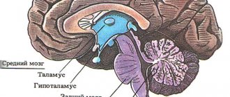
Intermediate
Participates in coordinating the work of organs, regulating body movement, maintaining temperature, metabolism, and emotional background.
Thalamus
. The main task is to sort information. It works like a relay - it processes and sends data coming from receptors and pathways to the brain. The thalamus affects the level of consciousness, attention, sleep, wakefulness. Supports speech functioning.
Epithalamus
. Interaction with other structures occurs through melatonin, a hormone produced by the pineal gland in the dark (therefore, it is not recommended to sleep in the light). A derivative of serotonin - the “happiness hormone”. Melatonin is a participant in the regulation of circadian rhythms, being a natural sleep aid, it affects memory and cognitive processes. It affects the localization of skin pigments (not to be confused with melanin), puberty, and suppresses the growth of a number of cells, including cancer cells. Through connections with the basal ganglia, the epithalamus participates in the optimization of motor activity, and through connections with the limbic system, in the regulation of emotions.
Subthalamus
. Controls the body's muscle responses.
Hypothalamus
. Forms a functional complex with the pituitary gland and directs its work. The complex controls the endocrine system. The hormones it produces help cope with distress and maintain homeostasis.
Thirst and hunger centers are located in the hypothalamus. The department coordinates emotions, human behavior, sleep, wakefulness, and thermoregulation. Endorphins similar in action to opiates are found here, which help to endure pain.
Finite brain
Hemispheres
They act together with subcortical structures and the brain stem. Main destination:
- Organization of interaction of an organism with the environment through its behavior.
- Consolidation of the body.
Corpus callosum
The corpus callosum received attention after operations to dissect it in the treatment of epilepsy. The operations relieved seizures while changing a person’s personality. It was found that the hemispheres are adapted to work independently. However, to coordinate activities, information exchange between them is necessary. The corpus callosum is the main transmitter of information.
Striatum
- Reduces muscle tone.
- Contributes to the coordination of internal organ function and behavior.
- Participates in the formation of conditioned reflexes.
The olfactory brain contains centers that control the sense of smell.
Cerebral cortex
Head of mental processes. Controls sensory and motor functions. Consists of 4 layers.
The ancient layer is responsible for elementary responses (for example, aggression) characteristic of humans and animals.
The old layer is involved in the formation of attachment and laying the foundations of altruism. Thanks to the layer we are happy or angry.
The intermediate layer is a formation of a transitional type, since the modification of old formations into new ones is carried out gradually. Ensures the activity of the new and old cortex.
The neocortex concentrates information from subcortical structures and the brainstem. Thanks to it, living beings think, talk, remember, and create.
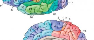
5 cerebral lobes
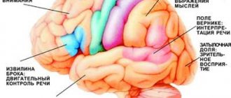
The occipital lobe is the central section of the visual analyzer. Provides visual pattern recognition.
Parietal lobe:
- controls movements;
- orients in time and space;
- provides perception of information from skin receptors.
Thanks to the temporal lobe, living things perceive a variety of sounds.
The frontal lobe regulates voluntary processes, movements, motor speech, abstract thinking, writing, self-criticism, and coordinates the work of other areas of the cortex.
The insula is responsible for the formation of consciousness, the formation of an emotional response and the maintenance of homeostasis.
Brain of a newborn, children, child, human: structure, anatomy
The brain of a newborn baby is shorter and wider than that of school-age children and adults. It is devoid of all tertiary and a number of secondary grooves. By the end of the child’s first year of life, the brain increases in size by 2–2.6 times. By 3 years it increases 3 times. From birth to adulthood, brain weight increases 4 times, and body weight increases 21 times.
The mass of the right hemisphere is most often greater than the mass of the left hemisphere. After birth, the parietal and frontal lobes develop most intensively. And because of this, the overall configuration of the brain changes. Unlike the adult brain, in a newborn the neurons of different layers are located closely next to each other, because of this, the radial striation of the cortex may be absent. Single neurons may be located in the subcortical white matter. In the substantia nigra of the brain stem, neurons do not yet have the pigment melanin, which usually appears by 3–4 years of age. Up to 3–6 months of extrauterine life, the outer embryonic layer, which is called “Obersteiner’s layer,” remains in the cerebellar cortex. Obersteiner's layer consists of medulloblasts and spongioblasts. The surface of the inferior olives of the fetal medulla oblongata is smooth. After the birth of a child, the olives acquire elevations and then noticeably increase in size with age. Almost always in newborns, immature cellular elements are found in the subependymal parts of the ventricular system of the lateral ventricles, the presence of which erroneously resembles manifestations of Virchow's local encephalitis. Immature cells are located in the subependymal layer diffusely or in the form of separate foci. Sometimes they can be traced along the blood vessels over a significant extent of the white matter. By 3–6 months of a child’s life, these cells gradually disappear. The presence of a large number of immature cells in the subependymal parts of the ventricular system is an additional morphological sign of fetal prematurity.
Praxis, praxis analyzer, praxis center
Praxis is the ability to perform purposeful motor acts. Praxis is formed during human life, starting from infancy, and is ensured by a complex functional system of the brain involving the cortical fields of the parietal lobe (inferior parietal lobe) and the frontal lobe, especially the left hemisphere in right-handed people. For normal praxis, the preservation of the kinesthetic and kinetic basis of movements, visual-spatial orientation, programming processes and control of purposeful actions is necessary. The defeat of the praxic system at one level or another is manifested by such a type of pathology as apraxia. The term "praxis" comes from the Greek word "praxis", which means "action". Apraxia is a violation of purposeful action in the absence of muscle paralysis and the preservation of its elementary movements.
How does a child's brain mass change with age?
If you monitor how the mass of a child’s brain changes depending on age, you will notice the following picture. If the child’s age is from 3 to 8 days, body length is 49 – 50 cm, then the brain weight will be 336 grams. At 1 month, the child’s height is 52 cm, brain weight is 360 grams. At 3 months, the child’s height is 56 cm, brain weight is 520 grams. At 6 months, with a height of 62 cm, GM weight is 670 g. At 9 months, with a height of 67 cm, GM weight is 760 g. At 1 year of age, the child’s height is 73 cm, brain weight is 960 g. At 1.5 years, with a height of 79 cm, the weight of the GM is 1045 g. At 2 years old, with a height of 85 cm, the weight of the GM is 1070 g. At 3 years old, the child’s height is 89 cm, and the brain weight is 1150 g. At 5 years old, height is 106 cm, weight is 1240 g. At 10 years old, height 132 cm, brain weight 1300 g. At 12 years old, height 145 cm, brain weight 1370 grams.
Physiology, brain function
Physiologically, all the work of the brain is built on the principles of hierarchy, integrity, systematicity and plasticity. These are the principles of functioning that carry out all conditioned and unconditioned reflexes. They contribute to the flow of conscious mental activity of a person. The principle of hierarchy is that phylogenetically younger parts of the brain carry out higher-order control, complementing but not replacing the function of phylogenetically more ancient parts. As a result of this, the body’s capabilities in more subtle differentiation of each stimulus by each analyzer are expanded, and a more adequate perception of the overall picture of the world is achieved based on the correlation of the results of the activities of many analyzers.
The highest form of expression of the hierarchical principle is the process of corticalization of functions. The principle of hierarchy is combined with the principles of integrity and systematicity, which consist in the fact that the brain functions as a single whole with the entire nervous system, while receiving afferent impulses, carries out its analysis and synthesis, forms a flow of efferent impulses that determine the adequate activity of all peripheral organs . As a result, a stable system is formed that provides continuous information communication: center - periphery - environment - periphery - center. Plasticity refers to the functional variability of nerve centers, which is clearly manifested in the process of compensation for impaired brain functions.
Irradiation of excitation plays an important role in the normal functioning of the brain. The feedback mechanism consists of closing the input and output of the same element or system. The dominant mechanism regulates the relationships between nerve centers.
Occipital lobe
At the base of the brain, visual and auditory fibers intersect. Therefore, the visual zone of the left hemisphere receives impulses from the retina of the right and left eyes. Moreover, if the area is damaged, the person will not experience complete blindness - disturbances are observed only in the left eye.
The back of the head is also necessary to ensure the normal functioning of the visual speech center - with its help we recognize written words and letters and read.
Diseases, disorders, brain lesions
Diseases, disorders, and brain lesions are varied. In further articles, we will focus on pathologies such as brain tumor, brain cyst (including arachnoid, retrocerebellar, liquor), trauma, concussion or bruise of the brain, brain cancer, hydrocephalus (hydropsis), vascular atherosclerosis, aneurysm, encephalopathy, demyelination, ischemia, ischemic or hemorrhagic stroke, heart attack, atrophy, spasm or vasoconstriction, glioblastoma, meningioma, dysfunction, dystonia, diffuse changes, hypoxia (oxygen starvation), encephalitis, inflammation, vascular diseases, atrophic changes. The clinical picture of such diseases depends on the type of pathology.
Brain treatment in Saratov, Russia
Sarklinik provides treatment for a number of diseases, diseases of the central and peripheral nervous system in Saratov, in Russia for children, adolescents, adults, boys, girls, guys, girls, men, women, brain treatment in Saratov . Hardware and non-hardware treatment methods make it possible to restore the functioning of the human nervous system.
Sign up for a consultation. There are contraindications. Specialist consultation is required.
Text: ® SARCLINIC | Sarclinic.com \ Sarlcinic.ru Photo: © pixologic / Photobank Photogenica / photogenica.ru The people depicted in the photo are models, do not suffer from the diseases described and/or all coincidences are excluded.
Related posts:
Claude H Claude syndrome, alternating peduncular syndrome
Zones of the cerebral cortex, functions, human cerebral cortex: structure, activity
Causes of urinary incontinence in women and men, treatment in Saratov
Mobius syndrome, treatment of Mobius syndrome, congenital facial palsy, Poland syndrome
Agnosia: visual, auditory agnosia, treatment of agnosia
Comments ()
Motor area
The left and right hemispheres contain the motor cortex, which is necessary to ensure the activity of the striated muscles. In the left hemisphere, the activity of the right side of the body, coordination of precise movements, and orientation on the ground are controlled. The internal organs send their impulses to this zone.
If the motor cortex is damaged, the following problems will occur:
- disturbances in the functioning of the cardiovascular system and respiratory organs;
- paresis of limbs;
- ataxia.