Anyone at least once in their life has heard caustic expressions about the convolutions in the head and their relationship with intelligence, but few people know that, contrary to popular belief, a frequent and far from original phrase is that “the number of convolutions in a person’s brain is the same in him.” mind” is completely unfounded. So is the number of brain convolutions an indicator of any characteristics of the human body, and is there a certain “ideal” number of them? Is there any difference between the normal number of grooves in the brain of a woman and a man? This article will provide answers to these questions.
What are convolutions
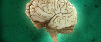
The brain is the most complex organ of our body. It is he who directs all processes in our body, thanks to him we recognize ourselves as individuals. The brain is one of the least studied organs of the human body. This is due to its complex structure and the individual characteristics of each individual person.
The surface of the brain is called the cerebral cortex, it is covered with grooves and convolutions. Consists of more than 100 billion nerve cells - neurons. Convolutions and grooves in humans are formed during the period of intrauterine embryonic development, their number does not change with age. They are needed so that the embryo's brain can fit into a small skull. A large skull in a baby can cause problems during childbirth.
At birth, the weight of the human brain is 300 grams, and the weight of an adult is about 1500 g. The area of the adult brain is approximately 2200 cm2, and if not for its special folded structure, it simply would not fit into the narrow cranium.
During an external examination, it is impossible to see the entire area of the cerebral cortex, because two-thirds is located deep in the grooves, and only a third is on the outer side of the hemispheres.
Magnetic resonance imaging (MRI) in St. Petersburg
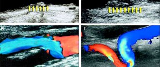
Localization of the pathological focus with MRI of the brain begins with determining the location of the lesion in relation to the tentorium of the cerebellum. Therefore, formations above the tentorium are classified as supratentorial, and everything below is classified as infratentorial.
MRI of the brain. Midsagittal section. tentorium cerebellum (arrow).
Above the tentorium are the cerebral hemispheres. Each hemisphere of the brain consists of four lobes - frontal, parietal, occipital and temporal. If the pathology is located in the hemisphere, then it is necessary to decide which lobe it belongs to. To do this, you first need to find the grooves that serve as the boundaries of the lobes. The central sulcus (sulc.centralis) is better visible in the sagittal plane. It is located in the middle between the parallel precentral and postcentral sulci. There are many options for the structure and course of the furrow. Usually it has a significant extent and goes in the anterior-inferior direction from the interhemispheric fissure to the Sylvian fissure, which it does not always reach. The lower end of the furrow either continues in its main direction or bends back. The central sulcus may be interrupted along the way. In the transverse plane on the upper sections the groove has the greatest extent, reaching almost to the interhemispheric fissure. The lower the cut, the shorter the central groove on it. At the level of the lateral ventricles it is barely visible. The central sulcus separates the frontal and parietal lobes.
MRI of the brain. Lateral sagittal section. Central sulcus (arrow).
MRI of the brain. Axial slice. Central sulcus (arrows).
MRI of the brain. Axial section at the level of the roof of the lateral ventricles. Central sulcus (arrows).
MRI of the brain. Borders of the frontal and parietal lobes in the axial plane.
Another important groove is the Sylvian fissure (fissura cerebri lateralis). On sagittal sections it goes from bottom to top in the anteroposterior direction (Fig. 32). In the axial plane, the Sylvian fissure itself also deviates backward, while its branches are directed perpendicularly towards the interhemispheric fissure. The Sylvian fissure separates the frontal and parietal lobes from the temporal lobe.
MRI of the brain. Lateral sagittal section. Sylvian fissure (arrows).
MRI of the brain. Axial section at the level of the third ventricle. Sylvian fissure (arrows).
MRI of the brain. Borders of the frontal, parietal, temporal and occipital lobes on a sagittal section.
To delimit the parietal lobe, you also need to find the parieto-occipital sulcus (sulc. parietooccipitalis). This groove in the sagittal plane can be traced on the median and medial sections. It extends from the surface of the brain downwards, has a considerable extent and is often segmented. In the transverse plane, the parieto-occipital sulcus extends almost perpendicular to the interhemispheric fissure (Fig. 36) and gives off many small branches. Thus, the boundaries of the parietal lobe are with the frontal lobe - the central sulcus, with the occipital - the parieto-occipital sulcus, with the temporal - the Sylvian fissure and the superior temporal sulcus (angular gyrus).
MRI of the brain. Medial sagittal section. Parieto-occipital sulcus (arrow).
MRI of the brain. Axial slice. Parieto-occipital sulcus (arrow).
MRI of the brain. Borders of the parietal lobe on the medial sagittal section.
The next important dividing groove is the collateral groove (sulc.collateralis). On sagittal sections, it is visible as the inferolateral border of the parahippocampal gyrus, in the region of the pole of the temporal lobe (Fig. 38). It is easier to see in the axial plane in sections at the level of the midbrain (Fig. 39). When the axial plane of the slices is tilted backward, it is visible simultaneously with the temporo-occipital groove. The temporo-occipital groove (sulc. temporooccipitalis) on lateral sagittal sections runs sinuously backward along the border of the brain with the temporal bone and then bends upward (Fig. 40). On axial sections at the level of the Varoliev bridge, it is located in the anteroposterior direction. Thus, the border of the temporal lobe (Fig. 41) with the frontal and parietal lobes is the Sylvian fissure, with the occipital lobe - the temporo-occipital sulcus and the collateral sulcus.
MRI of the brain. Sagittal section. Collateral groove (arrow).
MRI of the brain. Axial slice. Collateral groove (arrows).
MRI of the brain. Axial section at the level of the Varoliev bridge. Temporo-occipital sulcus (arrows).
MRI of the brain. Axial section at the level of the cerebral peduncles. Borders of the temporal lobe.
To determine the boundaries of the occipital lobe, we already have all the landmarks. The border with the parietal lobe is the medially located parieto-occipital sulcus, and the border with the temporal lobe is the laterally located temporo-occipital sulcus.
MRI of the brain. Coronal section. Border sulci (SPO - parieto-occipital sulcus, STO - temporo-occipital sulcus, SCol - collateral sulcus).
MRI of the brain. Borders of the occipital lobe on the medial sagittal section.
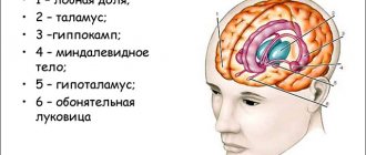
Usually localization by lobes is sufficient to describe hemispheric pathologies. In some cases, when reference to gyri or functional areas is required, we recommend using the appropriate atlases (A.V. Kholin, 2005). With centrally (axially) located space-occupying formations, the ventricles of the brain and the subcortical (basal) nuclei located around them may be involved. The optic thalamus, hypothalamus, subthalamus, and epithalamus belong to the diencephalon, a component of the brain stem.
MRI of the brain. Axial slice. Lateral ventricles and subcortical nuclei (NC - caudate nucleus, NL - lenticular nucleus, Th - thalamus optic). Parts of the brainstem (lower midbrain, pons, and medulla oblongata) and the cerebellum are located infratentorially.
The midbrain only partially occupies the supratentorial space; a significant part of it passes through the hole in the tentorium into the posterior cranium. hole. The paired legs of the brain and roof (tectum) are always clearly visible from behind. The roof of the midbrain lies posterior to the aqueduct and consists of the quadrigeminal plate.
MRI of the brain. Midsagittal section. Brain stem (V3 - third ventricle, V4 - fourth ventricle, Q - plate quadrigeminal, Mes - midbrain, P - pons, C - cerebellum, M - medulla oblongata).
The boundary between the midbrain and the pons is the superior sulcus, and the border with the medulla oblongata is the inferior sulcus of the pons. The bridge has a characteristic protruding front part. The posterior surface of the pons is a continuation of the medulla oblongata. At the upper border of the bridge between its abdomen and the middle cerebellar peduncle, the trigeminal nerves (n. trigeminus, V pair) begin. They are clearly visible on transverse MR sections, as they run horizontally forward and have a thickness of about 5 mm. The trigeminal nerve is divided into 3 branches - optic (1), maxillary (2) and mandibular (3). They all go forward into Meckel's cavity to the trigeminal ganglion. From here the 3rd branch goes down through the foramen ovale, and the 1st and 2nd branches go through the cavernous sinus, along its lateral wall. Then, branch 1 enters the orbit through the superior foramen, and branch 2 exits the cranial cavity through the foramen rotundum. The III, IV and VI pairs of cranial nerves, which provide movement of the eyeball, are usually not visualized on MRI scans.
MRI of the brain. Axial slice. Trigeminal nerves (arrow).
The facial nerve (n. facialis, VII pair) and the vestibular-cochlear nerve (n. vestibulocochlearis, VIII pair) exit their trunk together, the facial nerve is slightly more medial, and go in one bundle, crossing the pontocerebellar cistern, and go into the internal auditory opening temporal bone. In the internal auditory canal, the vestibular branch runs in the posterior superior and inferior quadrants, the cochlear branch in the inferior, and the facial nerve in the anterior superior. The VII nerve enters the labyrinth (labyrinthine segment), runs inside the temporal bone to the geniculate body, turns back and passes under the lateral semicircular canal (tympanic segment) and exits the temporal bone through the stylomastoid foramen (foramen stylomastoideum). Next, the nerve goes to the salivary gland, where it divides into terminal branches. On MRI scans in sections 3-5 mm thick, the VII and VIII nerves are not separated and are designated as the auditory nerve. With thinner sections, the course of each nerve can be visualized separately.
MRI of the brain. Axial slice. Auditory nerve.
The medulla oblongata begins from the lower border of the pons. At the level of the foramen magnum it passes into the spinal cord. From it depart from the IX to XII pairs of cranial nerves, of which the initial part of the hypoglossal nerve (n. hypoglossus, XII pair) and, in the form of a single complex, IX, X, XI pairs are sometimes visible on transverse MRI. The IV ventricle runs from the aqueduct above to the foramen of Majendie below. It is located between the brainstem anteriorly and the velum and cerebellar peduncles posteriorly. Posterior to the pons and medulla oblongata is the cerebellum. It is connected to the brain stem by the superior, middle and inferior cerebellar peduncles. The cerebellum consists of a midline vermis and paired hemispheres.
MRI of the brain. Axial slice. Cerebellum (CV - cerebellar vermis, CH - cerebellar hemisphere).
MRI in St. Petersburg, carried out by us, always clearly indicates the localization of the lesion in the report, which is necessary for comparison with the clinic and deciding on the possibility and scope of the operation.
Leave feedback.
MRI in St. Petersburg USA
How many convolutions does a person have?
No animal has as many convolutions as humans. Their number is influenced by two variables: the area of the cerebral cortex and its thickness.
It is impossible to count exactly how many there are. This happens due to the individual differences of all people. Some convolutions may be too small or almost invisible, and they are located differently in different males and females. Sometimes they are able to slightly change their size during life, so it is impossible to say how many convolutions a person has.
Does their number affect intelligence?
As a result of the research (when some scientists bequeathed their brains to their colleagues for the sake of science for comparison with the brain of an ordinary person), no special physiological differences were discovered. Human convolutions are formed during intrauterine development and the level of intelligence is not able to influence the increase in their number during a person’s life.
Does not affect intelligence or brain weight. Weight primarily depends on a person’s height and build. Thus, based on how many convolutions a person has in his brain, it is impossible to draw a conclusion about his intelligence.
Who has more convolutions: men or women?
It has long been known that a man's brain weighs more than a woman's. There are many jokes and prejudices in this regard. However, scientists have found that, despite being lighter, a woman's brain has more convolutions, essentially compensating for her lower weight. The volume of a woman's brain is smaller than that of a man, but in terms of area, thanks to the greater number of convolutions, it is the same.
It should be noted that the stronger sex has twenty percent less gray matter than the weaker sex. In men, neurons are located further apart. For this reason, the difference in the volumes of the craniums is compensated. However, despite the greater number of convolutions in the head of women, both sexes do not differ from each other in mental development.
Thus, intelligence does not depend on how many convolutions a person has. Despite the greater number of them in the minds of women, ladies are not mentally superior to men. It is simply impossible to know the exact number. Yes, this information does not make sense, because each of us is individual, and, therefore, there is no consensus on this issue. However, it is known that we do not use our brain to its full capacity. Consequently, how many convolutions a person has in his head will not matter.
Is there a connection between a person's gender and the number of convolutions?
The long-known fact that the male brain weighs more than the female brain has given rise to many ridiculous jokes and stereotypes. However, a worthy answer to the jokers was given by scientists who found that the female brain, as opposed to the male brain, has a more complex structure with a significantly larger number of convolutions, which compensates for less weight. For the same reason, neurons in men's heads are located at a greater distance. Thus, the area of the human brain, regardless of the gender of its owner, is equal.
Studies have found that the gray matter content in men's heads is 20% less. In accordance with this, the difference in the number of convolutions or the mass of the brain does not give a single advantage to the genders: in the level of intelligence, both sexes do not differ.
The human brain is the largest element of the central nervous system, which determines the complexity of its structure. It is he who makes a person himself, gives him the miracle of consciousness. Naturally, scientists have long been interested in whether there is a connection between the appearance of the brain and the kind of personality it makes its owner. For now, we can say for sure: neither its mass, nor how many convolutions a person has in the brain, defines an individual as smart or stupid. The grooves in the gray matter are just folds of a huge organ squeezed into the human skull. Attempts to calculate their average number are pointless, because for each person this number is individual, and in structure and appearance they can be both deep and barely visible to the eye, which makes the counting process impossible.