Osteochondrosis is a chronic degenerative dystrophic disease in which gradual destruction of the cartilage tissue of the intervertebral discs occurs. They lose their elasticity and shock-absorbing ability, and stop protecting the radicular nerves extending from the spinal cord from compression. A decrease in the height of the intervertebral discs leads to instability of the position of the vertebral bodies, their constant displacement, which can cause the development of spinal canal stenosis.
Strain syndrome is excessive tension of the muscle fibers in the area of the affected area of the spinal column. In this way, the body tries to compensate for the decrease in the height of the intervertebral disc and eliminate compression from the radicular nerve. These signs are important in the differential diagnosis of osteochondrosis with other possible pathologies.
Tension syndrome in osteochondrosis helps the neurologist to clarify the degree of damage to the cartilage tissue of the disc and compression of the radicular nerve. There are several symptoms of tension: Lasegue, Neri, Matskevich, Vaserman, Sekar, Bekhterev and Dejerine. We will examine some of them in detail in the proposed material.
If you find at least one of them in yourself, then do not hesitate to contact a neurologist. Such a manifestation indicates that there is serious damage to the nerve fiber. Without timely treatment, this condition can provoke atrophy of the radicular nerve and trigger a cascade process of innervation disruption. In this case, not only the muscles of the back frame can be damaged, but also the lower limbs, vascular bed, and internal organs of the abdominal cavity and pelvis.
If you have symptoms of tension, then in Moscow you can make an appointment with a neurologist at our manual therapy clinic. The initial appointment is free for each patient. During the consultation, an experienced doctor will check whether you really have a positive symptom of tension due to osteochondrosis. Once an accurate diagnosis is made, an individual treatment plan will be developed.
Positive symptoms of tension in neurology
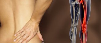
Let's look at some symptoms of tension in neurology. The most common symptom of root tension is Lasegue's syndrome. It indicates an ongoing muscle spasm that accompanies vertebrogenic pain syndrome. The nerve roots are blocked by spasmed muscles. When making sudden movements of the leg or body with a positive Lasegue tension symptom, a nerve fiber may rupture. Therefore, the patient is shown bed rest and complete physical rest until the muscle spasm passes.
Bilateral symptoms of tension in neurology with osteochondrosis appear when large nerves responsible for the innervation of the lower extremities are damaged. For example, bilateral Lassègue's syndrome may be present with sciatica (inflammation of the sciatic nerve). The same sign may indicate piriformis syndrome, plexopathy of the lumbosacral and coccygeal nerve plexuses.
With positive Lasegue syndrome in osteochondrosis, the following clinical signs are present:
- when trying to raise the upper leg on the side of the lesion of the radicular nerves, the patient experiences a severe exacerbation of the pain syndrome;
- when the position of the body in space changes, a sensation of pulsation occurs in the leg, associated with compression of a large nerve;
- there is no possibility to move the leg freely in the lateral direction.
When determining Lasègue tension syndrome, the physician must determine at what angle the patient can lift the leg upward. The condition of the limb in a flexed state is also assessed. If the pain when bending the knee joint decreases, this indicates that the symptom is positive.
Symptoms of tension in lumbar osteochondrosis
Let's consider other symptoms of tension in lumbar osteochondrosis, one of which is called planting syndrome. It indicates the etiology of pain in the spinal column. It can also be used to assess the condition of the radicular nerves. In order to evaluate the symptoms of tension in osteochondrosis of the lumbar region, it is necessary:
- ask the patient to take off his shoes and sit on the couch so that his back is tilted back and his legs remain straight and parallel to the floor;
- the patient should remain in this reclining state for approximately 20–30 seconds;
- the appearance of complaints of acute pain in the knee joint and falling on the back due to exceeding the pain threshold in the lumbar spine indicates that the landing syndrome is positive.
While the patient is in a reclining position on the couch, it is important to conduct a palpation examination of the area in which pain is detected. Reflex tension in certain areas of the muscular frame of the back and lower back indicates that destruction of individual intervertebral discs has occurred.
In the event that the patient can remain in a reclining position with straightened legs for a long time, the diagnosis of lumboischialgia is not confirmed. You should look for the cause of pain in other parts of the musculoskeletal system. Most often this is deforming osteoarthritis of the iliosacral joints or spondyloarthrosis of the coccygeal-sacral joint. With these pathologies, tension syndromes remain negative.
Causes and mechanism of development of osteochondrosis
To understand why your back hurts, you need to understand how our spine is structured, how it works, what functions it performs and what factors can lead to its damage.
The human spine consists of 32-34 vertebrae (7 cervical, 12 thoracic, 5 lumbar, 5 sacral, 3-5 coccygeal), between which there is an intervertebral disc consisting of cartilage tissue. In the middle of the intervertebral disc there is the nucleus pulposus - a semi-liquid formation in the form of a “ball”, which performs the function of shock absorption and is surrounded by dense cartilage tissue (fibrous ring). The spinal canal, which contains the spinal cord and the nerves extending from it, runs through the entire spine. This entire structure is surrounded by muscles and ligaments. The main functions of the spine are musculoskeletal, shock-absorbing, and protective.
Imagine the Ostankino TV tower, which is held in a vertical position thanks to a whole system of cables stretched from the base to the top. Likewise, our spine is held in the desired position by a group of stabilizer muscles, which normally evenly distribute the load on the spine and joints. Unlike the Ostankino TV tower, our spine is more complex; it can bend in different directions and even twist, all this is possible due to the presence of an intervertebral disc, muscles and ligaments.
Every day a person makes some monotonous, repetitive movements associated with work or leisure activities. If the same muscles work for a long time, they become overstrained and spasm, while other muscles at this time do not experience any stress at all and atrophy. This leads to a change in the “geometry” of the body, the load on the intervertebral disc is redistributed, spasmed muscles tighten the vertebrae, and nutrition deteriorates. With monotonous hard physical labor, the same processes occur. In addition, the intervertebral disc does not have blood vessels, and its nutrition is provided by the surrounding muscles, and during movement in the intervertebral joint, nutritious synovial fluid enters it.
Cartilage tissue consists of 80-85% water, so the drinking regime is of great importance. During the day, a person should drink at least 2 liters of clean water. If not enough water enters the body, then dehydration (drying out) of the intervertebral disc occurs, the cartilage cracks and collapses.
In my practice, I have long noted that stress, anxiety, and worries often contribute to the occurrence of back pain. Our body perceives any stressful situation as danger. At the same time, the sympathetic part of the nervous system is activated, the adrenal glands “inject” stress hormones into the blood, blood pressure rises, the heartbeat quickens, and muscles tense. In nature, if an animal is frightened of something, it runs or defends itself, accordingly, stress hormones burn and the muscles, after working, relax. Man is a social being; he begins to worry more often and move less, so there is no relaxation. As a result, pain in the spine, headache, motor tics and more occurs.
In the literature you can find different formulations of osteochondrosis, but their essence is the same. Osteochondrosis is a “breakdown” of the motor segment, destruction, degeneration of cartilage tissue. The reason is an incorrect motor stereotype and, as a consequence, a malnutrition of the cartilage.
The progression of osteochondrosis is promoted by: a sedentary lifestyle, heavy physical activity, heavy lifting, obesity, carrying a bag on one shoulder, high-heeled shoes, too soft a mattress for sleeping, frequent hypothermia, bad habits, hereditary predisposition, stressful situations, insufficient drinking regime and other.
Tripod symptom for osteochondrosis
Another reliable sign of the development of osteochondrosis is tripod syndrome. It allows you to distinguish between psychogenic pain in the back and that reflected in the development of pathologies of the internal organs of the chest and abdominal cavity. Using the tripod syndrome, the doctor can assess the condition of the spinal column, the muscular frame of the back, nerve fiber, tendon and ligament apparatus.
With vertebrogenic pain syndrome, characteristic clinical signs are present. This is muscle tension and spasm, the inability to perform certain actions, a feeling of stiffness and much more.
A specific sign of the development of osteochondrosis and an attack of vertebrogenic radicular pain is tripod syndrome. In order to recognize this tension syndrome, the doctor must pay attention to the position of the patient’s body when trying to rise from a lying position, when sitting on a chair, when getting out of bed, etc.
Distinctive signs of positive tripod tension syndrome:
- lying on his back, the patient tries to raise his torso by resting his hands on a hard surface; if he sits down in a semi-recumbent position, he must support the weight of his torso by resting on his hands;
- when asked to get out of bed, the patient first turns over on his side or stomach and only after that, leaning on his hands, he rises, while trying not to use the muscles of the back and lower back as much as possible;
- the seating position on the chair is also specific - the torso is tilted back, the arms are moved behind the back and the body weight is transferred to them.
Tripod syndrome is a unifying term for all positions of the human body in which he tries to maximally unload the muscles of the back and lower back and at the same time transfer weight to his arms.
IV. Treatment of radiculitis (radiculopathies)
IMPORTANT! Treatment is aimed, first of all, at eliminating the causes leading to the disease!
The main method of treatment is complex therapy, which is aimed at:
- to eliminate muscle spasms and pain,
- to eliminate compression of nerve roots, blood vessels,
- to activate metabolic processes in the affected area, improve trophism and microcirculation,
- for the prevention of repeated exacerbations of radiculitis and other spinal diseases
- central analgesics that help to avoid or reduce chronic pain syndrome associated not with problems in the spine, but with pathological restructuring of the pain perception system.
The most effective complex treatment , which includes:
- restriction of physical activity during an exacerbation should not be long; according to research, early expansion of physical activity helps prevent chronic pain;
- during an exacerbation, a method of drug therapy using painkillers, anti-inflammatory non-steroidal drugs, muscle relaxants, corticosteroids, drugs that improve microcirculation in tissues;
- manual therapy (osteopathic techniques), reflexology, massage and skeletal traction;
- physiotherapeutic treatment methods - shockwave therapy, laser radiation, ultrasound therapy, etc.
- physical therapy, swimming
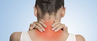
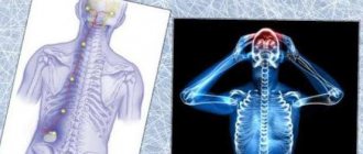
Symptoms of tension in cervical osteochondrosis
Osteochondrosis of the cervical spine also gives symptoms of tension, which manifest themselves in the collar area, upper chest, and shoulders. Determining the symptoms of tension in cervical osteochondrosis is quite simple - for this, the person needs to be asked to raise or move their arm to the side, and tilt their head.
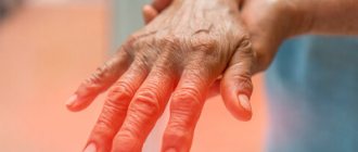
For example, with positive Bonnet tension syndrome, the patient cannot move the arm raised to shoulder level to the side and bring it back. In this case, the upper limb on the side where there is no damage to the radicular nerve moves freely in different directions. In Bonnet syndrome, sensory disturbances are also present. Local areas of decreased skin sensitivity are identified along the back surface of the shoulder and forearm. When palpating the cervical spine, shoulder, collar area and upper limb, pain is detected only at the site of localization of degenerative dystrophic destruction of the intervertebral disc.
The Wasserman-Matskevich symptom is a combination of two clinical signs that appear with severe muscle tension in the neck and collar area. To determine them, the patient is asked to lie on his back on a hard surface. Then he should take turns raising his arms up in front of him and abducting them in different directions at different angles. With positive tension syndrome, raising your arm up in front of you will be accompanied by severe pain in the area of the shoulder blade and forearm.
Neri's tension syndrome may indicate inflammation or compression of the dorsal roots of the spinal cord. To determine it, the patient must stand up straight with his legs straight. Then the doctor asks him to tilt his head as far as possible towards his chest so that his chin touches his chest. If it is impossible to do this on their own, the doctor helps the patient until the chin touches the sternum or until the patient says that he has severe pain in the lumbar or collar area. This tension syndrome is present with radiculitis, myelitis of the roots, herniated intervertebral disc in the cervical spine.
Neri's tension syndrome can be positive when osteophytes grow in the area of the cervical and cervicothoracic spine, osteochondrosis with radicular syndrome, displacement of vertebral bodies, destruction of intervertebral joints with compression of the roots, etc.
Also, with cervical osteochondrosis, Dejerine's syndrome is of great diagnostic importance. It indicates damage to the membranes of the spinal cord. If a person experiences pain only during a certain type of physical activity, for example, when turning the head to the side, then this indicates a positive symptom of Dejerine’s tension.
Diagnosis and treatment of chronic low back pain (a practitioner's view)
For decades, in domestic clinical practice, spinal osteochondrosis was considered a universal cause of back pain. Meanwhile, degenerative-dystrophic changes in the spine detected using spondylography, computed tomography (CT) or magnetic resonance imaging (MRI), which were considered markers of osteochondrosis, poorly correlate with the clinical picture and are often found in people who do not suffer from back pain [4]. Thus, the degenerative process in the spine itself can only be considered a prerequisite for back pain, but not its direct cause. Unfortunately, a simplified understanding of the problem of spinal osteochondrosis hinders attempts to elucidate the true causes of back pain and the development of differentiated approaches to its treatment [5]. This article discusses the issues of diagnosis and differentiated treatment of various syndromes that arise against the background of degenerative changes in the lumbosacral region. Classification Back pain can be caused by pathology of the spinal structures (vertebrogenic pain) and have another origin (for example, viscerogenic pain). According to the mechanism of development, back pain can be nociceptive, neuropathic, psychogenic and mixed. In most cases, back pain is nociceptive in nature and is associated with irritation of pain receptors in muscles, joints or ligaments. When a nerve root or ganglion is involved in the pathological process, neuropathic pain occurs. However, it must be remembered that signs of damage to the spinal roots are detected in only 5% of patients with back pain. According to the course, there are acute pain, which resolves within 6 weeks, subacute, lasting from 6 to 12 weeks, and chronic, lasting more than 12 weeks. [6]. Reasons for the formation of pain syndrome The high frequency of lesions of the lumbar spine is due to the peculiarities of its biomechanics - a large range of movements carried out in various planes, significant physical stress on the vertebrae and discs in this area, which significantly accelerates degenerative processes. In addition, the formation of pain syndrome occurs due to structural and functional changes in the tendon-ligamentous apparatus with gradually developing hypertrophy of the ligaments, the occurrence of persistent muscle spasm, which in turn leads to changes in biomechanics, posture and causes secondary changes in the musculoskeletal system. It follows that degenerative changes in the spine are only the beginning of a complex cascade process. The development of degenerative changes is facilitated by repeated injuries, excessive static or dynamic load, and hereditary predisposition. As a result, dehydration of the intervertebral discs occurs, they lose their shock-absorbing function and become more sensitive to mechanical stress. The fibrous ring, located along the periphery of the disc, becomes thinner, cracks appear in it, along which the central part of the disc - the nucleus pulposus - moves to the periphery, forming a protrusion (protrusion) [5]. Due to injury or intense stress, the protrusion can increase abruptly, which leads to protrusion of the nucleus pulposus and part of the fibrous ring into the spinal canal, which is already called a disc herniation. The hernia is usually a solid formation that maintains connection with the body of the disc, but sometimes its fragments can fall out into the spinal canal, then the hernia is called sequestered. Pain during a disc herniation first appears due to irritation of the pain receptors of the outer layers of the fibrous ring and the posterior longitudinal ligament, information from which enters the spinal cord through the branches of the sinuvertebral nerve. A reflex spasm of segmental muscles occurs, which is protective in nature and leads to immobilization of the affected segment (myofixation), but over time it loses its protective role and becomes an independent factor maintaining pain [7]. Shifting towards the spinal canal or intervertebral foramen, the hernia can compress the adjacent spinal root and the corresponding spinal ganglion, which leads to the occurrence of radicular syndrome (radiculopathy). Damage to the root can be caused not only by its mechanical compression, but also by inflammation, edema and demyelination [8]. In addition, at an earlier stage of the degenerative process, a gradual decrease in the height of the disc disrupts the functioning of the entire spinal motion segment (SMS), which includes 2 adjacent vertebrae connected by the intervertebral disc in the front and two facet joints in the back, with the surrounding muscles and ligaments. The articular facets in the intervertebral joints shift relative to each other, which can lead to subluxation and displacement of the vertebrae (spondylolisthesis). The resulting instability of the SMS increases the sensitivity of the spine to injury or sudden movements and accelerates degenerative changes, primarily arthrosis of the facet joints. These changes often remain asymptomatic, but with injury or overuse they can become a source of pain [7, 8]. An important role in the formation of pain is played by the development of reversible blockade of the SMS, which is clinically manifested by limited mobility of the spine, local and referred pain, and degenerative changes in connective tissue structures [9]. Over the years, the mechanical stability of the PDS and the entire spine is restored due to marginal growths (osteophytes), fibrosis of the discs and capsule, ankylosis of the facet joints, and thickening of the ligaments. These changes complete the “degenerative cascade” in the spine and sometimes lead to spontaneous subsidence of pain. But at the same time they can cause spinal canal stenosis. In addition, osteophytes directed towards the spinal canal can injure the roots, thereby causing persistent pain [5]. Thus, at various stages of the degenerative process in the spine, various factors play a leading role in the development of pain syndrome: disc herniation, instability or blockade of the SMS, arthrosis of the facet joints and spinal canal stenosis. In each of these cases, the pain syndrome has a clinical uniqueness, different time dynamics, prognosis and requires a special approach to treatment [9]. In most cases, pain in the back and lower extremities is nociceptive, local or referred, only in a relatively small number of cases it is explained by damage to the spinal root or ganglion and is neuropathic. Clinical manifestations Discogenic pain syndrome Pathology of the intervertebral disc is the cause of approximately 30% of cases of back pain. This is the main cause of chronic back pain in young people (30–50 years) [2]. In most cases, the last 2 discs are affected: L4–L5 and L5–S1, less commonly L3–L4. For convenience, we will call any protrusion of an intervertebral disc more than 3 mm a hernia. Hernias can vary in location. Posterior hernias can be divided into lateral (displaced towards the intervertebral foramen and infringe on the root contained in it), paramedian (or mediolateral), median (perforate the posterior half of the fibrous ring and the posterior longitudinal ligament). Pain syndromes are usually caused by posterior hernias [6]. At the initial stage of the formation of a disc herniation, local reflex pain occurs, which can radiate to the sacroiliac joint, sacrum, coccyx, scrotum or perineum; at a later stage, radicular pain appears. The relationship between the affected root and the location of the hernia depends not only on the level of the hernia, but also on the direction of its protrusion. Lumbar disc herniations are most often paramedian and put pressure on the root that exits through the intervertebral foramen to a level below. For example, with a hernia of L4–L5, the L5 root is most often affected. Thus, based on clinical data, we can say with certainty which of the roots has been compressed, but we cannot determine the location of the hernia. With a herniated disc, pain often occurs with sudden movement, bending, lifting something heavy, or falling. The pain intensifies with movement, straining, lifting weights, staying in one position for a long time, coughing and sneezing and weakens with rest, especially in a position on the healthy side with the affected leg bent. On examination, there is a slight forward tilt of the body and limited movement, especially to the front and to the painful side. There is pronounced tension in the paravertebral muscles, which decreases in the supine position. Symptoms of tension are not specific to a radicular lesion; they can occur when the paravertebral muscles or posterior muscles of the thigh and lower leg are tense, but with their help it is possible to assess the severity and dynamics of the pain syndrome. Lasègue's symptom is checked by slowly raising the patient's straight leg upward until pain appears in the leg along the sciatic nerve. When the L5–S1 roots are compressed, pain appears when lifting the leg up to 30–40o, and when bending the leg at the knee and hip joints, it goes away. If pain occurs when raising the leg above 70o, then most likely it is not associated with compression of the root. If the pain does not go away when bending the leg at the knee and hip joints, this may indicate a pathology of the hip joint or the psychogenic nature of the pain. To confirm the radicular nature of Lasègue's symptom, the leg is raised to a level above which pain occurs, and then the foot is forced to flex at the ankle joint, which in case of radiculopathy causes pain to radiate along the affected root. When the vessels supplying the spinal nerve root are compressed, ischemic damage occurs, which can lead to the development of foot paresis. In most cases, paresis regresses with conservative therapy. Radiculopathy can also be the result of compression of the root by the articular facet due to arthropathy of the intervertebral joints, osteophyte or hypertrophied ligamentum flavum, which is especially common in elderly patients. Unlike pain from hernias, pain from radiculopathy is relieved by sitting. Lasègue's symptom is often negative. Non-radicular lumbar sciatica is more common than radicular sciatica and can be due to various reasons. Such pain may be discogenic in nature. Discogenic pain, not associated with root compression, is most pronounced in the lower back, but often spreads to the buttock and thigh. It is characterized by a number of signs that distinguish it from reflex pain of other origins. Discogenic pain can be constant or intermittent and usually worsens with bending and sitting. During extension exercises, pain can be reported that is relieved in the leg, but intensifies in the lumbar region [6, 7]. Confirmation of the discogenic nature of pain is possible with discography performed under fluoroscopic control - stimulation of the affected disc induces pain with irradiation characteristic of the patient. Painful dysfunction of the PMS Painful dysfunction of the PDS can also contribute to the appearance of non-radicular lumbar sciatica. It occurs as a result of uniform moderate protrusion of the disc, which leads to a decrease in its height and a change in the relative position of the main elements of the SDS. This can cause instability or pathological fixation of the segment, leading to chronic pain. Instability can also be caused by spondylolisthesis and other spinal diseases. Instability of the MJ leads to excessive stress on the facet joints and muscles. The pain associated with it is usually bilateral, intensifies with prolonged stay in one position, bending over, or lifting heavy objects. Movements in the spine are usually not limited, but painful (especially extension). Lumbar pain sometimes radiates to the sacroiliac joint and iliac wing, but not to the buttock or thigh. The diagnosis can be confirmed using functional radiography of the spine. Arthrosis of the intervertebral (facet) joints Arthrosis of the intervertebral (facet) joints (spondyloarthrosis) is the cause of back pain in approximately 20% of cases. In the elderly (over 65 years of age), spondyloarthrosis is the most common cause of chronic back pain, accounting for up to 40% of cases [6]. Spondyloarthrosis can occur: – as a result of overload of the posterior sections of the SMS (for example, due to a violation of the statics of the spine); – with widespread osteoarthritis affecting the joints of the spine and limbs; – due to degeneration and a decrease in the height of the disc, leading to a change in the relationship of the articular processes, and can be accompanied by functional blockade of the joints, subluxation in the joints with pinching of the articular capsule, inflammation of the articular tissues [9]. Clinically, spondyloarthrosis manifests itself as bilateral pain, which, unlike discogenic pain, is usually localized paravertebrally and not along the midline. The pain is most pronounced in the lower back, but often radiates to the sacroiliac joint, buttock, thigh, and possibly a more distal spread of pain - up to the foot. As a rule, it is intermittent and increases with prolonged standing and extension, but decreases or at least does not increase with bending forward, sitting and walking, as well as in the supine position [10]. X-rays for spondyloarthrosis reveal narrowing of the interarticular spaces, subchondral sclerosis, deformation and hypertrophy of the articular facets due to osteochondral growths, but often such changes are not accompanied by any symptoms. Therefore, the most reliable sign is a reduction in pain during bilateral blockade of the intervertebral joints or the medial fibers of the dorsal branches of the spinal nerves innervating them with a local anesthetic [5]. Sacroiliac joint syndrome Sacroiliac joint syndrome (sacroiliitis) is manifested by pain in the joint area, radiating to the groin, greater trochanter, and buttock. Examination may reveal tenderness in the articulation area upon palpation, lateral pressure on the pelvis, abduction of the hip against resistance, hyperextension or external rotation of the hip. The pain intensifies when walking, bending, or sitting or standing for a long time. The development of the syndrome is facilitated by shortening of the leg, injury, and pregnancy. According to some reports, sacroiliac joint syndrome is detected in approximately 15% of cases of chronic back pain. But at the same time, it should be taken into account that the sacroiliac joint often serves as a projection of reflected pain during a disc herniation (especially with compression of the S1 root). The most significant diagnostic test that allows you to associate back pain with articular pathology is the disappearance of pain after blockade of the articulation, carried out under fluoroscopic control. True sacroiliitis, accompanied by characteristic radiographic changes, is one of the main signs of ankylosing spondylitis [4]. Spinal canal stenosis Spinal canal stenosis can be congenital, acquired or combined. Acquired stenosis is a consequence of spondylolisthesis, herniated intervertebral discs, formation of posterior osteophytes, ligament hypertrophy, spondyloarthrosis with hypertrophy of the articular facets, spinal trauma, hypertrophy and ossification of the posterior longitudinal ligament. Spinal canal stenosis at the thoracic and lumbar level is diagnosed if the anteroposterior diameter of the spinal canal is less than 12 mm. Stenosis of the lumbar spinal canal leads to compression of the roots of the cauda equina and the vessels feeding it. Its main clinical manifestation is intermittent claudication. It is expressed in the appearance of bilateral pain, numbness, paresthesia, weakness in the muscles of the lower leg, and, less commonly, the thigh muscles when walking or standing for a long time, which decrease within a few minutes if the patient bends forward or sits down. Spinal stenosis accounts for about 5% of cases of back pain [6]. Myofascial syndrome Myofascial syndrome in the paravertebral muscles can develop against the background of a degenerative process in the spine or independently of it due to prolonged stay in a non-physiological position, chronic microtrauma, overload, overstretching or compression of muscles, trauma, prolonged immobilization. Myofascial syndrome is based on the formation of trigger points in the muscles, the irritation of which causes not only local, but also referred pain (in an area remote from this point). Myofascial syndrome in the gluteus minimus and piriformis muscles can mimic vertebrogenic non-radicular lumboischialgia. The trigger point corresponds to a zone of local muscle tightness, palpation of which not only reproduces the pain experienced by the patient, but also causes local muscle contraction. Inactivation of active myofascial zones by injuring local anesthetics in them, followed by passive muscle stretching, leads to a sore syndrome. The syndrome of the pear -shaped muscle syndrome of the pear -shaped muscle is rare and can have both a vertebrogenic and non -frying nature. It is characterized by compression of the sciatic nerve with an inflamed or shortened pear -shaped muscle or its tendon. Compression is enhanced by internal rotation of the thigh. The syndrome is manifested by pain in the gluteal region, radiant on the posterior surface of the thigh and lower leg. Mild weakness of the flexors of the lower leg and paresis of the foot, a decrease in sensitivity in the lower part of the lower leg and along the outer edge of the foot, the decrease or loss of the Achilles of the reflex, and autonomic disorders on the lower leg and foot. In the diagnosis, the fact that the nerve is compressed below the place of discharge of the nerve branches to the medium and small gluteal muscles and the muscle pulling the wide fascia of the thigh, so the electromyography reveals the intactness of these muscles, but the denial of the large gluteal muscle and other leg muscles. Blockade of muscles, special exercises, ultrasound or phonophoresis with hydrocortisone reduce muscle spasm and lead to weakening of pain [4]. Psychogenic back in the back of anxiety, depression or other mental changes most often only enhance the pain of vertebrogenic origin, but sometimes they are a direct cause of chronic back and legs. In these conditions, the pain, as a rule, is unfilled, diffuse. During examination, weakness or numbness is also detected in the entire limbs and do not fit into the anatomical zones of innervation, pain is superficial (painful skin) or noted in unusual places (for example, above the sacrum). To identify psychogenic pain, they resort to special tests. For example, the patient is asked to stand with knees into a chair and bend forward, in this position the hips of the thigh relax, and a patient with a vertebrogenic pain syndrome can bend quite low, while a patient with psychogenic pain cannot even a slight slope. When checking the symptom of Lasheg in the sitting position, patients with psychogenic pain, who showed the inability to raise a leg by several degrees, can completely straighten the sore leg. It should be borne in mind that the presence of signs of psychogenic pain does not always exclude a real physical disease (for example, a disc of a disk), because Organic and psychogenic factors can be combined [9]. Diagnosis when examining a patient with pain in the lower back, first of all, it is necessary to exclude diseases requiring specific therapy, such as tumors, infectious and inflammatory processes, osteoporosis, etc., such signs as the beginning of acute pain are especially alerted to 15 or after 50 years; constant pain that does not depend on the position of the body; history of malignant neoplasms; reduction of immunity and tendency to infections; the occurrence of pain against the background of fever; the presence of signs of spinal cord damage; prolonged stiffness in the morning; Changes in blood and urine tests. In the presence of these signs, a thorough additional examination is necessary. The main task of radiography of the spine is to exclude traumatic or pathological fractures, congenital anomalies, infectious or inflammatory diseases, primary or metastatic tumors. It is shown to all patients with injury, persistent pain syndrome, as well as when directing to physio or manual therapy. Often radiography is necessary to convince the patient of the benignity of his disease. Lumbization, sacralization, Spina Bifida are usually not the immediate cause of back pain. They are not the cause of the pain and hernia of the shomorle (the hernia of the disk in the body of the above or the underlying vertebra), but they can serve as an indicator of the degenerative process in the spine. A compression fracture without indications of injury may indicate metabolic osteoporosis, infection or tumor. However, a tumor or spondylitis is detected on an X -ray only if there is a destruction of more than 50% of bone tissue. Therefore, if clinical data forces you to suspect a tumor or spondylitis, and the radiography of the spine has not detected pathology, then radioisotopic scintigraphy, CT or MRI are indicated. CT better detects the pathology of bone tissue and allows you to diagnose the hernia of the disk and stenosis of the vertebral canal. MRI is more sensitive to changes in soft tissues, in particular, it reveals a change in the change in the disk structure (by changing the intensity of the signal from it), the localization and size of the disc hernia, it allows you to study the condition of the spinal cord and horse tail, the area of the intervertebral hole and more sensitive to infectious and more sensitive and more sensitive. Tumor lesions of the spine, pathology of the membranes of the spinal cord, aortic aneurysm. On an MRI, patients should be directed in the presence of signs of compression of the spinal cord or horse tail. When interpreting the data of KT and MRI, it is important to consider that about 2/3 of people who have never experienced back pain, these research methods reveal degenerative changes in the spine, often at several levels. Thus, the detection of a disk hernia, manifestations of spondylosis and stenosis of the spinal canal do not mean that they are responsible for the patients in the patient. A prerequisite for the diagnosis of a particular version of vertebroneurological pathology is the compliance of clinical and paraclinical data. Treatment of chronic back pain in the low efficiency of chronic therapy is largely explained by an undifferentiated approach that does not take into account its direct causes. In addition to competent diagnosis, the maximum individualized therapeutic tactics is important. It is necessary to identify factors contributing to the pain chronic: vertebrogenic, peripheral (neurodistrofic changes in muscles and connective tissue structures), psychological, somatic, and in accordance with them to develop a rehabilitation program. Treatment should primarily include non -drug methods: measures to reduce body weight, therapeutic gymnastics, massage. It is necessary not only to strengthen the muscles of the back and abdominal press, which will ensure the correct distribution of the load on the spine, but also teach the patient to avoid provoking movements, change his motor stereotype, and make posture correction. In practice, manual therapy, physiotherapeutic, balneological, reflexology methods are widely used. Although all methods can lead to short -term improvement, their long -term effectiveness remains in question, since it cannot be proved in controlled tests. Drug therapy includes such groups of drugs as non -steroidal anti -inflammatory drugs (NSAIDs), muscle relaxants, chondroprotectors, antidepressants, therapeutic blockade by local anesthetics, and is indicated with exacerbations of chronic back pain. One of the most important tasks of its therapy is the quick and safe relief of the pain syndrome. From the standpoint of evidence -based medicine, the "gold standard" of the treatment of pathological conditions manifested by acute pain is NSAID. The main mechanism of their action is associated with the inhibition of various isoforms of cyclooxygenase (COD) - enzymes that provide biotransformation of arachidonic acid into prostaglandins. COG-1 plays an important role in the functioning of the mucous membrane of the stomach, kidneys, platelets, vascular endothelium. Its suppression significantly mediates the development of side effects of NSAIDs. TsOG-2 is induced directly in the inflammation zone and is involved in the development of the inflammatory process and pain. Braking TsOG-2 underlies the analgesic and anti-inflammatory effect of NSAIDs. In addition, the blockade of the Central Military District in the central nervous system, apparently, can additionally block the transmission of pain impulses at the level of the spinal cord [11]. According to the action on various isoenzyme NSAIDs, it is customary to divide into 3 main groups: - non -selective TSOG inhibitors - approximately suppress the activity of both isoforms of the COO (diclofenac, indomethacin, ibuprofen, ketoprofen, and Lornexics); - mainly selective (nimesulide (Nise®), meloxicam); - Highly sequencetic (celloxib). When using selective COX-2 inhibitors, the risk of developing complications, primarily from the gastrointestinal tract, is lower than with traditional NSAIDs that act on both COX-1 and COX-2. Selective TSO-2 inhibitors should be considered as drugs of choice for poor tolerance of traditional NSAIDs (taking into account risk factors), the presence of stomach and duodenal ulcer in the ulcer disease, as well as, if necessary, prolonged use of NSAIDs. The frequency of side effects depends on the pharmacokinetic and pharmacodynamic features of the action. The use of COXIBs (for example, Celecoxib), selectively acting on the COO-2, significantly reduces the risk of side effects from the gastrointestinal tract, but at the same time, it may increase the risk of cardiovascular complications, especially with prolonged use [12]. When choosing NSAIDs, one should be guided by the optimal ratio of the effectiveness and safety of the drug proven in clinical studies. Such qualities are possessed by the drug Nimesulide (Nise®), which in therapeutic doses selectively acts on the COO-2. The history of the use of Nimesulide has more than 25 years, and during this time the drug has established itself as a relatively safe remedy. In a number of comparative studies, it was shown that nimesulide less often causes gastrointestinal complications than such a traditional NSAID as diclofenac, not inferior to it in effectiveness [13–15]. The group for the study of the effectiveness of Nimesulide also noted other advantages of this drug, which allow it to use it in clinical practice: a pronounced anti -inflammatory and analgesic effect in the presence of a rapid start of action (30 minutes after admission) [16]. In addition, it was shown that due to the lower frequency of gastrointestinal complications and, accordingly, lower costs of their treatment, the use of nimesulide from a pharmacoeconomic point of view is more profitable than the use of other drugs with a relatively low risk of gastrointestinal complications (for example, COXibes ) [17]. All these properties are possessed by Nise®. Elderly patients need to control the risk factors of the gastrointestinal tract. Patients with a high risk of gastrointestinal tract (a history of peptic ulcer, the presence of diseases of the cardiovascular system, taking corticosteroids and anticoagulants) in combination with NSAIDs to protect the gastrointestinal tract to prescribe a prototon pump (omeeprzole (omez®) 20 mg/day). It should be remembered that with parenteral or rectal use of NSAIDs, the risk of ulcers and erosion is not significantly reduced. When dyspepsia or ulcers occur, the abolition of NSAIDs and the appointment of an inhibitor of the proton pump are indicated. Thus, we can say that Nimesulide exceeds the “gold standard” of anti -inflammatory drugs (diclofenac) regarding safety. Nise® begins to operate quickly and can be used 100 mg 2 r/day (maximum daily dose-200 mg/day), not inferior in relation to the efficiency and tolerance to other widely used chief-2 selective NSAIDs. Literature 1. Quintero S., Manusov Eg the Disability EvalUtion and Low Back Pain // Prim Care. 2012 SEP. Vol. 39 (3). R. 553–559. doi: 10.1016/j.pop.2012.06.011. 2. Strong Ja, Xie W., Bataille FJ, Zhang JM Preplinical Studies of Low Back Pain // Mol Pain. 2013 Mar 28. Vol. 9. R. 17. Doi: 10.1186/1744-8069-9-17. 3. Gerster JC Medical Treatment of Low Back Pain According to Evidence-Based Medicine. PRAXIS (BERN 1994). 2000 APR 6. Vol. 89 (15). R. 619–623. 4. McCullock Ja, Transfeldt ee Macnab's Backache. Baltimore. Williams & Wilkins, 1997. 5. Levin O.S. Diagnosis and treatment of neurological manifestations of spine osteochondrosis // Consilium medicum. 2004. T. 06. No. 8. 6. Levin O.S., Stulman D.R. Neurology: Practical Handbook. 7th ed., Additional. and processed M.: MRISPRES-Inform, 2011. S. 183–200. 7. Popelansky Ya.Yu. Orthopedic neurology (vertebroneurology). M.: MOLSPress-Inform, 2003. 8. Bogduk N., McGuirk B. Medical Management of Acute and Chronic Low Back Pain. Amsterdam: Elsevier, 2002. 9. Waddel G. The Back Pain Revolution. Edinburg. Churchill LivingStone, 1998. 10. Hall H. Back Pain. J. H. Noseworthy (eds.). Neurological therapeutics: principles and practice. London: Martin Dunitz, 2003. 11. Henry D., Lim LL, Garcia Rodriguez la et al. VariaBility in Risk of GastroinTestinal Complications with Individual Non -Sereidal Anti - Inflammatory Drugs: A Results of a Collaborative Meta - Angysis // BMJ. 1996. Vol. 312. P. 1563–1566. 12. Shah AA, Thjodleifsson B., Murray Fe et al. Selective Inhibition of Cox -2 in Humans is Associated with Less Gastrointestinal Injury: Comparison of Nimesulide and Naproxen // GUT. 2001. Vol. 48. P. 339–346. 13. Dreiser RL, Riemenfeld D. Nimesulide in the Treatment of OsteoartHritis: Double - Blind Studies in Comparison with Piroxicam, Ketoproprofen and Plags // Drugs. 1993. Vol. 46. (S.1). P. 191–195. 14. Porto A., almeda H., cunha mj et al. Double - Bind Study Evaluating by Endoscopy The Toleability of Nimesulide and Diclofenac on the Gastric Mucosa in Osteoarthritic Patients // EUR J. RHEUMATOL. Inflamm. 1994. Vol. 14. P. 39–50. 15. Singla ak, Chawla M., Singh A. Nimesulide: SOME PHARMACEUTICAL and PHARMACOLOGICAL ASPECTS // J.Pharm. Pharmacol., 2000. Vol. 52. P. 467–486. 16. Rainsford KD Nimesulide - A Multifactorial Approach To InflaMMATION and Pain: Scientific and Clinical Consensus // Current Medical Research and Opinion. 2006 Jun. Vol. 22 (6). R. 1161–1170. 17. Feldman M., McMhon at Do Do COX - 2 Inhibitors Provide Benefits Similar to Those of Traditional Nonsteroidal - Inflammatory Drugs with Less Gastrointestinal Toxicity? //Ann. Intern. Med., 2000. Vol. 132. P. 134-143.
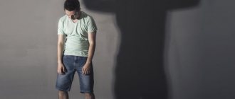
What to do if tension pain syndromes appear
Painful tension syndromes indicate that compression damage to the structures of the autonomic nervous system occurs. This may be the spinal cord, its dural membranes or radicular nerves. In any case, muscle spasm occurs, which is not relieved by the use of non-steroidal anti-inflammatory drugs. Pain from muscle tension can only be relieved with the help of muscle relaxants. But this measure does not always give a positive result.
What to do if tension pain syndromes appear? It is necessary to conduct a thorough differential diagnosis. This should be done by a neurologist, vertebrologist or orthopedist. These doctors determine what pathological change has occurred in the tissues of the spinal column. Then, after an accurate diagnosis is made, treatment is carried out.
It is most effective to treat osteochondrosis and its complications using manual therapy methods. The following techniques are used:
- osteopathy allows you to restore normal processes of microcirculation of blood and lymphatic fluid, due to which the physiological structure of the tissue is restored;
- massage improves the elasticity and permeability of muscle tissue, increases its saturation with oxygen and glucose, which has a positive effect on the diffuse nutrition of cartilaginous intervertebral discs;
- therapeutic exercises and kinesiotherapy regulate the tone of the muscular frame of the back and restore its performance;
- reflexology allows you to start the process of regeneration of all damaged tissues by using the hidden reserves of the human body.
Physiotherapy, laser treatment and much more are also used. If you need effective and safe treatment for tension pain syndromes due to osteochondrosis of the spinal column, then you can contact our manual therapy clinic in Moscow. Here you will be offered a free initial examination. During it, a preliminary diagnosis will be made and recommendations for subsequent examination and treatment will be given.