The life of a modern person is associated with many unfavorable factors - stress, conflicts, poor environment, low-quality products, and little physical activity. The result of their influence is the development in the body of various functional and organic disorders, sometimes very severe. Such, for example, as neuritis .
This inflammatory disease of the peripheral nerves is characterized by paroxysmal pain, decreased or complete loss of sensitivity, as well as movement disorders including paresis and paralysis.
With mononeuritis, only one peripheral nerve is affected (ophthalmic, facial, radial, etc.), with inflammation of the nerves from one plexus, we talk about plexitis .
Neuritis is dangerous due to its complications. Particularly severe consequences of the disease include paresis, paralysis, and muscle atrophy.
1 Neuritis. Diagnosis and treatment
2 Neuritis. Diagnosis and treatment
3 Neuritis. Diagnosis and treatment
Symptoms
With neuritis, the nerve fiber is damaged, and therefore the functions suffer, the main ones being sensitive, motor and trophic, therefore the following symptoms are clinically noted:
- In the phase of irritation of the nerve fiber: pain, burning, feeling of “crawling goosebumps”, feeling “as if many needles are being pricked”.
- In the phase of loss of functions: decreased muscle strength, muscle atrophy, decreased reflexes, decreased sensitivity, tissue atrophy including skin, subcutaneous fat, muscles, bone tissue.
Surgery
Certain surgical procedures can help relieve neuralgia pain when the condition does not respond to treatment.
Examples of surgical procedures that may help treat neuralgia include:
Microvascular decompression : Helps remove an enlarged blood vessel affecting a nerve. The procedure involves placing a soft pad between the blood vessel and the affected nerve.
Stereotactic surgery : This is a non-invasive procedure that directs highly concentrated beams into the root of the damaged nerve. The radiation disrupts the transmission of pain signals to the brain.
Percutaneous balloon compression : This involves inserting a small balloon into the affected nerve. The balloon is inflated, causing controlled, deliberate nerve damage. This procedure prevents the affected nerve from sending pain signals to the brain. However, the effects of the procedure usually wear off after 1-2 years.
Causes
The main causes of neuritis:
- infectious (viruses, bacteria, their toxins);
- exposure to toxic factors
- metabolic disorders (diabetes mellitus, deficiency of vitamins B1 and B6);
- allergic reactions;
- injuries;
- oncological diseases;
- hereditary diseases;
- diseases of the spine leading to mechanical compression of the nerve trunk (intervertebral hernia, tunnel syndromes, etc.).
The mechanism of nerve damage when exposed to various unfavorable factors is associated with the launch of a cascade of inflammatory reactions. Swelling of the nerve trunk occurs, damage to the myelin sheath and subsequently to the axial cylinder, which in turn leads to the death of the nerve cell.
Make an appointment
Disease prevention
To prevent optic neuritis, it is recommended to give up bad habits, promptly treat infectious diseases, avoid eye and head injuries, and visit specialized doctors in the presence of chronic pathologies.
Article sources:
- Retrobulbar optic neuritis. Kukhtik S.Yu., Popova M.Yu., Tantsurova K.S. Bulletin of the Council of Young Scientists and Specialists of the Chelyabinsk Region, 2016
- Visualization of the optic nerve in the diagnosis and monitoring of retrobulbar neuritis. Yuryeva T.N., Burlakova E.V., Khudonogov A.A., Ayueva E.K., Sukharchuk O.V. Acta Biomedica Scientifica, 2011. p. 133-136
- Modern view on the problem of optic neuritis (systematic review). Krivosheeva M.S., Ioileva E.E. Saratov Scientific and Medical Journal, 2021. p. 602-605
- Results of treatment of optic neuritis. Latypova E.A. Saratov Scientific and Medical Journal, 2021. p. 875-879
Clinical picture of some individual nerves
- Trigeminal nerve – pain is sharp, piercing, in series, along one or several branches of the nerve.
- Facial nerve – muscle weakness on one side of the face. It is difficult to close the eye, the corner of the mouth on the affected side is drooping. When liquid food or drink is taken, everything comes out of the mouth.
- Diaphragmatic – feeling of lack of air, shortness of breath, hiccups.
- Median – impaired flexion of the hand, fingers I, II and III and decreased sensitivity on the palmar surface.
- Ulnar – weakness of the flexors of the IV, V and partly III fingers, decreased sensitivity on the palmar surface of the above fingers.
- Radial – impaired extension of the hand and fingers, decreased sensitivity in half of the back of the hand (I and II fingers). It is difficult to move the thumb away.
- External cutaneous nerve of the thigh (Roth-Bernhardt disease) – pain, numbness and burning along the outer surface of the thigh.
- Femoral – impairment of leg extension at the knee joint and hip flexion. Pain and sensory disturbances on the lower 2/3 of the anterior surface of the thigh, the anterior inner surface of the lower leg.
- Sciatic – pain along the back of the thigh and lower leg, weakness of the flexors and extensors of the foot.
- Olfactory – anosmia on one side (lack of sense of smell). When a nerve is irritated, foreign odors may appear.
- Visual – decreased acuity and loss of visual fields. The phenomena of nerve irritation manifest themselves in the form of photopsia (sensation of light, flames, sparks, etc.).
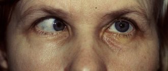
- Oculomotor – drooping of the upper eyelid (ptosis), limited mobility of the eyeball, dilated pupil, diplopia (double vision).
- Block - restriction of the mobility of the eyeball downwards and outwards.
- Auditory – hearing loss, often accompanied by a feeling of noise or ringing in the ear.
- Glossopharyngeal - twitching pain in the tonsils, root of the tongue, pharynx and taste disturbances in the back third of the tongue, impaired salivation and swallowing.
- Wandering – manifested by disturbances in swallowing and speech. On the affected side, the soft palate is lowered, the uvula is deviated to the healthy side. There are also disturbances in the functioning of internal organs - bradycardia, shortness of breath, motility disorders of the esophagus, stomach and intestines (spasms), etc.
- Additional – difficulty turning the head in the healthy direction, the head is tilted towards the affected nerve, the shoulder is lowered.
- Tibial – the foot is extended, but the patient cannot bend it. Cannot stand on toes. Sensitivity is reduced along the back of the lower leg and on the sole.
- Peroneal – the impossibility of standing on the heels and straightening the foot, it hangs down. Sensitivity is reduced on the outer surface of the lower leg and the back of the foot.
- Intercostal nerves – pain in the intercostal space, often radiating into the chest, simulating pain in the heart, chest, lungs, and stomach. Often, pain in the paravertebral muscles is detected at the level of the thoracic spine.
Possible complications
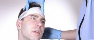
Doctors call facial paralysis the first complication that appears in the absence of adequate treatment. This means that a person who does not receive medical care in a timely manner is deprived of the opportunity to express his emotions through facial expressions on one side of his face. This condition can no longer be corrected, which will certainly affect the quality of life. Distortion of facial expressions will lead to the development of depression and constant dissatisfaction with one’s appearance. Not every patient can come to terms with irreversible changes in their appearance without deep distress.
One of the most unpleasant manifestations of paralysis is the inability to close the eyelids on the injured side of the face. In this case, the eye will have to be regularly instilled with artificial tears to prevent the cornea from drying out, since natural hydration through blinking becomes unavailable for this eye.
Diagnosis of neuritis
The symptoms of neuritis are in many ways similar to the clinical manifestations of various diseases, including non-neurological ones. Therefore, the doctor should conduct a thorough differential diagnosis to make an accurate diagnosis.
Primary diagnosis consists of a thorough collection of patient complaints, identification of possible preceding factors, and direct examination of the patient. Most symptoms of neuritis are specific, so depending on their severity, the doctor can make a preliminary diagnosis.
Stimulation and needle electroneuromyography are used to diagnose peripheral nerve damage. This study makes it possible to answer the questions - which nerve is damaged, in what place it is damaged, what percentage of nerve damage, as well as give a prognosis for its recovery and monitor its recovery.
Diagnostic methods
Electroneurography is used to measure the speed of passage of nerve impulses through the fibers of peripheral nerves from their exit point to the nerve endings in ligaments and muscles. The technique allows you to determine the damaged nerve, determine the location and extent of damage, and identify the severity of the process.
Electromyography – used to study the bioelectrical activity of muscles. This method allows us to answer the question: what is the problem - damage to the nerve or damage to the muscle itself? EMG allows for differential diagnosis of neuropathy with muscle pathology (myasthenia, myotonia, myoplegia, polymyositis).
Ultrasound is a method for diagnosing damage to peripheral nerves. Evaluates changes in nerve diameter, continuity, and deterioration of sound conduction. Ultrasound clearly shows swelling of the nerve trunk and surrounding tissues
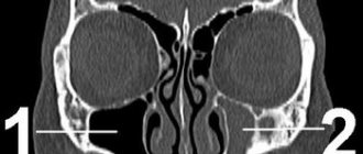
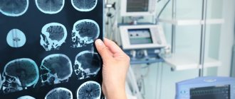
MRI – visualizes nerves and soft tissue structure, detects malignancies and provides information about muscle atrophy and nerve damage. With the help of MRI diagnostics, it has become possible to detect nerve damage in areas that are difficult to examine using electrodiagnostics or ultrasound.
Make an appointment
Classification and stages of disease development
Depending on the affected area:
- Intrabulbar (intraocular) neuritis, optic papillitis. This is an inflammation of the intraocular part (disc) of the optic nerve. More often develops in children.
- Retrobulbar neuritis. Damage to the nerve section lying between the eyeball and the optic chiasm. Forms of retrobulbar neuritisSource: Retrobulbar optic neuritis. Kukhtik S.Yu., Popova M.Yu., Tantsurova K.S. Bulletin of the Council of Young Scientists and Specialists of the Chelyabinsk Region, 2021: axial - a bundle of axons passing in the optic nerve is involved in the pathological process;
- peripheral - inflammation covers the nerve sheaths and spreads deep into the nerve trunk with the formation of a large amount of exudate under the sheaths;
- transversal - the process affects all layers of the optic nerve.
According to the etiology of the lesion:
- infectious:
- parainfectious (post-vaccination, after acute respiratory viral infections);
- demyelinating;
- ischemic;
- toxic;
- autoimmune.
According to the severity of the flow:
- spicy;
- chronic.
According to the prevalence of the lesion:
- mononeuritis – inflammation of one nerve (the vast majority of cases);
- polyneuritis – involvement of both optic nerves in the process (less than 1% of cases).
Treatment of neuritis
Treatment must be comprehensive. It is necessary to fight both the damaging factor and restore the damaged nerve trunk.
In treatment I use the following groups of drugs:
- Vascular;
- Anti-inflammatory;
- B vitamins;
- Improving the conduction of impulses along the nerve, etc.
Non-drug treatment methods are also used:
- Acupuncture - exposure to biologically active points of the skin leads to signal transmission along the nerve trunk to the spinal cord and brain, resulting in a complex cascade of reactions, which includes improved blood circulation, release of biologically active substances, and hormonal response, which in turn accelerates restoration of nerve fiber.
- Physiotherapy is designed to locally influence reflex zones. The main effects achieved after physiotherapy are: analgesic, anti-inflammatory, antispasmodic, fibrinolytic.
- Massage (improves microcirculation, reduces swelling).
- Therapeutic exercise (by working out muscle groups using a feedback system, the recovery period is accelerated).
To relieve pain and accelerate the recovery of nerve fibers, local injection techniques are used - therapeutic and diagnostic blockades, pharmacopuncture - with various mixtures of drug solutions, if necessary, together with ultrasound navigation. Due to the local administration of drugs (vitamins, anti-inflammatory drugs, anesthetics), it is possible to quickly relieve pain, reduce swelling and the inflammatory reaction, and restore the nerve trunk.
Causes of inflammatory damage to the trigeminal nerve
Factors contributing to inflammation of the trigeminal nerve are:
- surgical interventions on the jaw bones;
- fractures of the base of the skull, lower and upper jaws;
- tumors;
- complex tooth extraction;
- hypothermia;
- surgery on the maxillary sinus;
- improperly administered anesthesia;
- incorrectly performed dental prosthetics;
- metabolic disorders;
- the presence of foreign bodies that irritate the nerve trunk or injure nerve endings;
- bacterial or viral infection;
- various types of intoxication of the body;
- hypovitaminosis;
- weakening of the immune system.