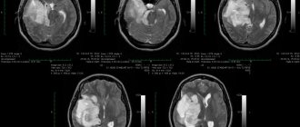
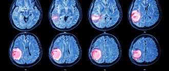
Brain meningioma (ICD 10 code – D32.0) is a neoplasm that originates from the arachnoid (arachnoid) membrane of the brain. Brain meningioma has a clear outline morphologically and looks like a horseshoe-shaped or spherical node, most often fused with the dura mater of the brain.
At the Yusupov Hospital, qualified specialists use the most advanced technologies and time-tested effective methods to treat meningioma: radiation therapy, stereotactic radiosurgery, high-quality removal of brain meningioma. Recovery after surgery is carried out in the rehabilitation department of the Yusupov Hospital under the careful supervision of competent rehabilitation doctors and attentive medical staff.
What can cause a brain tumor?
Until now, doctors do not know exactly what causes a brain tumor. Only a few risk factors for the occurrence of this group of diseases have been established.
Here are the main factors that cause a brain tumor:
- Irradiation. Radiation exposure leads to changes in DNA in cells, followed by uncontrolled growth. In the past, children were treated for ringworm with low-dose radiation. As it turned out, their risk of brain tumors subsequently increased in adulthood.
- Genetic diseases . There are several genetic reasons why a brain tumor develops. This may be neurofibromatosis type 1 or 2, Bourneville disease, Hippel-Lindau disease, Li-Fraumeni syndrome. Some other hereditary pathologies also provoke neoplasms in the central nervous system. Most of them are quite rare.
- Immunodeficiency conditions . The risk of some tumors is increased due to decreased immunity. Immunodeficiencies can be either congenital or acquired (for example, due to infection with the human immunodeficiency virus).
Some doctors suggest that factors that cause brain tumors include cell phone use, certain viral diseases, exposure to electromagnetic fields, and taking the sugar substitute aspartame. But there is no convincing evidence for this. So the influence of these factors remains at the level of assumptions.
Send a request for treatment
Symptoms of benign brain tumors
With a benign brain tumor, symptoms may be general or focal.
Common signs include:
- Nausea and vomiting
- Headache
- Visual impairment
- Loss of coordination
- Epilepsy
- Drowsiness
- Depression of consciousness, up to coma
With a benign brain tumor, the symptoms that appear first can be quite varied. Sometimes these are focal symptoms. In other cases, the first sign is an epileptic seizure or headache.
Any, including benign, brain tumors can cause focal symptoms. This may be a speech, writing, or reading disorder. Sometimes mental disorders develop. Often, benign brain tumors are accompanied by a decrease in memory and intelligence; they can cause oculomotor disorders or hearing loss. In some cases, the mobility of the limbs or the sensitivity of the skin is impaired.
Symptoms
Clinical manifestations are divided into general and focal. The latter manifest themselves depending on which part of the brain the pathological formation is formed.
General symptoms
General signs of brain cancer (or a benign tumor) arise due to increased pressure inside the skull, compression of neighboring tissues by the tumor body and the negative influence of its metabolites. Common symptoms include:
- dizziness, headache, discomfort in the head;
- dyspeptic manifestations not related to food intake or poisoning;
- loss of appetite, weight loss;
- blurred vision;
- changing the psycho-emotional behavior habitual for a person;
- memory problems, weakening of mental activity;
- signs of epilepsy.
It's important to understand. That general symptoms are not specific. If they bother you regularly, you need to urgently visit a doctor.
The pathological process in the early stages, as a rule, occurs latently or its signs are weak. Often neoplasms are found by chance, when examining a patient for another disease or for preventive purposes.
Focal symptoms
These are specific signs that arise as a result of damage to the cerebral structures of the central nervous system. The clinical picture depends on the location of the pathological focus:
- Glioma of the optic nerve is formed from cells of its trunk. Tissue growth is slow, accompanied by infiltration, but does not grow into the dura mater. The neoplasm is considered benign; it can form along the entire length of the nerve, but is more often located in the orbital part. Symptoms of optic nerve glioma: first, visual acuity decreases (as the nerve and elements of the eye are damaged), then bulging eyes develop.
- Diffuse glioma of the brainstem is highly malignant. Since diffuse dysplasia is formed in the area where the nerves responsible for the functioning of the limbs are located, when they are damaged, the coordination of the arms and legs is disrupted, up to paresis and paralysis. Clinical signs of diffuse pontine glioma are significant even with small sizes of dysplasia. There are many nerve cords passing through the bridge, so the clinic can be more diverse.
- Glioma of the frontal lobe of the brain is manifested by apathy, mental changes, the person becomes nervous, hot-tempered, and sudden mood changes are characteristic. The later stages of the disease are characterized by the onset of paralysis.
- Glioma of the temporal region causes impaired coordination of movements, memory deteriorates, and diction suffers. In addition, temporal lobe glioma negatively affects the sense of smell.
- Glioma of the cerebellum and cerebellar vermis leads to movement abnormalities. The gait is disturbed, it seems that the person is drunk. This is because it becomes difficult for the patient to maintain balance, his arms and legs do not obey, and maintaining the body in space becomes problematic.
- Glioma of the parietal lobe is accompanied by disturbances in fine motor skills, so a person’s handwriting changes, and over time it becomes difficult to write. Tactile sensations become dull.
- Glioma of the quadrigeminal plate or quadrigeminal plate causes visual disturbances, abnormalities in eye movement, and hearing impairment.
- Glioma of the corpus callosum is accompanied by visual hallucinations (the same ones occur in the presence of a tumor in the occipital lobe), violations of logic, as well as many other signs: changes in sensitivity, muscle weakness, and so on.
- Pontine glioma (diffuse or common) leads to disruption of conduction functions, since the nuclei of the cranial nerves are localized in this area. Therefore, the clinical picture will be quite broad. A violation of sensory perception, movements, and so on is recorded.
- Glioma of the cerebral ventricle leads to an increase in intracranial pressure due to disruption of the outflow of cerebrospinal fluid. Hydrocephalus may develop.
Examination for benign brain tumors
A benign brain tumor cannot be diagnosed by symptoms. Because focal neurological symptoms correspond to the level of brain damage. But it is not specific - that is, it does not indicate a tumor process. Many other diseases can lead to damage to the structures of the central nervous system - infections, injuries, degenerative or autoimmune processes.
Therefore, instrumental diagnostics remains the main method for diagnosing the disease. A benign brain tumor is detected on MRI. From the image you can determine not only its location and size, but also suggest the histological type. Although it is possible to definitively understand whether a brain tumor is benign or malignant, a stereotactic biopsy will help. During the procedure, the doctor obtains a section of the brain, which he sends for histological examination.
A biopsy may be surgical. In this case, diagnosis and treatment are carried out simultaneously. The doctor sends a fragment of the removed tumor for examination. He then receives the results and, if necessary, expands the scope of the surgical intervention.
Another diagnostic method is labial puncture. The doctor inserts a needle between the lumbar vertebrae and takes cerebrospinal fluid for analysis. It sometimes allows you to detect tumor cells. But not all tumors can be diagnosed in this way. Lymphoma or ependymoma can be detected using a lumbar puncture.
Diagnostics
The most informative and accurate methods for diagnosing meningioma are computed tomography (CT) and magnetic resonance imaging (MRI). As a rule, these studies are carried out with contrast. CT and MRI can determine the size of the tumor, its location, the extent of damage to surrounding tissues and possible complications.
Magnetic resonance spectroscopy (MRS) is used to determine the chemical profile and nature of the neoplasm. Positron emission tomography (PET) allows identifying foci of recurrent meningioma.
An auxiliary method that allows you to determine the nature of the blood supply to the tumor is angiography. This study is often used in the process of preoperative preparation.
Make an appointment
How are benign brain tumors treated?
Patients often ask whether a benign brain tumor can be cured. Complete cure is possible with surgery. But only in the case when the tumor is located outside the functionally important areas of the brain. Otherwise, partial removal is performed to avoid causing neurological deficits.
If a benign brain tumor is diagnosed, treatment is usually done without chemotherapy or radiation. Removal alone is highly likely to cure the pathology completely. Benign brain tumors rarely recur after treatment. This mainly happens with incomplete removal.
There are other ways to treat a benign brain tumor. In German clinics, a cyber knife or gamma knife is often used for this. Stereotactic radiosurgery allows you to irradiate the tumor with a high dose of radiation. As a result, tumor cells die. The effectiveness is comparable to surgical treatment. In this case, it is possible to treat tumors in hard-to-reach localizations.
In some cases, chemotherapy or radiation therapy may be used in addition to the main treatment. Chemotherapy may be given through a shunt left after surgery. Because intravenous or parenteral administration is ineffective - the blood-brain barrier prevents the drugs from working.
Rehabilitation programs and recovery periods
To talk about timing, you need to have an idea of the complexity of the initial condition, surgical intervention and its results. If the process is launched on time and goes well, it usually takes about 3-6 months.
Among the most popular methods:
- physiotherapy. It is aimed at symptomatic treatment, helps relieve swelling and pain;
- massage. Prescribed for paresis of the limbs. Improves blood supply, ensures the outflow of blood and lymph, increases neuromuscular conductivity and sensitivity;
- Exercise therapy. Restores lost functions, forms reflex connections, eliminates vestibular disorders.
All rehabilitation measures are carried out by specialists. But the motivation of the patient himself, his desire to work and get results is very important.
Prognosis for benign brain tumors
With a benign brain tumor, the consequences depend on a number of factors:
- How early is the tumor detected?
- What dimensions does it have?
- What is the histological type of the tumor?
- Where is it located
- How long ago did the first neurological symptoms appear, etc.
In addition, with a benign brain tumor, the prognosis largely depends on the quality of the treatment provided. The most modern methods of therapy are used in Germany. When used, if successful, the life expectancy of a patient who has undergone treatment will be the same as that of a person who has never had cancer of the central nervous system.
Patients often ask how long they live after surgery for a benign brain tumor. Potentially, life expectancy is the same as the average population. But there is a small risk of relapse. It is especially high in the case of incomplete removal of the tumor, which is possible if it is located in hard-to-reach places.
Send a request for treatment
Relapses
If a benign, clearly circumscribed meningioma that has not grown into the surrounding tissue is detected in a patient, surgical intervention most often ensures a complete recovery.
However, it must be remembered that after removal of even benign meningiomas, relapses can occur. Recurrences of atypical meningiomas are recorded in almost 40% of cases, of malignant ones - in 80%.
The development of relapses within five years after surgery also depends on the location of the tumor.
Relapses are least likely to occur with meningioma localized in the cranial vault, more often in the area of the sella turcica and the body of the sphenoid bone. The most common tumors that recur are those affecting the sphenoid bone and cavernous sinus.
Treatment of brain tumors in Germany
To receive the most modern treatment in Germany, use the services of Booking Health. We will select the best clinic for you, where you can receive the highest quality medical services at an affordable price. Leading clinics that accept foreign patients for treatment of brain tumors:
- University Hospital Münster, Department of Neurosurgery
- Charité University Hospital Berlin, Department of Adult and Pediatric Neurosurgery
- University Hospital Freiburg, Department of Adult and Pediatric Neurosurgery
- University Clinic named after. Goethe Frankfurt am Main, Department of Adult and Pediatric Neurosurgery
- University Hospital Erlangen, Department of Adult and Pediatric Neurosurgery
Modern treatment in Germany will help:
- Completely get rid of a benign tumor
- Cure cancer without surgery using stereotactic radiosurgery
- Avoid complications and neurological deficits after treatment
- Take a course of postoperative recovery to normalize all impaired functions of the central nervous system
We will provide visa support, planning a trip to Germany, and translation of documentation. An interpreter will be provided to you. We also organize airport-clinic-airport transfers. All you have to do is focus on your health, and Booking Health will take care of all other concerns.


