The human body has 12 systems: central nervous system (CNS), respiratory system, cardiovascular, hematopoietic, digestive, excretory (including urinary system and skin), reproductive system, endocrine, musculoskeletal, lymphatic, immune, peripheral nervous system . There are no important or unimportant systems. Each one is needed and each one is important. If one of them suffers in the body, then over time all the others will be involved in the process.
central nervous system
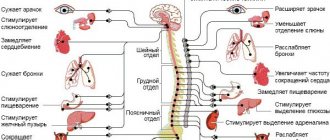
The central nervous system is a system that provides control over all life activities in the human body. In our body there are billions, trillions of nerve cells that exist on their own and, unfortunately, do not reproduce. In 3-4 years, a person can completely (in the sense of cell health and their full function) restore the liver, heart, our blood is renewed after 4 months. But with brain cells it's different. Over the course of life, they do not become larger; these cells can only be fully nourished and improve the intercellular space between them, and, if possible, cleanse them of various kinds of toxins. Therefore, if we kill a child from childhood with environmental poisons, then this will happen for the rest of his life. For example: let’s take you to a kindergarten where the walls are being painted, or to a school that hasn’t been completely renovated. Or we’ll send you to swim in a pool with chlorinated water 3 times a week. Nobody perceives neurons as living cells. After all, it is very important that we understand: we are controlled by the brain. Example: we think it would be nice to buy cottage cheese. In fact, there is a deficiency of calcium in the body, and neurons cannot live without calcium, so they send you to the store to buy it.
In order to understand what a neuron needs, you need to study the life of a cell. For its vital functions it requires: 28 amino acids, 15 minerals, 12 vitamins, fatty acids, enzymes, water and oxygen.
In medicine, the nervous system is divided between two doctors: one part is controlled by neurologists, the other by psychiatrists. As if behavior is something special. Behavior is the life of a neuron. The feeling of fear is nothing more than a lack of oxygen experienced by a neuron. Stress, adrenaline was released, blood vessels spasmed, there was a lack of blood supply to the brain, hence the lack of oxygen. A feeling of fear is formed.
Why can the brain get sick? Let's remember 12 causes of diseases.
Structure of the central nervous system
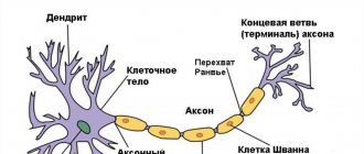
The CNS consists of the brain and spinal cord, each of which has white and gray matter. White matter consists of pathways, myelinated and unmyelinated axons. Myelin is white, which gives the corresponding shade to the tissue. Gray matter consists of neuron cell bodies. It can be located in the nervous system in the form of a tube (spinal cord); nuclei, or ganglia (clusters of neuron bodies in the thickness of the white matter), as well as the cortex (gray matter on the surface of the white matter).
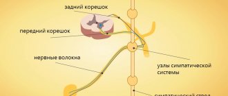
The spinal cord is located in the spinal canal and its mass is 40 g. On its lateral surface, the dorsal roots, carrying afferent (sensitive, to the brain) information, enter from the back, and the anterior roots, carrying efferent (motor, from the brain) information, exit from the front. The section of the spinal cord corresponding to each pair of roots is called a segment. The segments are named according to where the roots exit the spine. The spinal cord has 8 cervical, 12 thoracic, 5 lumbar, 5 sacral and 1 coccygeal segments. In general, the number of spinal cord segments corresponds to the number of vertebrae. Exceptions are the cervical region, where there are 8 segments per 7 vertebrae; and coccygeal, where there is 1 segment for 3-4 vertebrae (Fig. 1).
Rice. 1. Structure and location of spinal cord segments.
In a cross section of the spinal cord, there is gray matter in the center surrounded by white matter. The gray matter has the shape of a butterfly, in the center of which is the spinal foramen, filled with cerebrospinal fluid (CSF). The butterfly consists of approximately 13 million neurons and has anterior and posterior horns (Fig. 2b, 3). The middle horns are also well defined in the middle sections of the spinal cord. Sensitive (sensory) information to interneurons (interneurons) enters the dorsal horns along the dorsal root. The anterior horns contain motoneurons (motor neurons) that send motor information to the muscles; it is their axons that form the anterior root. The middle horns contain neurons of the central sections of the ANS.
The spinal cord works on a reflex principle. A reflex is a stereotypical response of the body to any (external or internal) influence. The simplest reflex is monosynaptic. To implement it, two neurons are enough. An example of such a reflex is the knee reflex. When the receptor is irritated, the impulse is transmitted along the dendrite to the body of the neuron located in the nerve ganglion near the spinal cord. The axon of this neuron enters the spinal cord through the dorsal roots and forms a synapse with the motor neuron in the anterior horn. The axon of the motor neuron exits through the anterior roots and goes to the effector organ, where it changes the activity of the organ itself (Fig. 50a). The polysynaptic reflex includes an additional link in the form of one or more interneurons between the ganglion and motor neurons. Interneurons can additionally process information, compare it with other stimuli and the internal state of the body, making a decision about how to respond to the stimulus.
Rice. 2. Reflex arc (a) and histological section (b) of the spinal cord.
Rice. 3. Diagram of the structure of a section of the spinal cord.
The white matter of the spinal cord includes pathways. It is divided by the butterfly into anterior, posterior and lateral funiculi (Fig. 3).
In the posterior cords there are ascending tracts through which information is transmitted from the PNS to the spinal cord and further to the brain. In the anterior horns of the spinal cord there are descending tracts through which information goes from the brain to the spinal cord, and from the latter to the PNS. In the lateral horns, the ascending tracts are located posteriorly, and the descending tracts are located anteriorly.
The brain is located in the skull and consists of 5 sections. Its average mass is 1.5 kg and it contains up to 100 billion neurons. There are 12 pairs of cranial nerves (cranial nerves) that arise from the brain.
The medulla oblongata is the junction of the spinal cord and the brain. Its length is approximately 25 mm. In the lower part of the medulla oblongata one can still distinguish a butterfly; in the upper parts the bodies of neurons are collected into nuclei. The IX-XII pairs of the cranial nerves (Fig. 5) depart from the medulla oblongata and the corresponding nuclei lie in it. These nerves control movement and sensation in the throat, tongue, and neck. The medulla oblongata contains the largest center of the parasympathetic nervous system, which, through the X nerve (vagus, vagus nerve), controls the activity of all internal organs. The medulla oblongata contains centers for regulating breathing and vital reflexes, such as sneezing and coughing. Here is the olive kernel, which is responsible for balance. All tracts from the spinal cord to the brain pass through the medulla oblongata.
The hindbrain consists of the pons and cerebellum. The pons serves as a continuation of the medulla oblongata. It contains a lot of white matter that connects the cerebellum to the rest of the brain. This white substance forms a ridge on the underside of the bridge, making it easy to distinguish. The pons, together with the medulla oblongata, form the bottom of the 4th ventricle of the brain (a continuation and expansion of the spinal canal). V-VIII ChMN depart from the bridge. Here lie the auditory and vestibular nuclei, nuclei that innervate sensitivity and facial muscles (including facial muscles). The pons contains the locus coeruleus, which is responsible for regulating sleep.
Rice. 4. Main parts of the brain.
Rice. 5. Cranial nerves. I-olfactory, II-visual, III-oculomotor, IV-trochlear, V-trigeminal, VI-abducens, VII-facial, VIII-vestibular-cochlear, IX-glossopharyngeal, X-vagus, XI-accessory, XII-hyoid.
The cerebellum is well developed in humans due to upright posture and fine motor skills of the hands. This part of the brain is responsible for maintaining posture, balance, motor learning, and some motor reflexes. The cerebellum has a cortical structure. The cerebellar cortex consists of three layers and is divided into two hemispheres by the vermis. Under the cortex there is white matter, among which there are 3 pairs of cerebellar nuclei. To carry out its functions, it receives information from the vestibular apparatus, olive and other parts of the human motor system.
Rice. 6. External structure (a) and histological section (b) of the cerebellar cortex.
The midbrain consists of the cerebral peduncles and the roof (Fig. 7). The aqueduct of Sylvius passes through the center of the spinal cord and connects the third and fourth ventricles. The third and fourth pairs of cranial nerves depart from the midbrain. These nerves control the movements of the eyeballs. The third nerve contains parasympathetic fibers that control pupil width. The midbrain contains elements of the motor system: the red nucleus and the substantia nigra. On the roof of the brain is the quadrigeminal region. The colliculus receives visual information, and the inferior colliculus receives auditory information. This is necessary for the implementation of the orientation reflex.
Rice. 7. Appearance (a) and section (b) of the midbrain.
The medulla oblongata, pons, and midbrain together form the brainstem. The reticular formation runs through the entire trunk, regulating the overall level of brain activity.
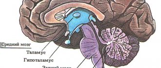
The diencephalon consists of the thalamus, hypothalamus, pituitary gland and pineal gland. The third ventricle of the brain is located here. It serves as the origin of the second cranial nerve. The pituitary gland is the gland through which the nervous system controls the humoral one. The pineal gland is also a gland that regulates circadian rhythms. The thalamus filters information entering the cortex and removes unimportant repetitive sensory stimuli (heartbeat, gastrointestinal function, nose in the field of view, touch of clothing, etc.) In addition, the thalamus contains nuclei of the limbic system (forms mood), motor and associative kernels. The hypothalamus controls the activity of the pituitary gland and also regulates the internal state of the body. It contains the centers of hunger, thirst, sexual behavior, pleasure, displeasure, etc. Thus, the main function of the hypothalamus is to maintain homeostasis of the entire organism.
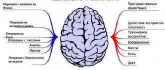
The telencephalon (forebrain) consists of the cerebral cortex and basal ganglia (nuclei). The first and second ventricles of the brain are located symmetrically under the cortex. Its area is about 220 cm2, it forms grooves and convolutions (Fig. 8). It consists of 6 layers. The hemispheres are connected to each other by the corpus callosum - a ridge of white matter. The cerebral cortex processes sensory information, the formation of voluntary movements, memory and higher nervous activity. The first cranial nerve approaches the olfactory bulbs. The basal ganglia are nuclei of gray matter located in the thickness of the white matter. They play an important role in voluntary movements, motor learning and the formation of emotions.
Rice. 8. Structure (a) and histological sections (b, c) of the cerebral cortex.
Nutrition and the central nervous system
Mental disorders are often corrected using inquisitorial methods. A person has a lack of essential amino acids, vitamins, minerals, and he is persuaded to remember how he was born, how he passed through the birth canal. There is probably a rational grain in this, but I would like to remember the Russian fairy tale about Ivan Tsarevich. Before completing Baba Yaga’s task, he told her: “Grandma, first feed the young man, give him something to drink, take a bath in the bathhouse, and then ask about the matter.” This is a 100% working algorithm. It is impossible to suppress the activity of small single-celled living creatures called neurons. If they are hungry and want to eat, if they are tired and want to sleep, if there is dirty water between them, and they are forced to absorb some toxic substances from this water, they begin to hallucinate. What is delirium tremens? This is poisoning of intercellular water with alcohol substitutes. Cells begin to take toxic substances from the intercellular space, and a person begins to see miracles. The scary thing is that food has now also become toxic. And the second reason is artificial, pickled, over-sweetened, over-salted, enzyme-free food destroyed by chemicals. When the brain doesn't receive anything, it starts to get nervous. And he can behave in any way: from fear to depression.
Parasites, viruses and the central nervous system
A large number of “interventionists” live in the brain. Various viruses: cytomegalovirus, herpes virus, papillomavirus. Toxoplasma enters the human body, for example, through cat scratches, resulting in the formation of toxoplasmosis gumma. If someone is diagnosed with epilepsy, can you somehow relate this to the fact that they have worms? Hardly. If a child has epilepsy, will you go to a helminthologist? You 100% won't go. And in vain. It is very important to establish the direction in which to go. It is necessary to carry out some specific actions: antiparasitic programs, or at least be examined for the presence of toxoplasma, cytomegalovirus.
Cerebral cortex
The surface of the human cerebral cortex is about 1500 cm2, which is many times greater than the inner surface of the skull. This large surface of the cortex was formed due to the development of a large number of grooves and convolutions, as a result of which most of the cortex (about 70%) is concentrated in the grooves. The largest grooves of the cerebral hemispheres are the central one, which runs across both hemispheres, and the temporal one, which separates the temporal lobe from the rest. The cerebral cortex, despite its small thickness (1.5–3 mm), has a very complex structure. It has six main layers, which differ in the structure, shape and size of neurons and connections. The cortex contains the centers of all sensory (receptor) systems, representatives of all organs and parts of the body. In this regard, centripetal nerve impulses from all internal organs or parts of the body approach the cortex, and it can control their work. Through the cerebral cortex, conditioned reflexes are closed, through which the body constantly, throughout life, very accurately adapts to the changing conditions of existence, to the environment.
Ecology and the central nervous system
Can the brain of a painter who works all his life with oils and paints, or a miner, be normal? Even theoretically it cannot. What should he do? One thing is necessary: cleanse the body every three months, drink water and eat right. And who will take care of the nervous system of teachers? For people in this profession, it is in constant tension. If it were possible to reach doctors who deal with occupational pathology, then for each profession it would be possible to choose a saving factor for the central nervous system.
Maintaining the Central Nervous System
Cellular nutrition for the central nervous system is amino acids. An excellent product from NSP is Peptovit. This is the best product for the brain. In second place are fatty acids - Omega-3 and Lecithin. In third place are B vitamins and folic acid. They are contained in the following products: Nutri-Calm, Super Complex, Mega-Hel, and in combination with calcium - Osteo Plus. In fourth place will always be enzymes, or better yet coenzymes - Coenzyme-QlO. This is brain ATP. Without this coenzyme, brain energy is not released. After 40 years, its production drops sharply. And only in fifth place is something that improves blood circulation - Ginkgo/Gotu Kola or Gotu Kola.
This is what the brain needs to receive to function optimally.
Maltseva M.V. – Philosophy of health
Complete recording of material on the topic “Central nervous system. Optimal working conditions" can be listened to below:
Multiple sclerosis
Multiple sclerosis
is a progressive chronic disease. Characterized by the formation of dispersed foci of demyelination in the brain and spinal cord (white matter). In this case, the growth of glia occurs with the formation of foci of sclerosis. It is a common disease of the nervous system. It usually appears between the ages of 20 and 40, more often in men. Symptoms in this condition sometimes get worse and sometimes get better.
Differences and multiple localization of lesions of the brain and spinal cord determine the variety of clinical manifestations of the disease: deliberate Tremor, nystagmus, scanned speech, a sharp increase in tendon reflexes, spastic paralysis, visual impairment. The course of the disease is different. There may be an acute and severe course (acute forms of the disease) with the rapid development of blindness and cerebellar disorders, and possibly a mild course with minor dysfunction of the central nervous system and its rapid recovery.
The causes of the disease remain unclear, but it appears that the causative agents are viruses. 80% of patients have antiviral antibodies in their blood, but the spectrum of these antibodies is quite wide. The role of autoimmunization does not exclude the development and progression of the disease. Evidence of immune aggression against myelin cells and oligodendroglia was obtained.
- How to recover
- Structure and functions of the central nervous system
- Tick-borne encephalitis
The morphogenesis of sclerotic plaques in multiple sclerosis has been well studied. Firstly, fresh foci of demyelination appear around the veins, which are combined with remyelination processes. The vessels in the lesions dilate and are surrounded by an infiltrate of lymphoid and plasma cells. In response to destruction, glial cells proliferate, and macrophages phagocytose the products of myelin decomposition. The end of these changes is sclerosis.
Pathological anatomy
externally, the superficial parts of the brain and spinal cord change little. In areas of the brain and spinal cord, a large number of gray plates were found scattered throughout the white matter (sometimes they have a pink or yellowish tint), with clear contours, up to several centimeters in diameter. There are always a lot of plates. They can merge with each other, covering large areas. They are especially common around the ventricles of the brain, in the spine and bone marrow, brain stem and optic mounds, in the white matter of the cerebellum; smaller plaques in the cerebral hemispheres. In the spinal cord, injuries can be located symmetrically. The optic nerves, chiasm, and visual pathways are often affected.
At microscopic
The study reveals foci of demyelination at the initial
stage
.
Usually around blood vessels, especially veins and venules (perivenous demyelination).
The vessels are usually surrounded by lymphocytes and mononuclear cells, and the axons are relatively preserved. Using special stains on myelin, it can be established that at first the myelin pods swell and change their coloring properties. Their contours are uneven, spherical thickenings appear along the fibers. Fragmentation and disintegration of the myelin pods then occur. The products of myelin degradation are absorbed by microglial cells, which turn into granular balls.
Important Intercellular substance: structure and functions
Axonal changes may be found in cool lesions: improved silver impregnation, uneven thickness, bloating; Severe axon destruction is rarely observed.
With progression of the disease (late stage)
Small foci of perivascular demyelination merge, proliferation of microglial cells and lipid cells appear. As a result of a productive glial reaction, typical plaques are formed, in which oligodendrocytes are rare or completely absent.
At exacerbation of the disease
Against the background of old injuries, typical plaques and fresh foci of demyelination appear.
Cause of death.
Most often, patients die from pneumonia.