The structure and location of the frontal lobe of the brain
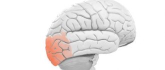
The frontal lobe is actually made up of two paired lobes and makes up two-thirds of the human brain. The frontal lobe is part of the cerebral cortex, and the paired lobes are known as the left and right frontal cortex. As its name suggests, the frontal lobe is located near the front of the head under the frontal bone of the skull.
All mammals have a frontal lobe, although it varies in size. Primates have the largest frontal lobes than other mammals.
The right and left hemispheres of the brain control opposite sides of the body. The frontal lobe is no exception. Thus, the left frontal lobe controls the muscles on the right side of the body. Likewise, the right frontal lobe controls the muscles on the left side of the body.
brain
The human brain is an organ weighing 1.3-1.4 kg, located inside the cranium. The human brain is made up of more than one hundred billion neuron cells that form the gray matter, or cortex, the vast outer layer of the brain. Neuronal processes (kind of like wires) are the axons that make up the white matter of the brain. Axons connect neurons to each other through dendrites. The adult brain consumes about 20% of all the energy that the body needs, while the child's brain consumes about 50%.
- How does the human brain process information?
- Functions of the right and left hemispheres of the brain
- Emotions
- The structure of the human brain, the trinity of the brain
- white and gray matter
- prefrontal cortex
- hippocampus
- islet of Reil
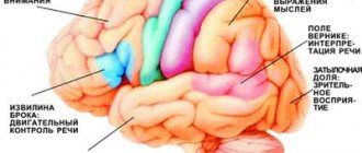
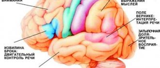
- Broca's area
How does the human brain process information?
Today it is considered proven that the human brain can simultaneously process on average about 7 bits of information[2]. These can be individual sounds or visual signals, shades of emotions or thoughts distinguished by consciousness. The minimum time required to distinguish one signal from another is 1/18 of a second. Thus, the perceptual limit is 126 bits per second. Conventionally, we can calculate that over the course of a 70-year life, a person processes 185 billion bits of information, including every thought, memory, and action. Information is recorded in the brain through the formation of neural networks (a kind of routes).
Functions of the right and left hemispheres of the brain
In the human brain there is a kind of “division of labor” between the hemispheres. The hemispheres work in parallel. For example, the left is responsible for the perception of audio information, and the right is responsible for visual information. The hemispheres are connected by fibers called the corpus callosum .
As you can see from the picture, all operations in the market are performed by the left hemisphere. Naturally, in order to make a profit from the market, the question arises of achieving maximum productivity of the left hemisphere. There are several simple ways to develop the hemispheres. The simplest of them is to increase the amount of work on which the hemisphere is oriented. For example, to develop logic, you need to solve mathematical problems, solve crosswords, and to develop imagination, visit an art gallery, etc. As soon as you pressed the mouse with your right hand, the signal came to you from the left hemisphere.[] Processing of emotional information occurs in the right hemisphere.
Emotions
Behind all sinful acts is the neurotransmitter Dopamine, the work of which determines the pleasure we receive.
[4]. Cheating, passion, lust, excitement, bad habits, gambling, alcoholism, motivation - all this is somehow connected with the work of dopamine in the brain. Dopamine transmits information from neuron to neuron. Dopamine affects many areas of our lives: motivation, memory, cognition, sleep, mood, etc.
Interestingly, dopamine increases during stressful situations.
People with low dopamine in the striatum and prefrontal cortex are less motivated than people with higher dopamine. This has been proven by experiments on rats [].
Structure of the human brain
trinity of the brain
The idea of the Triune Brain was proposed in the 60s by American neuroscientist Paul MacLean[]. In accordance with it, the brain is conventionally divided into three parts:
- R-complex (ancient, reptilian brain). Consists of the brainstem and cerebellum. The reptilian brain controls muscles, balance, and autonomic functions such as breathing and heartbeat. It is responsible for unconscious behavior aimed at survival and responds directly to certain stimuli.
- Limbic system (brain of ancient mammals). The section consists of sections located around the brain stem: amygdala, hypothalamus, hippocampus. The limbic system is responsible for emotions and feelings.
- Neocortex (new cortex or brain of new mammals). This part is found only in mammals. The necortex is a thin layer made up of 6 layers of neuronal cells that surround the rest of the brain. The neocortex is responsible for higher order thinking.
white and gray matter
Gray matter is formed by the cell bodies of neurons. White matter is axons. The white and gray matter of the brain are responsible for memory and thinking, logic, feelings and muscle contractions.
prefrontal cortex
This part of the brain is also called the frontal lobe. It is the development of the prefrontal cortex that distinguishes humans from animals. The prefrontal cortex of the human brain is responsible for logic, self-control, determination and concentration.
Throughout almost all of human evolutionary history, this part of the brain was responsible for physical actions: walking, running, grasping, etc. (primary self-control). But over the course of evolution, the prefrontal cortex grew in size and connections with other parts of the brain expanded. Now the cortex inclines a person to do what is more difficult, to leave the comfort zone. If you force yourself to give up sweets, get off the couch and go for a run, this is the result of the work of the frontal lobes. You run and don't eat sweets because you have logical reasons for this, which are processed in this part of the brain. Willpower in the brain:
Damage to the prefrontal cortex leads to loss of willpower. In psychology, there is a well-known case of Phineas Gage (1848), whose personality changed dramatically after brain damage. He began to swear, he became impulsive, he began to treat his friends with disrespect, he began to reject restrictions and advice, he comes up with a lot of plans and instantly loses interest in them.
left frontal lobe - responsible for positive emotions
“Left-sided children”, i.e. those whose left side is initially more active than the right are more positive, smile more often, etc. Such babies explore the world around them more actively. It is also interesting that the left part of the cortex is responsible for “I will” tasks, for example, getting up from the couch and going for a run.
right frontal - responsible for negative emotions. Damage to the right hemisphere (switching off the right lobe) can cause euphoria.
Experiment: While viewing pleasant pictures, a pulsed tomograph detects changes in the brain's glucose consumption and records them as bright spots in photographs of the left side of the brain. The right part of the cortex is responsible for “I won’t” tasks, such as allowing you to cope with the urge to smoke a cigarette, eat cake, etc.
The center of the prefrontal cortex “monitors” a person’s goals and aspirations. Decides what you really want.
cerebellar amygdala - protective emotional reactions (including “ego barrier”). Located deep in the brain. MM. in humans is not very different from the MM of lower mammals and works unconsciously.
Turns on the control center that mobilizes the body in response to fear.
nucleus basalis - responsible for the habits we rely on in everyday life.
The medial temporal lobe is responsible for the cognitive lobes.
hippocampus
The hippocampus is a structure in the medial temporal region of the brain that looks like a pair of horseshoes.
The hippocampus allows you to absorb and remember new information. Research by scientists has shown that the size of the hippocampus is directly related to a person’s level of self-esteem and a sense of control over their own life. Damage to the hippocampus may cause seizures
listening to music involves: the auditory cortex, the thalamus, the anterior part of the parietal cortex.
islet of Reil
The insula of Reil is one of the key areas of the brain that analyzes the physiological state of the body and transforms the results of this analysis into subjective sensations that force us to act, for example, to speak or wash the car. The anterior part of the insula of Reille converts body signals into Emotions. MRI studies of the brain have shown that smells, tastes, touch, pain and fatigue excite the insula of Reille [7].
Broca's area
Broca's area is the area that controls the speech organs. In right-handers, Broca's area is located in the left hemisphere, in left-handers - in the right.
Brain reward system
When the brain notices the possibility of a reward, it releases the neurotransmitter dopamine. Dopamine is the basis of the human reinforcement (reward) system. Dopamine itself does not cause happiness - rather, it excites (This was proven in 2001 by scientist Brian Knutson). The release of dopamine gives agility, vigor, passion - in general, motivates. Dopamine motivates action, but does not cause happiness. Tempting food, the smell of coffee - everything we desire - everything triggers the reinforcement system. Dopamine is the basis of all human addictions (alcoholism, nicotine, gaming, gambling addiction, etc.) Lack of dopamine leads to depression. Parkinson's disease results in a lack of dopamine.
Brain differences between men and women
The brains of men and women are different[3]:
Men have better motor function and spatial function, are better at concentrating on one thought, and process visual stimuli better. Women have better memory, are more socially adapted and are better at multitasking. Women are better at recognizing others' moods and showing more empathy. These differences are due to different connections in the brain (see picture)
Human brain aging
Over the years, brain function deteriorates.
Thinking slows down and memory deteriorates. This is due to the fact that neurons communicate with each other no longer so quickly. The concentration of neurotransmitters and the number of dendrites decreases, and because of this, nerve cells are less able to pick up signals from neighbors. It is becoming increasingly difficult to retain information for a long time. Older people take longer to process information than younger people. However, the brain can be trained. Studies have shown that 10 one-hour sessions per week, during which people train their memory or practice reasoning, significantly improve cognitive abilities [7].
At the same time, during the period of 35-50 years the brain is especially elastic. A person organizes information accumulated over many years of life. By this time, glial cells (brain glue), a white substance covering axons that provides communication between cells, are growing in the brain. The amount of white matter is maximum in the period of 45-50 years. This explains why at this age people reason better than those who are younger or older.
Sources
: [1] Happiness, lessons from the new science.
Richard Layard. [2] Flow. Psychology of optimal experience. Mihaly Csikszentmihalyi. [3] Economist[4] Article about dopamine[5] Dopamine and motivation[6] Brain and trading (01/11/2014) [7] Brain online: man in the Internet era. Gary Small, Gigi Worgan [8] Willpower. How to develop and strengthen. Kelly McGonigal. [9] https://en.wikipedia.org/wiki/Triune_brain [10] Kurtis Face, trading based on intuition Links: Is it possible to increase your abilities by controlling your brain? Quotes from Savelyev S.V. (01/12/2014) How to learn trading taking into account the characteristics of the brain (04/12/2014) Neuropsychopharmacologists spoke about the differences in the brains of healthy people and gamblers SHOULD YOU NOT SLEEP between sessions? (8.2.2015) Brain. History of development. Briefly. Fundamentally. (03/16/2015)
Functions of the frontal lobe of the brain
The brain is a complex organ with billions of cells called neurons that work together. The frontal lobe works alongside other areas of the brain and controls the functions of the brain as a whole. Memory formation, for example, depends on many areas of the brain.
What's more, the brain can "repair" itself to compensate for damage. This does not mean that the frontal lobe can recover from all injuries, but other areas of the brain can change in response to head trauma.
The frontal lobes play a key role in future planning, including self-management and decision making. Some functions of the frontal lobe include:
- Speech
: Broca's area is an area in the frontal lobe that helps verbalize thoughts. Damage to this area affects the ability to speak and understand speech. - Motor function
: The frontal lobe cortex helps coordinate voluntary movements, including walking and running. - Comparing objects
: The frontal lobe helps categorize objects and compare them. - Memory formation
: Almost every region of the brain plays an important role in memory, so the frontal lobe is not unique, but it plays a key role in the formation of long-term memories. - Personality formation
: The complex interplay of impulse control, memory, and other tasks helps shape the basic characteristics of a person. Damage to the frontal lobe can radically change personality. - Reward and motivation
: Most of the brain's dopamine-sensitive neurons are located in the frontal lobe. Dopamine is a brain chemical that helps maintain feelings of reward and motivation. - Management of attention
, including
selective attention
: When the frontal lobes cannot manage attention, attention deficit hyperactivity disorder (ADHD) may develop.
What fields are included?
Fields and subfields are responsible for specific functions that are generalized under the frontal lobes. Because The polymorphism of the brain is enormous; the combination of the sizes of different fields makes up a person’s individuality. Why do they say that over time a person changes. Throughout life, neurons die, and the remaining ones form new connections. This introduces an imbalance in the quantitative ratio of connections between different fields that are responsible for different functions.
Not only do different people have different margin sizes, but some people may not have these margins at all. Polymorphism was identified by Soviet researchers S.A. Sarkisov, I.N. Filimonov, Yu.G. Shevchenko. They showed that the individual ways in which the cerebral cortex is structured within one ethnic group are so great that no common features can be seen.
- Field 8 - located in the posterior parts of the middle and superior frontal gyri. Has a center for voluntary eye movements
- Area 9 - dorsolateral prefrontal cortex
- Area 10 - Anterior prefrontal cortex
- Area 11 - olfactory area
- Area 12 - control of the basal ganglia
- Field 32 - Receptor area of emotional experiences
- Area 44 - Broca's Center (processing information about the location of the body relative to other bodies)
- Field 45 - music and motor center
- Field 46 - motor analyzer of head and eye rotation
- Field 47 - nuclear zone of singing, speech motor component Subfield 47.1
- Subfield 47.2
- Subfield 47.3
- Subfield 47.4
- Subfield 47.5
Consequences of damage to the frontal lobe of the brain
One of the most notorious head injuries occurred to railroad worker Phineas Gage. Gage survived an iron spike piercing his frontal lobe. Although Gage survived, he lost an eye and suffered a personality disorder. Gage changed dramatically, the once meek worker became aggressive and out of control.
It is not possible to accurately predict the outcome of any frontal lobe injury, and such injuries can develop very differently in each individual. In general, damage to the frontal lobe due to a blow to the head, stroke, tumor, and disease can cause symptoms such as:
- speech problems;
- personality change;
- poor coordination;
- difficulties with impulse control;
- planning problems.
Structural features
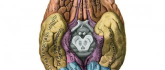
The frontal lobes are a part of the brain that is responsible for communication skills (the ability to talk, think, remember). The anatomical structure of the frontal lobe suggests a clear limitation from the temporal regions by the lateral grooves. On the frontal side, the frontal part is separated from the parietal area by the central sulcus. The lower edge of the section is limited by the Sylvian fissure.
The main gyri located in the frontal lobe include the vertical precentral and superior, middle, inferior, running horizontally. The gyri are separated by grooves. During wakefulness, neurons and neurotransmitters in this area are more active than during sleep.
The area of the prefrontal cortex is functionally connected and actively interacts with parts of the limbic nervous system - brain structures located on both sides of the thalamus. The limbic system is involved in regulating the sense of smell, the functioning of internal organs, the expression of emotions, sleep and wakefulness, and memory.
The hypothalamus, part of the limbic system, controls the autonomic nervous system with the help of hormones, due to which a person feels thirst and hunger, wakes up and falls asleep in accordance with nature’s biological clock, and experiences sexual attraction to members of the opposite sex. The hippocampus is involved in the process of forming the base of long-term memory. This structure is responsible for the perception, analysis and storage of spatial information.
The frontal lobe is considered the cortical region of the limbic system. In the frontal part of the brain there are association zones where information received from outside is processed and compared with data stored in memory. The anatomy of this brain structure involves division into sections:
- Motor.
- Premotor.
- Dorsolateral prefrontal.
- Medial prefrontal.
- Orbitofrontal. It is the intersection of neural pathways through which information is transmitted to the associative zones of the cortex - a place of close interaction between the prefrontal and limbic systems.
The prefrontal region regulates complex behaviors and patterns, ensuring the interconnection of motivational, emotional and thought processes. The department participates in assessing the environment, taking into account the circumstances, formulating the order of planned actions, analyzing the consequences of planned actions, and developing typical behavior patterns for specific situations.
Treatment of frontal lobe damage
Treatment for frontal lobe damage is aimed at eliminating the cause of the injury. Your doctor may prescribe medications for an infection, perform surgery, or prescribe medications to reduce your risk of stroke.
Depending on the cause of the injury, treatment is prescribed that may help. For example, if a frontal injury occurs after a stroke, it is important to adopt a healthy diet and physical activity to reduce the risk of future stroke.
The drugs may be useful for people who have problems with attention and motivation.
Treatment of frontal lobe injuries requires ongoing care. Recovery from injury is often a lengthy process. Progress can come suddenly and cannot be completely predicted. Recovery is closely related to supportive care and a healthy lifestyle.
Symptoms of the lesion
Symptoms of the lesion are revealed in such a way that the selected functions are no longer adequately performed. The main thing is not to confuse some symptoms with laziness or imposed thoughts on this matter, although this is part of frontal lobe diseases.
- Uncontrollable grasping reflexes (Schuster reflex)
- Uncontrolled grasping reflexes when the skin of the hand is irritated at the base of the fingers (Yanishevsky-Bekhterev Reflex)
- Extension of the toes due to irritation of the skin of the foot (Hermann's sign)
- Maintaining an awkward arm position (Barre's sign)
- Constantly rubbing your nose (Duff's sign)
- Speech Impairment
- Loss of motivation
- Inability to concentrate
- Memory impairment
The following injuries and illnesses may cause these symptoms:
- Alzheimer's disease
- Frontotemporal dementia
- Traumatic brain injuries
- Strokes
- Oncological diseases
With such diseases and symptoms, a person may not be recognizable. A person may lose motivation, and his sense of defining personal boundaries becomes blurred. Impulsive behavior associated with the satisfaction of biological needs is possible. Because disruption of the frontal lobes (inhibitory) opens the boundaries to biological behavior controlled by the limbic system.
Literature
- Collins A., Koechlin E. Reasoning, learning, and creativity: frontal lobe function and human decision-making //PLoS biology. – 2012. – T. 10. – No. 3. – P. e1001293.
- Chayer C., Freedman M. Frontal lobe functions // Current neurology and neuroscience reports. – 2001. – T. 1. – No. 6. – pp. 547-552.
- Kayser AS et al. Dopamine, corticostriatal connectivity, and intertemporal choice // Journal of Neuroscience. – 2012. – T. 32. – No. 27. – pp. 9402-9409.
- Panagiotaropoulos TI et al. Neuronal discharges and gamma oscillations explicitly reflect visual consciousness in the lateral prefrontal cortex // Neuron. – 2012. – T. 74. – No. 5. – pp. 924-935.
- Zelikowsky M. et al. Prefrontal microcircuit underlies contextual learning after hippocampal loss // Proceedings of the National Academy of Sciences. – 2013. – T. 110. – No. 24. – pp. 9938-9943.
- Flinker A. et al. Redefining the role of Broca's area in speech //Proceedings of the National Academy of Sciences. – 2015. – T. 112. – No. 9. – pp. 2871-2875.
What is Share Dilution?
Share dilution is a decrease in the share relative to the increased authorized capital of the organization while maintaining its nominal value or with a disproportionate increase in the nominal value of the share relative to the increase in the authorized capital.
For example, two individuals (Ivanov and Petrov) decided to create a limited liability company on a parity basis (50% to each) and with the minimum authorized capital (10,000 rubles or 100%) necessary to create the company. In total, they created an LLC in which everyone owns a share equal to 5,000 rubles or 50% of the authorized capital.
Important Capgras syndrome (double syndrome). What is Capgras syndrome?
After some time, they needed, for example, to obtain a license to sell alcohol (the minimum authorized capital is 1,000,000), but Ivanov could not invest anything, and Petrov only 485,000. They decided to attract Sidorov, who invested the missing 505,000.
Total: the authorized capital increased from 10,000 to 1,000,000 (now it is 100%), while Sidorov’s share was 505,000 or 50.5%, although Petrov’s share increased to 490,000, but relative to the new amount of authorized capital decreased to 49 % (the share was diluted from 50%), and Ivanov’s share remained in the same nominal amount of 5,000, but relative to the new amount of the authorized capital it became only 0.5% (diluted 100 times), instead of the previous 50%.
The above example demonstrates both blur options mentioned at the beginning, but of course the reasons and technical implementation may differ. This example is also suitable for joint-stock companies, where instead of shares the percentage will be the number of shares.
