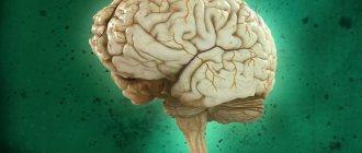
The human brain is a unique, complex system. Due to it, the body can not only coordinate the work of all organs and force them to work as a single whole. The activity of highly organized cerebral structures allows a person to be a social being, capable of learning and cognition, to show multi-level emotions, and to have his own judgment about what is happening around him. To a greater extent, this is determined by the significant development of the cerebral cortex and a number of subcortical formations that make up the concept of the forebrain (or prosencephalon).
The forebrain is the most rostral cerebral section and includes in its structure two hemispheres with the gray matter of the cortex, subcortical structures and nerve fibers-conductors.
The functions of the forebrain include higher mental activity, complex reflex acts, the ability to cognition, features of a person’s emotional reactions and his socialization. Moreover, the cerebral structures of the anterior regions can determine what characteristics of character and temperament will be inherent in an individual.
During the process of ontogenesis, the forebrain is formed at the stage of three brain vesicles already at 3-4 weeks of embryonic development . This occurs by separating it from the midbrain (mesencephalon).
Closest to the spinal cord is the brain vesicle, which gives rise to the hindbrain (rhombencephalon). The latter, in turn, differentiates into two sections - metencephalon (from which the pons and cerebellum are formed) and myeltncephalon (forming the medulla oblongata). In some sources, the metencephalon is mistakenly called the forebrain, given that the anatomy of this formation determines its location anterior to the medulla oblongata. It would be more accurate to consider the metencephalon as the anterior part of the hindbrain.
By the end of the 4th week of gestation, the intermediate brain (diencephalon) and telencephalon (telencephalon), as well as the cavity of the third ventricle, are formed from the anterior medullary bladder prosencephalon.
Brain structure
It is customary to divide human intelligence into five main parts: the cerebral hemispheres, the cerebellum, the medulla oblongata, the median and the pons. In some textbooks you can find a different classification. It states that the brain consists of the forebrain, midbrain, hindbrain, and brainstem. Its composition is quite simple. It’s even funny that such an important organ consists of nothing but water, minerals, lipids and proteins. Today we will talk in more detail about the structure and functions of the forebrain.
to contents ^
Telencephalon (telencephalon)
The anatomy of the telencephalon considers it as the most anterior cerebral section. It is also called the big brain. Its structure includes:
- two hemispheres covered with cortex;
- corpus callosum (corpus callosum);
- amygdala and striatum;
- olfactory brain.
Each hemisphere is separated from each other by a longitudinal fissure, and they are connected by the corpus callosum, the anterior and posterior commissures and the fornix commissures. At the level of the telencephalon, multilevel reflexes are closed, including the central autonomic arcs.
Cortex
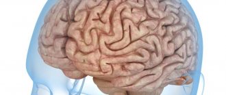
The cerebral cortex developed already at the later stages of the evolution of the animal world. It is she who plays an extremely important role in the development of higher mental functions, despite the fact that such activity can only be carried out with the coordinated work of the entire brain as a whole.
The cortex accounts for more than 45% of the total volume of the entire hemisphere . Its surface can exceed 1500 square centimeters.
However, the cortex is not homogeneous. When considering the structure of the cortex, its evolution should be taken into account. This factor divides the cortex into:
- paleocortex (or ancient cortex);
- arheocortex (old cortex);
- neocortex (new cortex).
The paleocortex has a single-layer structure . At the same time, the nerve cells are not yet completely separated from the subcortical formations. The ancient cortical layer is responsible for primitive emotional reactions, which are characteristic not only of humans, but are also present in animals at a lower stage of evolution, starting with reptiles. Manifestations of the work of the ancient bark include:
- ritual behavior;
- manifestation of aggression;
- protection of one's own territory;
- subordination to social hierarchy;
- dispassionate and submissive actions.
The archeocortex also consists of a single layer of neurons, but it is already completely separated from the subcortical formations. It is located along the lower edge of the neocortex. It is already present in birds and mammals.
The old bark performs the following functions:
- provides emotional reactions: affection, fun, fear, anger, others;
- concentrates attention;
- stimulates development and own activity;
- promotes altruistic behavior.
The limbic system, which is the substrate of emotions, partly attention and memory, as well as the regulator of circadian rhythms and autonomic reactions, is largely represented by the paleocortex. This is important when determining the unity of these processes. For example, often an individual’s subjectively colored experiences (emotions) are accompanied by vegetative manifestations (lacrimation, changes in skin color, increased heart rate, and so on).
The neocortex or new cortex consists of 6 layers. It accounts for more than 95% of the area of the entire cortical surface. Impulses from subcortical formations and the trunk arrive here. It is at this level that complex conditioned reflexes are closed and the higher mental functions of the forebrain are carried out.
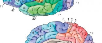
The folded structure of the neocortical tissue with the presence of grooves and convolutions is explained by an increase in the volume of cortical tissue while the overall size of the brain and cranium remains unchanged.
Forebrain and its structure
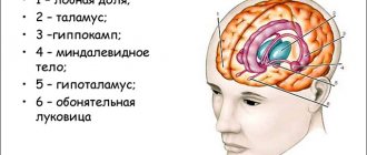
The forebrain is quite complex. Everyone knows it very well, and when we mention this organ, a picture of two hemispheres immediately comes to mind. It's right. Gray matter is divided into sections: the cerebral hemispheres and the diencephalon. If we talk about a more detailed division and go deeper into this topic, we can single out: the basal ganglia, the cerebrum, the hippocampus and the limbic system - a complex that consists of structures responsible for visceral, motivational and emotional sensations. Such a fairly extensive structure of the human forebrain will be of little interest to a person far from medical science, so in this article we will refer to the first classification and talk about the structure of which in more detail.
Useful to know: The medulla oblongata, what functions it is responsible for and what diseases it suffers from
to contents ^
Components and its functions
Hemispheres of the brain. Some of the important components that are separated by the posteroanterior cavity. The parts are connected by the corpus callosum - this is a white wall. The upper ball itself is covered with a shell of neurons and gray matter, arranged in columns in several layers. The surface of the hemispheres has the form of folds, convolutions and depressions, which are called grooves. It is these depressions that divide the brain into the temporal, frontal, parietal and occipital parts. They are named after the bones they are adjacent to. In neurons, the analysis of nerve connections that come from outside is carried out, these are visual, auditory, and neurons responsible for muscle activity. Gustatory and olfactory neurons are located in the temporal lobe, while behavioral neurons are located in the anterior gray matter. The central zone is responsible for human activity.
The main feature of the hemispheres is that they differ significantly from each other. For right-handed people, the neurons responsible for speech are located in the left hemisphere, and the right hemisphere is responsible for actions, logical chains, recognition of faces, songs, paintings and other things. Under the influence of external stimuli, experience is created and accumulated. In the hemispheres, to summarize and say briefly, the main centers are formed that interact with the most complex patterns of behavior, instincts and memory.
The diencephalon consists of three parts: lower, upper and central. Everyone has heard the word thalamus at least once - this is precisely the upper part of the diencephalon. It, in turn, is made up of the ventricle and paired formations. This is where all information from outside comes in, the initial assessment takes place and then passes further into the cortex of the human intellect. The hypothalamus is the lower part, which performs the function of metabolism and regulates brain energy. In the centers of the hypothalamus there are nuclei that are responsible for various sensations. In combination with the components of gray matter in impulses supplied for motor activity.
Useful to know: Functions and structure of the cerebral pons, its description
to contents ^
Lesson 48. Structure and functions of the forebrain
Topic: Brain. Structure and functions of the forebrain
Target:
continue to develop knowledge about the parts of the brain, show the leading role of the cerebral hemispheres in behavior; introduce methods of brain research; develop skills in working with tables, text, and student messages.
During the classes
- Organizing time
- Updating knowledge
Slides 2-3
- Blitz survey. Question answer.
- Total number of pairs of cranial nerves (12).
- The white matter of the brain is represented by... (fiber pathways
)
- The cerebellar cortex, subcortical nuclei, and cerebral cortex are represented by... (gray matter of the brain
)
- The part of the brain responsible for coordinating voluntary movements and maintaining position in space, as well as regulating muscle tone and balance - ... (cerebellum
)
- The part of the brain responsible for orientation reflexes to visual and auditory stimuli, as well as taking part in the regulation of muscle tone and body posture - ... (midbrain
)
- The structure of the hindbrain, which connects the spinal cord and the overlying parts of the brain, is responsible for food and protective reflexes, as well as regulating the activity of the respiratory, cardiovascular and digestive systems - ... (medulla
)
- Structures included in the hindbrain - ... (medulla oblongata, pons, cerebellum
)
- They have a cortex made of gray matter of the brain - ... (cerebellum, cerebral hemispheres
)
- If damaged, instant death occurs - ... (medulla
)
Slide 4
– Explain cause and effect relationships.
A) Why can you accidentally swallow an inappropriate object?
B) There is a person in the clinic who has a tumor in one of the parts of the brain. The man wants to take the glass, but misses. After a few struggles, he grabs the glass and crushes it, squeezing too hard. In what part of the brain does he have a tumor?
C) After the surgical removal of some part of the brain, the dog stopped responding to its name, the sight and smell of food, and recognizing friends and foes. What part of her brain was removed?
- Learning new material
Slide 5
– “The brain is the most perfect and complex creation of earthly nature” (I.P. Pavlov)
Modern science, based on the doctrine of reflexes, has accurate data on brain activity. A person can use his knowledge of the nervous system and control the processes occurring in it. Let's talk about methods for studying brain functions. / Students present prepared messages
/
Slide 6 –
The brain connects together the functioning of the organs and systems of the human body, coordinating their activities. “Looking after” the body, the brain makes it possible to process information received through the senses, directs movements, is responsible for attention, memory and coordination, “deciphers” and forms speech. Considering that the brain is perhaps the most complex organ of the human body, the most advanced research methods are required to analyze the state of its functioning.
Timely diagnosis can identify both the diseases themselves and the causes of their occurrence, making effective treatment possible.
Slide 7
– EchoEG (echoencephalography) – ultrasound diagnosis of brain pathologies, performed using an oscilloscope. The device detects the reflected ultrasound and displays the returned signal on the display.
Slide 8
– REG (rheoencephalography) – the method is used to diagnose brain tumors, head injuries, epilepsy, migraines. REG helps to identify local lesions and determine the degree of filling of the brain with blood.
Slide 9
– Doppler ultrasound (Doppler ultrasound) – the study is carried out to assess blood flow in large and medium-sized vessels of the head and neck.
Slide 10
– MRI (magnetic resonance imaging) of cerebral vessels is a rather complex, but highly informative examination method based on the mechanism of nuclear magnetic resonance.
Slide 11
– MRA (magnetic resonance angiography) – a method for studying the vascular bed makes it possible to construct a three-dimensional reconstruction of the vascular network in the area under consideration, allows you to isolate for study certain nerve vessels and trunks from the projection of parts of the brain.
Slide 12
– Craniography is the name given to performing radiography of the skull in 2 projections. The method is well suited for detecting birth defects and bone fractures in the skull.
Slide 13
– EEG (electroencephalography) – the method consists of recording fluctuations in the electrical potentials of the brain using an electroencephalograph. Electrodes attached to the head read the biocurrents of the brain, recorded on paper or displayed on a display.
Slides 14-15
– PET (positron emission tomography) – based on the use of radiopharmaceuticals. Using PET, you can distinguish a benign tumor from a malignant one and diagnose the consequences of brain injuries and diseases.
Slide 16
– CT (computed tomography) involves measuring the intensity of x-rays passing through brain tissue. The method makes it possible to identify congenital malformations and determine the location and nature of pathologies of the brain system.
Today, medicine is introducing new methods and technologies for the early diagnosis of central nervous system diseases, which makes it possible to effectively treat serious diseases and their consequences.
Slide 17 –
Now let’s turn our attention to the forebrain, which, as you already know, consists of two parts: the diencephalon and the cerebral hemispheres. The diencephalon consists of the visual thalamus (thalamus) and the subthalamic region (hypothalamus).
Slide 18
– Thalamus, sometimes – visual hillocks (Latin Thalamus; from ancient Greek θάλαμος “room, chamber, compartment”) – a section of the brain, which is a large mass of gray matter located in the upper part of the thalamic region of the diencephalon of chordates, including humans. It was first described by the ancient Roman physician and anatomist Galen.
Slide 19
– The thalamus is located deeper than the structures of the cerebrum, in particular the cortex or mantle. Below the thalamus are the structures of the midbrain.
The thalamus performs several important physiological functions. It is responsible for transmitting sensory and motor information from the senses (except information from the olfactory organs) to the corresponding areas of the cerebral cortex. The thalamus plays an important role in regulating the level of consciousness, processes of sleep and wakefulness, and concentration.
Slide 20
– The thalamus is a kind of gate through which basic information about the surrounding world and the state of the body enters the cortex and reaches consciousness. The thalamus consists of approximately 40 pairs of nuclei, which are functionally divided into specific, nonspecific and associative.
The thalamus acts as a “filter”, selecting the most significant information for the body, which enters the cerebral cortex.
Slide 21
– Hypothalamus (Latin hypothalamus, from Greek ὑπό - “under” and θάλαμος - “room, chamber, compartment, thalamus”) is a small area in the diencephalon, which includes a large number of groups of cells (over 30 nuclei) that regulate neuroendocrine activity of the brain and homeostasis of the body.
Slide 22
– The hypothalamus is connected by nerve pathways to almost all parts of the central nervous system. It controls the release of pituitary hormones and is a central link between the nervous and endocrine systems and regulates functions such as the sensation of hunger and thirst, thermoregulation of the body, sexual behavior, sleep and wakefulness.
Slide 23
– Research in recent years shows that the hypothalamus plays an important role in the regulation of higher functions, such as memory and emotional state, and thereby participates in the formation of various aspects of behavior.
- Reinforcing the material learned
Slide 24
- Answer the questions:
What are the similarities and differences between the functions of the thalamus and hypothalamus?
What can happen if the thalamus and hypothalamus are disrupted?
When this area of the brain is damaged, excessive obesity occurs due to excessive fat consumption and the appearance of “ravenous hunger.” What part of the brain are we talking about? ( hypothalamus
)
- Summing up the lesson
Slide 25 – Homework:
Repeat §§ 45-46.