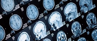
MRI of the brain is a new, high-precision diagnostic method based on obtaining layer-by-layer sections of the main organ of the human central nervous system and then converting them into a three-dimensional image on the monitor of a computer connected to the tomograph.
Magnetic resonance imaging is suitable for detecting a wide range of brain diseases. MRI is also used for preventive purposes, as it helps to diagnose diseases in the initial stages, when symptoms do not yet make themselves felt.
What will a brain scan show?
The effect of MRI is based on the effect of nuclear magnetic resonance. Electromagnetic waves, passing through the human body, affect hydrogen atoms, changing the frequency of their vibrations. The energy released by the nuclei in this process is captured by a tomograph and then converted into an image on a computer, which allows one to obtain a detailed picture of the anatomical structures of the brain under different planes - coronal, transverse and sagittal. The image of the diagnosed area is formed by combining many layer-by-layer sections with a thickness of 1-3 mm, which makes it possible to detect the slightest deviations and pathologies.
Appearance of the Philips Intera 1.5T tomograph
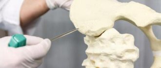
When performing magnetic resonance imaging of the brain, the slice thickness can be set at values of 1-3 mm, which makes it possible to accurately examine such small structures as the pituitary gland and optic nerves.
Benefits of MRI
The advantages of magnetic resonance imaging include:
- speed of the procedure and obtaining results;
- high accuracy;
- obtaining three-dimensional images of organs;
- safety for the patient;
- possibility of carrying out in late pregnancy and during breastfeeding;
- early diagnosis of diseases.
When performing MRI, unlike radiography, no harmful radiation is used. The magnetic waves created by the tomograph are absolutely safe for humans. MRI diagnostics can be performed as often as necessary.
Medical staff training
Training of medical staff of institutions that will directly use the equipment in their work is one of the important stages of commissioning medical equipment, be it a tomograph, ultrasound machine or something smaller, according to GOST 12.0.004-2015 “Organization of occupational safety training.” A detailed briefing is required, the fact of which is recorded in the contract for work on commissioning the equipment. However, one should not be confused that the equipment supplier provides instructions, and the actual training is carried out by companies that have a license for this. After instructions and training have been completed by the personnel regarding the technical use of the equipment, you can sign the act of putting the device into operation. From this moment the warranty period of the new equipment begins.
What does the user get when contacting?
The undoubted benefits of cooperation with us are positively assessed by all medical institutions; each client receives:
- assembly, installation and commissioning work by experienced qualified engineers;
- quality of work ensuring reliable, uninterrupted and accurate operation of medical equipment;
- full compliance with the requirements of the regulations when installing equipment;
- full reporting and preparation of documentation and necessary regulations on the work done;
- competent instruction and training of medical personnel in handling the installed equipment;
- the possibility of concluding a contract for the maintenance of your equipment by our service engineers.
Contraindications for MRI
Despite its high safety, MRI also has a number of contraindications.
Absolute contraindications include:
- the presence in the body of implants, pins, brackets made of magnetic metal;
- presence of a pacemaker, neurostimulator;
- weight exceeding the technical specifications of the tomograph;
- first trimester of pregnancy;
- inability to remain still due to neurological or psychiatric diseases.
Relative contraindications are:
- second and third trimester of pregnancy;
- children under 5 years of age;
- fear of being in a confined space.
If contrast agents are used during the procedure, the following may be contraindications:
- renal failure;
- pregnancy and lactation;
- allergy to contrast agents used.
If there are objective restrictions on magnetic resonance imaging, the patient may be prescribed other types of diagnostics that do not have the above restrictions.
Types of equipment
There are several types of equipment on which diagnostics are carried out. There are open and closed devices. Tomographs are also divided into:
- low-floor - power up to 1 Tesla, usually 0.3 - 0.4 T;
- high-field - power 1.5 - 3 Tesla.
Some experts also highlight 1 Tesla mid-field devices.
The higher the magnetic field strength, the better the quality of the resulting images. Open devices have low voltage, so choosing such a tomograph is not always advisable. Always consult your doctor before going for an examination so as not to waste extra money and time redoing the tomography using other equipment.
MRI and CT, which are more effective for diagnosing the brain
CT, like MRI, is a highly accurate diagnostic method that allows you to obtain a three-dimensional image of internal organs.
However, to diagnose diseases localized in the cranial cavity, MRI is more often used, since this technique allows one to obtain better images of soft tissues. Whereas CT has proven itself better for diagnosing skull bones, cerebral vessels, hemorrhages in the cranial cavity, as well as in cases where MRI cannot be performed for a number of reasons.
If the presence of tumors localized in the brain and its deep structures is suspected, MRI has a number of advantages and shows:
- exact boundaries, size, location of the tumor;
- helps to make a preliminary assessment of the malignancy of the tumor;
- allows you to identify tumors located at the border with bone tissue and poorly visualized by other diagnostic methods.
The decision to choose one or another method is made by the attending physician based on the patient’s medical history.
What does a brain MRI show?
Magnetic resonance imaging helps to see:
- benign and malignant tumors;
- areas of nervous tissue necrosis;
- areas of hemorrhage;
- vascular diseases;
- inflammatory processes of brain tissue;
- dystrophic changes;
- traumatic brain injuries.
Due to its high accuracy, MRI diagnostics can detect even the slightest pathological changes in brain structures.
What diseases can be detected by MRI of the brain?
MRI is used to detect the following diseases:
- ischemic, hemorrhagic stroke;
- thrombosis and vascular aneurysms;
- neoplasms;
- cysts;
- Alzheimer's disease;
- epilepsy;
- atherosclerosis;
- multiple sclerosis;
- inflammatory diseases: meningitis, encephalitis;
- abscesses;
- hemorrhages;
- Parkinson's disease;
- cysts, pituitary adenomas.
Application of contrast agent
When carrying out MRI diagnostics, contrast can be used, which makes it possible to visualize the deep structures of the brain in more detail, to determine clear boundaries of hemorrhages, neoplasms, and sclerotic changes.
For contrast, drugs containing the chemical element gadolinium are used. The drug is administered intravenously immediately before the study. Carrying through the bloodstream throughout the body, particles of the contrast agent settle in pathologically altered structures, staining them.
Contrasting can be used in the following cases:
- Suspicions of the presence of neoplasms. The contrast agent settles in the tumor tissue, which helps to accurately determine the boundaries and size of the tumor, as well as draw preliminary conclusions about the nature of the tumor. Benign tumors usually have regular sizes and clear boundaries, while malignant tumors grow into surrounding tissues and are irregular in shape with jagged edges.
- Pituitary gland studies. The pituitary gland is very small and is located deep in the structures of the brain. Contrasting allows not only to clearly see the organ itself, but also to identify the presence of pathologies and neoplasms.
- Diagnosis of demyelinating diseases. Contrasting helps to more clearly identify the boundaries and total volume of brain tissue damage.
Brain MRI with contrast
Contrast can be used to diagnose any brain pathologies, because provides a much clearer picture than MRI without contrast.
Contrast drugs are harmless to the body and are excreted through the kidneys. Contraindications for the use of contrast may include renal failure and previously observed allergic reactions.
results
The study is interpreted by a diagnostician - a radiologist. The conclusion is issued on the day of the examination, but a description of the images may take the doctor several hours.
The doctor examines the obtained images and compares them with normal values:
- size and location of organs, symmetry;
- presence/absence of abnormal formations;
- presence/absence of an aneurysm, free fluid, blood clots in the blood stream;
- signs of infection and inflammation;
- stagnation in the ducts;
- traumatic injuries.
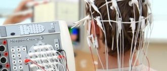
How does the procedure work?
MRI examination is completely painless. Immediately before the procedure, you must remove any metal items from yourself. The patient lies down on the tomograph couch, which is placed inside the device.
Brain MRI procedure
Before carrying out the procedure you need to know the following:
- High-resolution (high-field) magnetic resonance imaging scanners create a fairly high noise level. At our center, patients are provided with headphones to reduce discomfort from these sounds;
- the doctor conducting the study always remains in touch with the patient, the patient has an alarm button;
- The patient must remain absolutely still throughout the procedure to avoid motion artifacts.
The duration of an MRI of the brain is usually about 15 minutes. If you plan to use a contrast agent, the study will take a little longer - about 30 minutes.
Preparation
Many types of MRI examinations do not require special preparation, but there are exceptions.
Before an MRI of the abdominal cavity, it is advisable to follow the following diet:
- for two days, eliminate gas-forming foods, fresh fruits and vegetables;
- on the day of the procedure, avoid tea and coffee;
- It is advisable not to eat for at least 6 hours before the examination, and not to drink for 4 hours.
The doctor will recommend special medications for constipation and flatulence. To exclude artifacts from intestinal motility, two hours before the procedure you need to drink 2 tablets of No-shpa.
Before an MRI of the pelvis, it is advisable not to urinate for about two hours. If the study is carried out in women, it is recommended that it be carried out on days 5-12 of the cycle (immediately after menstruation), but this condition must be discussed with a doctor.
General recommendations
before MRI scan:
- remove all metal objects;
- leave jewelry and accessories at home;
- change into comfortable clothes;
- Leave valuables, money, and bank cards in a specially designated place or safe in front of the diagnostic room.
An approximate list of items that are not allowed in the diagnostic room:
- metal zippers on clothes, pins, hairpins;
- watch;
- Hearing Aids;
- pens;
- earrings, including piercings;
- glasses;
- removable dentures;
- pocket knives.
Decoding MRI of the brain
You can receive the test result immediately upon completion of the procedure.
It will take the doctor about 15-30 minutes to decipher the results. Recording images is possible both on a disk (it is provided free of charge), and on film or a flash drive, at the request of the patient.
The patient will be given a disc, as well as a doctor’s report, which will describe in detail the anatomical features of the area under study and possible pathological changes.
The resulting conclusion must be provided to the attending physician to determine a further treatment plan.
How to overcome fear of the procedure
There is no need to worry before an MRI - the examination is absolutely painless and safe. The radiologist remains in contact with the patient throughout the procedure and, if necessary, will answer any questions that arise.
If the patient experiences severe anxiety, then before the examination, you can take sedatives.
MRI is a modern type of diagnostics that makes it possible to accurately identify any pathological changes in the brain, even at the initial stages of their occurrence. Magnetic resonance imaging has virtually no contraindications and is a painless and safe technique used to diagnose a wide range of diseases.
| What does an MRI show? |
| How is an MRI of the brain done with contrast? |
| What does an MRI of the pituitary gland show? |
| MRI of the brain with contrast |
| MRI of the sella turcica and pituitary gland in Rostov-on-Don |
| MRI of the cerebellum of the brain |