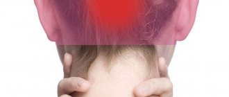
Symptoms
Almost all patients have at least 2 of the typical four symptoms:
- Fever;
- Headache;
- Stiff neck;
- Impaired consciousness with a score of less than 14 points on the Glasgow scale.
A hemorrhagic rash is typical for meningococcal infection (rarely for pneumococcal, staphylococcal) and is localized on the torso, arms, legs, and butt. A rash on the face, and especially the earlobe and conjunctiva, is an unfavorable prognostic sign.
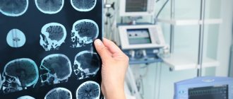
Diagnostics
Laboratory and instrumental studies:
- General urine analysis;
- Complete blood count with leukocyte formula;
- Biochemical studies: plasma glucose, blood urea and creatinine, blood electrolytes;
- Chest X-ray;
- HIV test;
- PCR – diagnostics;
- CT or MRI of the brain.
According to existing rules, a computed tomography (or MRI) scan of the brain should be performed before a lumbar puncture. These methods make it possible to exclude SAH, hematomas, abscesses and other space-occupying formations, as well as clarify the condition of the basal cisterns of the brain. Those. identify contraindications to lumbar puncture and clarify the diagnosis. But if CT or magnetic resonance imaging cannot be done within 1-2 hours from the moment the patient is admitted, then lumbar puncture should not be postponed until later.
CSF analysis
Analysis of cerebrospinal fluid (CSF) can confirm the diagnosis of meningitis.
| Table 1. The most typical changes in the cerebrospinal fluid in patients with meningitis | ||||
| Defined indicators | Normal values | Bacterial meningitis | Viral meningitis | Tuberculous meningitis |
| CSF pressure | 90-200 mm water. Art. | Often elevated | Normal or sometimes elevated | Often elevated |
| Number of cells | Lymphocytes, no more than 5 in 1 µl | Typically: 1000-10000 cells per 1 µl, neutrophilic leukocytes predominate | 20-200 cells in 1 µl, the content of lymphocytes is 60-90% of the total number of cells | 20-400 cells in 1 µl, the content of lymphocytes is 60-90% of the total number of cells |
| Pathogen | Not detected | Detected in 40-60% of cases | Not detected | Rarely detected |
| Protein concentration | 150-350 mg/l | above 1000 mg/l; | Within 1000 mg/l | above 1000 mg/l but there may be normal values |
| Glucose levels depend on blood glucose levels | 2.6-4.2 mmol/l. 55-60% of the concentration of glucose in the blood serum. | < 2 mmol/l; More precisely: less than 55-60% of the serum glucose concentration | > 2 mmol/l; More precisely: more than 55-60% of the glucose concentration in the blood serum | At the beginning of the disease it is not changed. After 1-2 weeks <2 mmol/l; More precisely: less than 55-60% of the serum glucose concentration |
| Lactate | 1.2-2.1 mmol/l | above 4.2 mmol/l | does not exceed 4.2 mmol/l. | varies |
Method of performing lumbar puncture
A lumbar puncture should be performed (preferably) before the person is given antibiotics. The puncture can be done in the intervertebral spaces L3-L4 (most often), L2-L3, L4-L5, and L5-S1. Doctors need to use 20 and 22 G needles. It is safer to perform the puncture when the patient is lying on his side. If it is not possible to obtain cerebrospinal fluid, the puncture is performed in a sitting position - higher cerebrospinal fluid pressure makes it easier to find anatomical landmarks for the puncture site.
Measurement of cerebrospinal fluid pressure should be carried out immediately after a successful puncture, and only after that the cerebrospinal fluid should be collected for laboratory tests. CSF pressure is measured with the patient lying on his side. Ideally, the cerebrospinal fluid should be collected in 4 sterile tubes:
– to determine the cellular composition of the cerebrospinal fluid;
– to determine microflora and its sensitivity to antibiotics;
– to determine the content of protein and glucose and other components;
– to determine the cellular composition of the cerebrospinal fluid (for comparison with the first tube).
To avoid diagnostic errors, laboratory tests should be carried out without delay.
Contraindications to lumbar puncture
- Lumbar puncture should not be performed if intracranial space-occupying formations (abscess, tumor, hematoma) are suspected.
- When signs of herniation appear - Cushing's syndrome (a combination of arterial hypertension and bradycardia), pathological pupillary reactions.
- Thrombocytopenia (platelet count <50×109/l).
- Coagulopathy, including those caused by taking medications.
- Inflammatory process in the puncture area.
If it was not possible to obtain cerebrospinal fluid (failure to do so, the patient’s refusal to undergo puncture, or contraindications to puncture), doctors should prescribe antibiotics to the patient. With bacterial meningitis, CSF pressure in most cases is higher than normal and there is a possibility of developing brain dislocation. Therefore, after a lumbar puncture, the person must be closely monitored (examination is carried out at least every 15-30 minutes during the first 4 hours).
If the neurological status after puncture progressively worsens, administration of osmotic agents (mannitol, sodium chloride 7.5%) should be started immediately. When to perform repeated lumbar punctures, the doctor decides individually. Some experts believe that in case of extremely severe meningitis, they should be performed every day. If the course of the disease is favorable, repeated lumbar punctures may not be performed.
Risk group
Children and the elderly are at risk for meningitis. In addition, this disease is often diagnosed in people with an absent spleen and immunodeficiency.
The spread of meningitis, like all other infectious diseases, is more active in crowded places, closed groups, for example, in kindergartens, schools, student dormitories, barracks, since it is easier to get meningitis if you are in the company of asymptomatic carriers.
Most often, meningitis is transmitted by airborne droplets. This mechanism of infection transmission is considered the most common and characteristic of meningitis of viral origin. The infectious agent is transmitted through coughing, sneezing, kissing and sexual contact.
Newborn babies can become infected from an infected mother through the birth canal. The likelihood of infection is especially high in children born by caesarean section. Bacterial and viral meningitis can be transmitted this way.
Infection with meningitis can occur through the oral-fecal route: through dirty food or untreated water.
In addition, meningitis can develop as a result of the bite of an insect or animal that carries an infectious agent.
Basic principles of management
They try to normalize body temperature, reduce headaches, and reduce the patient’s tension. For these purposes, non-narcotic analgesics, sedative therapy, and anti-nausea medications are used. Indications and implementation of artificial pulmonary ventilation follow the same principles as for patients with TBI.
In severe cases, due to impaired consciousness, vomiting, enteral food intake is often difficult. On the first day, less often, in the first 2 days, an infusion of saline solutions is used to replenish the loss of fluids and electrolytes. They try to make do with the minimum possible volumes - 1.5-2.5 liters per day. At the same time, hypotension must be corrected without delay. It is not recommended to use glucose solutions (if there is no hypoglycemia) derived from hydroxyethyl starch. For unstable hemodynamics, doctors use vasopressor infusions. It is better to give preference to norepinephrine (Norepinephrine) or phenylephrine (Mezaton).
Hyponatremia occurs in approximately 30% of patients with meningitis and can lead to increased cerebral edema. It must be eliminated immediately. As soon as the patient’s condition becomes stable, they switch to enteral feeding with nutritional mixtures. Any hyperglycemia that occurs should be promptly corrected. Seizures can dramatically increase intracranial pressure. In most patients with severe meningitis, the level of intracranial pressure is elevated.
With meningitis, an increase in ICP is often caused not so much by cerebral edema, but by excess production of cerebrospinal fluid. Therefore, lumbar punctures and furosemide injections are often effective when it comes to reducing ICP. If these actions fail to reduce ICP, cerebral herniation may occur; osmotherapy with mannitol or 3-7.5% sodium chloride solution should be used. In general, ICP quickly normalizes when antibacterial treatment is given. If the temperature drops, consciousness is clear, and focal neurological symptoms are not observed, then there is no need for decongestant therapy.
Personnel protection and patient isolation
To prevent airborne spread of infection, patients with meningococcal infection or meningitis of unknown etiology are isolated during the first 24 hours of antibiotic therapy. Infection of personnel can occur during tracheal intubation, CPR, and artificial ventilation. Standard precautions must be taken.
The need for prophylactic antibiotics should be considered for ICU staff if a patient is diagnosed with meningococcal meningitis. The likelihood of infection remains for 24 hours after antibiotics are prescribed. The chance of infection is higher among younger employees and people over 60 years old.
Use any of the following schemes:
1. Ciprofloxacin tablets, 500 mg twice a day. per day for 2 days;
2. Rifampicin tablets, 600 mg every 12 hours for 2 days.
Preparing for the examination
Preparation for cerebrospinal fluid analysis is as follows:
- Blood tests are taken (general, clotting tests).
- At the preliminary consultation, an anamnesis is collected. The patient needs to inform the doctor about past illnesses, the presence of chronic illnesses, and negative reactions to medications.
- It is necessary to donate cerebrospinal fluid on an empty stomach - eating is prohibited 12 hours before the procedure.
Before the examination, taking medications that thin the blood, as well as analgesics and non-steroidal anti-inflammatory drugs is not allowed.
Acute bacterial meningitis
Today, modern diagnostic and treatment methods are used, but the mortality rate for bacterial meningitis reaches approximately 20%. It should be remembered that only after 24 hours do changes in the cerebrospinal fluid become characteristic of purulent meningitis. Therefore, if meningitis is suspected, doctors immediately prescribe antibiotics to the person.
Pathogen identification
Identification of the type of pathogen and determination of its sensitivity to antibacterial agents, in most cases, occurs with a great delay - after 3-4 days. In cases where the ongoing antibacterial therapy gives the expected effect and the patient’s condition becomes better, after obtaining the results of sensitivity to antibiotics, as a rule, antibiotics are not changed. If a significant improvement in the patient’s condition has not occurred by the time the results of the bacteriological examination are obtained, the antibacterial drug that is most effective against the isolated pathogen is prescribed.
Treatment
Treatment of meningitis must be comprehensive and carried out by a qualified specialist in a hospital setting. The treatment regimen consists of bed rest, infusion therapy, antibiotic therapy, antiviral (antifungal) drugs and symptomatic treatment.
Antibiotics. In this case, penicillin, ampicillin, ceftriaxone or meropenem may be prescribed. Pyrazinamide and rifampicin will potentiate the effect of antibiotics.
Antifungal therapy: amphotericin B, 5-flucytosine.
Detoxification therapy: enterosgel, atoxil.
Symptomatic treatment: prescription of antiallergic and anti-inflammatory drugs, diuretics, painkillers.