Morphology and location in the body
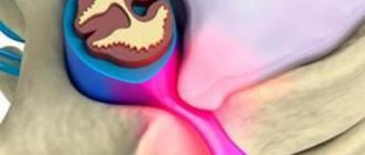
The spinal cord extends from the brain and is located in the spinal canal, which is formed by vertebral arches connected in a ring. The upper part is connected to the medulla oblongata of the brain. The spinal cord ends at the level of the first lumbar vertebra. Its lower part is reduced and has the appearance of a terminal filament with a cross section of up to 1 mm. In the lumbar and sacral spine, the filum terminale and roots of the lower spinal nerves form the “cauda equina.”
There are four sections of the spinal cord:
- cervical (8 segments);
- thoracic (12 segments);
- lumbar (5 segments);
- sacral (5 segments);
- coccygeal (from 1 to 3 segments).
Rice.
1. External structure and parts of the spinal cord. The length of the adult spinal cord varies from 40 to 45 cm, and the width - from 1 to 1.5 cm. The diameter is not the same in different parts of the spine. The average weight of the brain is 35 g.
Shells
The spinal cord resembles a cord. Between the spinal canal and the brain there is a space filled with fatty tissue, blood vessels, and cerebrospinal fluid.
The brain itself is protected by three membranes:
TOP 4 articles
who are reading along with this
The importance of the nervous system
Spinal cord: structure and functions
Brain structure and functions
Composition of blood - briefly and clearly
- soft - internal, tightly adjacent to the brain, consisting of loose connective tissue and containing blood vessels;
- arachnoid - middle, forming a soft cavity filled with cerebrospinal fluid and blood vessels;
- hard - upper strong, consisting of connective tissue with a rough outer and smooth inner surface.
Rice. 2. Sheaths of the spinal cord.
Spinal cord injury and its consequences
Treatment of spinal injury and spinal cord damage is a rather lengthy and serious process that does not always lead to a complete recovery of the patient. Some disorders appear immediately, and some can develop over time, appearing as symptoms of osteochondrosis, intervertebral hernias, and trigger the development of various arthrosis, stenosis, myelopathy and other diseases.
Anatomical structure of the spinal cord
The spinal cord, like the brain, despite its soft structure, is one of the most protected organs of the human body. It is covered with several membranes (soft layer, arachnoid and hard), the spaces are filled with cerebrospinal fluid. Thus, the brain is suspended in a liquid medium by thin cables, which allows it to withstand physical stress, shock from running and walking, stretching, and even shock.
The spinal cord is located in the spinal canal (a through hole in the spine) and is protected on all sides by bone structures, a strong muscular and ligamentous corset. The outer dense membrane of the brain does not touch the walls of the canal. Between them there is a gap called the epidural space, which is filled with fatty tissue, blood vessels, nerve roots and cerebrospinal fluid.
It is quite difficult to damage such a structure, even almost impossible in a calm and measured life. Therefore, even severe spinal injury usually occurs without damage to the spinal cord, although it provokes the development of chronic degenerative-dystrophic processes.
The consequences of damage to the spinal cord by fragments of spinal bone tissue can be very different and depend on the location of the damage (from temporary impairment of the motor abilities of the limbs to complete immobility or death). Sometimes there are cases when the initial problems can be quickly localized, and then the process of gradual death of brain cells, inflammation begins, oxygen starvation of the pathological area begins and further damage to healthy cells.
Causes and consequences of spinal cord injury
In everyday life, getting an injury that leads to a spinal fracture and spinal cord damage is quite difficult. But some extreme situations can provoke enormous stress, under which the body’s compensatory abilities are not effective enough.
Road traffic accidents. Car accidents are the most common cause of such severe injuries. In this case, not only the drivers themselves are at risk, but also pedestrians who find themselves at the scene of an accident at the wrong time.
Extreme sports. Some people deliberately put their lives in danger. These include skiers, rock climbers, mountain cyclists, roller skaters, skateboarders, divers, race car drivers, motorcycle drivers, etc.
Falls from heights. In this case, it does not matter at all from what distance and under what conditions the fall occurred - the risk of injury and damage is the same. Falling from scaffolding during construction work or childish jumping into water from a bridge or bungee jump can lead to the same consequences.
Domestic injuries and criminal cases. This section includes injuries sustained from an unsuccessful fall on ice in winter, from a stepladder or ordinary ladder, on a slippery floor (such injuries are more typical for older and elderly people, when coordination of movements is already impaired), as well as criminal stories when the integrity of the spine and spinal cord are damaged by knife or bullet wounds.
Injuries to the spinal column and spinal cord received in all of the above situations can have the most serious consequences. Of course, slight damage to the lining of the brain and leakage of cerebrospinal fluid may cause some temporary disturbances in motor abilities or nerve sensations, but nothing particularly terrible will result.
It is much more unpleasant when pathologies of muscle structures and internal organs do not disappear or kyphosis, scoliosis, vertebral instability, spondylolisthesis or retrolisthesis begin to develop, the treatment of which often requires surgical intervention. The occurrence and treatment of spinal neuromas are in most cases associated with injuries to the vertebrae and spinal cord. In some cases, the process of programmed death of spinal cord cells (apoptosis) may begin, in which clinical signs of damage appear progressively.
Complete rupture of the spine and spinal cord at the level of the cervical segment leads to instant death. Rupture of the lower sections gives a person a chance to live, since the lungs and heart are able to work independently of the spine, but the motor ability of the body will be blocked for some time by spinal shock.
Treatment of spinal cord injuries
The spinal cord reacts in a special way to serious injury - it shuts down (spinal shock). Until recently, doctors did not know how to deal with damaged area syndromes and bring the victim out of shock. To date, this condition has been studied quite well and after a few weeks the patient regains consciousness. Throughout spinal shock, the body is connected to intensive care devices that maintain breathing and heartbeat, as well as the tone of muscle joints.
After the shock has passed, the human body can be roughly divided into two halves: above the site of injury - controlled by the brain and below the site of injury - controlled autonomously using medical equipment. It is the lower part of the body and the spinal cord that will need to be treated.
If the spine and spinal cord are injured, the victim is immediately provided with medical assistance. Every minute of delay means even more damage to brain cells, their death and irreversible consequences. An urgent operation to remove splinters and fragments of bone structures from the spinal canal, restoration of the lining of the brain and decompression of the nerve roots provide a chance for faster and better further recovery of the body.
At the end of the emergency intervention, as far as possible, doctors restore blood circulation and fix the spine in a motionless state. The patient then remains in the intensive care unit for several weeks under constant observation.
Recovery after injury
Restorative processes of nerve cells in the spinal cord begin in proportion to the retreat of spinal shock. And only when the patient regains full consciousness will doctors be able to objectively assess his condition and chances of recovery. After the end of the resuscitation period, the patient’s condition is monitored by a neurologist and a spine doctor.
In most cases, initially not even damaged, but simply areas of the brain and nerve roots close to the injury and the corresponding body organs can work unstably, which manifests itself in loss of skin sensitivity or inability to move. Regeneration of nerve cells and roots is extremely slow, and the patient’s treatment will be lengthy. After a few months, motor ability and sensitivity of the limbs will gradually begin to return, and the functioning and control of internal organs will improve.
Those functions that have not recovered after one and a half years of treatment can be considered lost forever. But in some cases, brain cells regenerate, but the person is still unable to move or walk. This is explained by the body’s ability to “forget” long-unused muscle activity and atrophy of the muscles themselves.
Specially designed electrical stimulators and work on simulators helped overcome this problem. They allow you to “wake up” dormant connections, activate the work of channels and make them function again. After some time, the patient will be able to stand on his own and walk.
Further treatment for brain damage will take place in spinal rehabilitation centers and manual therapy clinics.
Physiotherapy, massages, swimming, exercise therapy and yoga, acupuncture and reflexology will help the body and human health recover faster. Author: K.M.N., Academician of the Russian Academy of Medical Sciences M.A. Bobyr
Internal structure
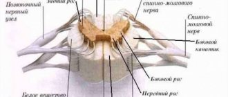
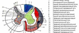
In cross section, the spinal cord has the shape of a butterfly. In the center is a hollow central canal, which surrounds two types of nerve matter:
- gray – accumulation of nerve cell bodies (neurons);
- white - a cluster of processes (axons) of nerve cells.
The gray matter is branched. Thickened anterior and elongated posterior horns extend in different directions. The thoracic region also has lateral horns. From the anterior horns, bundles of nerve fibers—anterior roots—extend in different directions. The posterior roots approach the posterior horns. These bundles form 31 pairs of spinal nerves.
On the outside, the gray matter is surrounded by dense white matter. Between the posterior horns, the white matter forms a narrow fold - the median fissure. On the other hand, between the anterior horns there is a wider fold with a small notch - the median groove.
Rice. 3. Transverse section of the spinal cord with outgoing bundles.
White and gray matter have different structures and play a specific role. Brief information about the structure and function of the spinal cord is presented in the table.
| Substance | Compound | Functions |
| White | Nerve fibers with a myelin (electrically insulating) sheath | Conduction of nerve impulses between different segments of the spinal cord, to the brain and back (conductive function) |
| Neuroglia (auxiliary cells of nerve tissue) | ||
| Blood vessels | ||
| Connective tissue | ||
| Gray | Bodies of motor and interneurons with processes without myelin sheath | Reflex function. The arcs of many reflexes associated with contractions of the muscles of the limbs and torso close in the gray matter. Regulation of the functioning of internal organs. The reflex function of the spinal cord is carried out under the control of the brain. |
| Myelin fibers | ||
| Neuroglia |
The spinal cord has two thickenings - in the cervical (13-15 mm) and lumbar (12 mm) regions. From here comes the largest number of nerves heading to the upper and lower extremities. The cervical thickening begins at the level of the 3-4 cervical vertebrae and ends at the second thoracic vertebra. The lumbar thickening begins at the level of the 9th–10th thoracic vertebra and ends at the 1st lumbar vertebra.
Spinal nerves
Two pairs of roots emerge from each segment of the brain: anterior and posterior. These are the beginnings of the spinal nerves connecting the central nervous system with organs.
The dorsal roots have thickenings (nodes) in which the bodies of sensory neurons lie.
Rice. 2. Spinal cord roots.
The axons of these neurons travel to the organs and form sensory nerves.
The anterior roots are formed from the axons of motor neurons, the bodies of which lie in the gray matter. From these roots originate motor nerves that transmit signals to the organs.
Functions
The spinal cord plays an important role in the functioning of the central nervous system and performs two functions:
- conductive - part of the long processes of neurons is responsible for transmitting signals to the brain (ascending pathways), part receives signals in the opposite direction (descending pathways);
- reflex - signals arrive from receptors in the spinal cord and are directly sent along the reflex arc to the working organs.
Thanks to the reflex function, the hand is pulled away from a hot object or the knee reflex is carried out.