What does it consist of?
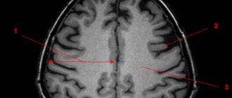
The volume between the basal ganglia and the cortex is completely filled with white matter. Consists of processes of neurons (axons). Collectively, they represent many nerve myelinated fibers. The presence of myelin determines the color of the fibers. They travel in different directions and conduct signals.
Nerve fibers are represented by three groups:
- Association fibers. Necessary for connecting parts of the cortex only in the area of 1 hemisphere. There are short and long ones. Their tasks are not the same: short ones connect convolutions located in the neighborhood, long ones connect distant areas.
- Commissural fibers. Responsible for connecting certain lobes of both hemispheres. Localized in brain adhesions. The basis of these fibers is represented by the corpus callosum. In addition, they monitor the compatibility of functions in the brain.
- Projection fibers. They are responsible for communication with other points of the central nervous system. Connects the bark to the formations below.
Intraoperative identification of functional areas of the cortex and brain pathways
Back in the 30s of the last century, the first steps were taken in determining the localization of the cortical functional organization during neurosurgical operations [46, 76].
Initially, only direct stimulation of the cortex was used during the removal of tumors and epileptogenic zones in conscious patients, which made it possible to assess the relationships between consciousness, motor acts and language production at the stages of surgical intervention. Later, in the 70s of the 20th century, monitoring of somatosensory evoked potentials (SSEPs) entered neurophysiological practice [73], and more recently, in the 90s, motor evoked potentials appeared, which made it possible to more accurately localize the functional areas of the cerebral cortex and pathways . In 1978, the phenomenon of phase reversal of the cortical component of SSEP in the region of the central gyri and the identification of the central sulcus was described [51], which has found wide application in the surgery of intracerebral tumors [28, 59, 63]. In 1937, W. Penfield [76] described a technique for direct stimulation of the cortex using rhythmic (50-60 Hz) bipolar impulses. This approach is mainly used to map the sensory and motor areas of speech in conscious patients, although it is also used in patients under general anesthesia [63]. Also currently used is the technique of stimulation in bursts (trains) of 4-5 pulses [90] for mapping and long-term monitoring of cortical-subcortical conductors [17].
Arousal surgery and direct electrical stimulation of the cortex and pathways are the “gold standard” for resection of gliomas located near speech areas and other functionally significant structures [36, 38, 42, 74]. During such operations, functional brain mapping is carried out by a neurosurgeon together with a neuropsychologist and neurophysiologist, the latter adjusting stimulation parameters while simultaneously monitoring the state of spontaneous cortical activity using an electrocorticogram (ECoG) to detect epileptic activity. This approach makes it possible to prevent the development of convulsive syndrome during prolonged rhythmic electrical stimulation. At the first stage, electrical stimulation reveals the localization of cortical zones of language functions, with each functionally important zone being marked [2, 11, 12].
The stimulation zone is considered functionally significant when speech disturbances are observed three times in a row after subsequent stimuli, and after the end of stimulation, restoration of speech (language) function is noted. The type of speech impairment is verified by a neuropsychologist, and the severity of speech impairment is assessed using the National Institutes of Health Stroke Scale [92]. The next stage is tumor resection taking into account mapping data, with periodic subcortical stimulation performed to search for functionally significant white matter pathways [2].
Thus, the use of intraoperative mapping and electrophysiological monitoring allows surgeons to remove the maximum volume of tumor with minimal impairment of neurological functions [31, 54, 61].
4. Neuropsychological methods of intraoperative research
The study of cognitive functions in dynamics (before and after surgery) is carried out using a well-selected set of neuropsychological tests, which must satisfy several criteria: 1) allow for the assessment of a number of cognitive functions, while the tests must be sensitive enough to determine the effect of the tumor and the treatment performed; 2) the testing procedure must be standardized to allow comparison of the results of a dynamic examination of patients; 3) the presence of standards for conducting tests will make it possible to objectively assess the performance of tests by patients; 4) tests must be sufficiently reliable and insensitive to repeated testing to obtain accurate data during a dynamic examination; 5) tests should have alternative forms for retesting; 6) to prevent fatigue, the total testing time should not exceed 30-40 minutes.
A set of neuropsychological testing is selected individually for each patient. Since tumors often impair memory, attention, speed of information processing, executive functions and speech, appropriate tests must be used during testing. Examples include tests for verbal associations (associations for a given letter of the alphabet and for a certain semantic category), finding similarities between two concepts (for example, “what is common between an apple and a banana”), a test of following route A and B (allows you to evaluate visual attention, distribution and switching of attention) [65], memorizing a list of words (allows you to assess auditory-verbal memory) [55, 60, 83], encryption test (allows you to assess the speed of information processing) [93], test for fine motor skills [53]. Depending on the location of the lesion, the study can be supplemented with the Wechsler Intelligence Test (WAIS-IV), which provides a summary assessment of verbal and nonverbal intelligence [93]. The Rey Complex Figure Test assesses visual constructive functions. Tests for memorizing non-verbalizable figures (Rey Visual Design Learning Test, Rey Complex Figure Test) [65, 89], tests for speech and non-speech working memory (Digit Span Forward, Digit Span Backward, Spatial Span Forward, Spatial Span Backward) are also used [ 65, 89], tests for speech research [22].
Intraoperative mapping uses a small set of relatively simple tests that can be performed during intraoperative awakening. Each test must include a sufficient number of samples to allow continuous testing of function over the course of brain stimulation. As an example, we present several tests used for intraoperative localization of motor and speech areas, most often determined during such operations. To localize the motor zone of the hand, a test for clenching and unclenching a fist is used; to localize the motor zone of the leg, a test for flexing and extending the leg at the knee joint is used. To assess speech functions, a picture naming test is used, and naming verbs is more sensitive to localizing Broca's area than naming nouns. Ordinal counting in forward and reverse order allows you to evaluate the fluency of speech and switching.
Thus, neuropsychological examination of cognitive functions is an important component of a comprehensive examination of patients with brain tumors in the pre- and postoperative period, determining both surgical tactics and subsequent rehabilitation measures.
Functions
Providing a safe environment for the functioning of the nuclei and other parts of the brain and conducting signals throughout the nervous system are the main tasks of white matter.
Constantly, uninterruptedly connecting all parts of the central nervous system is the main goal of the action of white matter. This ensures coordination of general life activities. A signal is transmitted through neural processes, which allows for a variety of human actions.
On the cerebral cortex, grooves and ridges that form convolutions can be clearly visible. The central sulcus divides the parietal and frontal lobes. On both sides of this groove are the temporal lobes. The furrows and convolutions separate the hemispheres, forming 4 lobes in each:
- Frontal lobes. They have undergone great changes in the process of evolution. They developed faster than others and have the largest mass. In them, the white matter must provide all motor processes. Here, thinking processes are started, the structure of speech and writing is regulated, and all complex forms of life support are controlled.
- Temporal lobes. They border on all other lobes. The functioning of the white matter in them is aimed at understanding speech and learning opportunities. Allows you to draw conclusions by receiving all kinds of information through hearing, sight, and smell.
- Parietal lobes. Responsible for pain, temperature, tactile sensitivity. They make possible the work of centers that have been brought to automaticity: eating, drinking, dressing. A three-dimensional understanding of the world around you and yourself in space is built.
- Occipital lobes. In this area, functions are aimed at remembering processed visual information. The form is evaluated.
Midbrain: structure and functions
2.1. General understanding of the anatomy of the midbrain
The midbrain, mesencephalon, develops from the midbrain. It distinguishes between the roof and legs of the brain.
The cavity of the midbrain is the cerebral aqueduct.
Rice. 5. General structure of the midbrain, cross section (drawing from the Internet).
Rice. 6. Midbrain - general view, sagittal section (drawing from the Internet).
Rice. 7. General diagram of the brainstem and midbrain (drawing from the Internet).
Its anterior border on the side of the ventral surface of the brain corresponds to the posterior border of the diencephalon and runs along the posterior surface of the optic tracts, the medial surface of the cerebral peduncles and along the anterior edge of the posterior perforated substance. The posterior border of the midbrain on the ventral surface is the upper edge of the pons.
On the dorsal surface of the brain, the upper border of the midbrain corresponds to the groove passing along the posterior edges (surfaces) of the thalamus and encircling the pineal body, the lower border corresponds to the level of exit of the roots of the trochlear nerve (n. trochlearis, IV pair of cranial nerves).
External structure
. The roof of the midbrain, tectum mesencephali, is a plate on which four mounds are located. This plate of the quadrigemina, quadrigemina, is located above the cerebral aqueduct. On a brain specimen, the roof of the midbrain can be seen only after the hemispheres have been removed. The roof mounds have the shape of hemispheres, which are separated from each other by two grooves intersecting at right angles. The longitudinal groove is located in the median plane and in its anterosuperior sections forms a bed for the pineal gland, and in the posteroinferior sections it serves as the place from which the frenulum of the superior medullary velum begins. A transverse groove separates the superior colliculi, colliculi superiores, from the inferior colliculi, colliculi inferiores.
From each of the mounds, thickenings in the form of a roller extend in the lateral direction - the handle of the mound. There is a handle of the superior colliculus, brachium colliculi superioris, which goes to the lateral geniculate body and partially passes into the thalamus, and partially continues the lateral root of the optic tract. The handle of the inferior colliculus, brachium colliculi inferioris, goes to the medial geniculate body, in the region of which it is lost, and a bundle emerges from the body itself, continuing into the medial root of the optic tract.
In humans, the superior colliculi and lateral geniculate bodies perform the function of subcortical centers of vision. The inferior colliculi and medial geniculate bodies are the subcortical centers of hearing.
The outside of the mounds is covered with a thin layer of white matter. In the thickness of the colliculi lies an accumulation of gray matter, which in the upper colliculus forms the gray and white layers of the upper colliculus, stratum griseum et album colliculi superioris, and in the lower colliculus - the nucleus of the lower colliculus, nucleus colliculi inferioris.
In addition to being connected to the geniculate bodies, the colliculi are also connected to each other by white matter fibers. A bundle of fibers connecting both inferior colliculi forms a commissure of the inferior colliculi, commissura colliculorum inferiorum. Between the superior colliculi there is also a commissure of the superior colliculi, commissura colliculorum superiorum.
The area corresponding to the connection of the midbrain and diencephalon is designated as the preopercular area
, area pretectalis. There are accumulations of gray matter that form the preopercular nuclei, nuclei pretectales. These nuclei have bilateral connections with the superior colliculus and parasympathetic nuclei of the oculomotor nerves. The bilateral nature of these connections ensures a friendly reaction of both pupils when one eye is illuminated.
The cerebral peduncles, pedunculi cerebri, are clearly visible at the base of the brain in the form of two thick white, longitudinally striated ridges that emerge from the pons and are directed forward, upward and laterally to the right and left hemispheres. The depression between the right and left cerebral peduncles above the upper edge of the pons is called the interpeduncular fossa, fossa interpeduncularis. The bottom of this fossa serves as a place where blood vessels penetrate the brain tissue. After removal of the choroid on brain preparations, a large number of small holes remain in the plate forming the bottom of the interpeduncular fossa. Therefore, this plate at the bottom of the interpeduncular fossa is called the posterior perforated substance, substantia perforata posterior. On the medial surface of each of the legs there is a longitudinal oculomotor groove, sulcus oculomotorius, from which the roots of the oculomotor nerve emerge, n. oculomotorius.
Internal structure.
In a cross-section of the midbrain, the substantia nigra (substantia nigra) is clearly distinguished by its dark color (due to the melanin contained in the neurons) in the cerebral peduncle. It extends in the cerebral peduncle from the pons to the diencephalon. Conventionally, the substantia nigra divides the cerebral peduncle into two sections: the dorsal section - the tegmentum of the midbrain, tegmentum mesencephali, and the ventral section - the base of the brain, basis pedunculi cerebri.
In addition, on a cross section of the midbrain, a cavity is visible, which is a narrow canal about 1.5 cm long. This canal is called the midbrain aqueduct, aqueductus mesencephali, connects the cavity of the third ventricle with the cavity of the fourth ventricle and contains cerebrospinal fluid. In its origin, the cerebral aqueduct is a derivative of the cavity of the middle cerebral bladder.
The tegmentum of the midbrain extends from the substantia nigra to the level of the cerebral aqueduct. The midbrain nuclei lie in the tegmentum and ascending pathways pass through. The most prominent nucleus is the red nucleus, nucleus ruber, within which there is a cranial small cell part and a caudal large cell part. The red nucleus has an elongated shape and extends from the level of the inferior colliculus to the thalamus. This nucleus received its name due to the presence of fine, abundant vascularization of its constituent structures. The red nucleus-spinal tract begins from the red nuclei and most of the fibers of the superior cerebellar peduncles end in them.
In the tectum of the brain, lateral and superior to the red nucleus, a bundle of fibers that are part of the medial lemniscus, lemniscus medialis, is visible. The nerve fibers that make up the medial lemniscus are formed by the so-called internal arcuate fibers, fibrae arcuatae internae. The latter are processes of cells of the nuclei of Gaulle and Burdach bundles (pathways of proprioceptive sensitivity) and are sent from the medulla oblongata to the nuclei of the thalamus along with fibers of general sensitivity (temperature, pain), forming the spinothalamic tract, tractus spinothalamicus.
Above and inward from the medial loop is the reticular formation, formatio reticularis.
Around the midbrain aqueduct there is a central gray matter, substantia grisea centralis, in which the nuclei of two pairs of cranial nerves are located in the area of the bottom of the aqueduct. At the level of the superior colliculi, near the midline, there is a paired nucleus of the oculomotor nerve, nucleus n. oculomotorii. Inwardly from it is localized the parasympathetic accessory nucleus of the oculomotor nerve, nucleus oculomotorius accessorius (Yakubovich-Edinger-Westphal nucleus). Fibers arising from the accessory nucleus innervate the constrictor pupillary muscle and the ciliary muscle. Here is also located one of the nuclei of the reticular formation - the intermediate nucleus, nucleus interstitialis (nucleus of Cajal). The processes of the cells of this nucleus participate in the formation of the anterior reticulospinal tract and the posterior longitudinal fasciculus.
At the level of the inferior colliculi in the ventral sections of the central gray matter lies the paired nucleus of the IV pair of the cranial nerves - the nucleus of the trochlear nerve, nucleus n. trochlearis.
In the lateral sections of the central gray matter throughout the entire midbrain there is the nucleus of the midbrain tract of the trigeminal nerve, nucleus mesencephalicus n. trigemini (V pair of cranial nerves). The processes of the cells of this nucleus form in the tegmentum of the midbrain fibers of the so-called trigeminal loop, lemniscus trigeminalis, which goes to the nuclei of the thalamus.
In the ventral parts of the tegmentum of the midbrain there are also decussations of the tegmentum, decussationes tegmenti. One of the decussations, the dorsal decussation of the tegmentum, is formed by fibers of the tegmental spinal tract, tractus tectospinalis. The other is the ventral decussation of the tegmentum, formed by the red nucleus-spinal tract, tractus rubrospinalis.
The base of the cerebral peduncle is formed by descending pathways. The innermost (medial) section of the pedicle is formed by the frontopontine tract, tractus frontopontinus (part of the common cortical-pontine tract). It occupies about a fifth of the base of the stem. The outermost (lateral) fifth of the base of the pedicle is occupied by the temporo-parieto-occipital-pontine tract, tractus occipito-temporo-parieto-pontinus. This tract is also part of the common corticopontine tract.
The middle part (3/5) of the base of the leg is occupied by pyramidal tracts. The corticonuclear fibers pass medially, and the corticospinal fibers pass laterally.
The midbrain contains structures that belong to the extrapyramidal system. This is the black substance, the red nucleus, the intermediate nucleus. The extrapyramidal system provides muscle tone and controls automatic, unconscious body movements.
White matter damage
Modern medical capabilities and the latest technologies make it possible to determine the pathology of white matter or a violation of its integrity in the early stages. This significantly increases the chance of coping with the problem.
White matter injury can be traumatic or pathological. Caused by any disease or congenital. In any case, this leads to serious conditions. It disrupts the coherence of the body.
Possible disturbances in speech, visual field, and swallowing reflex. Mental disorders may begin. The patient will no longer recognize people and objects. Each symptom corresponds to white matter damage in a specific area.
Prevention of operational disruptions
Physical activity, even in older people, affects the structure of white matter.
In addition, the load leads to densification of the white matter, which has a positive effect on increasing the speed of signal transmission.
A healthy lifestyle leads to improved brain function, which significantly improves the condition of the whole body. Intellectual activities along with physical activity, games in the fresh air, a variety of active recreation - all this will certainly help maintain memory and clarity of mind at any age.
The human brain contains white and gray matter of the hemispheres, which are necessary for the functioning of brain activity. We will look at what each of them is responsible for and what their fundamental differences are.
General plan of the structure of the human brain
Definition 1
The brain (Latin “cerebrum”, ancient Greek “encephalon”) is the main organ of the human central nervous system, which is a complex multi-level system, which is the highest component of autonomic control and ensures the functioning of all internal organs and the regulation of all life support processes , including higher nervous activity.
The structure of the brain consists of several sections, each of which provides certain functions of the body:
- telencephalon - two hemispheres of the cerebrum, which are connected to each other through the corpus callosum (corpus collosum), and the lateral ventricles;
- diencephalon (it consists of the thalamic region, hypothalamus and third ventricle);
- midbrain (consists of the plate of the quadrigeminal roof, aqueduct and cerebral peduncles);
- hindbrain (pons, cerebellum and part of the fourth ventricle);
- the medulla oblongata, located on the border with the spinal cord, and the second part of the fourth ventricle.
Are you an expert in this subject area? We invite you to become the author of the Directory Working Conditions
The structures of the brain are represented by two types of medulla:
- white matter;
- Gray matter.
In each part of the brain there are structures consisting of white and gray matter, the functions of which differ.
White matter of the brain
In medical science, it is customary to divide nerve fibers into three groups:
- Associative fibers, which, in turn, also come in different types - short and long, they are all concentrated in one hemisphere, but perform different functions. The short ones connect neighboring convolutions, and the long ones, accordingly, maintain the connection of more distant areas. The paths of associative fibers are as follows: the superior oblong fasciculus of the frontal lobe to the temporal, parietal and occipital cortex; hook-shaped bun and belt; inferior longitudinal fasciculus from the frontal lobe to the occipital cortex.
- Commissural fibers are responsible for the function of connecting the two hemispheres, as well as for the compatibility of their functions in brain activity. This group of fibers is represented by the anterior commissure, the commissure of the fornix and the corpus callosum.
- Projection fibers connect the cortex with other centers of the central nervous system, up to the spinal cord. There are several such types of fibers: some are responsible for motor impulses sent to the muscles of the human body, others lead to the nuclei of the cranial nerves, others lead from the thalamus to the cortex and back, and the last from the cortex to the nuclei of the bridge.
Functions of white matter of the brain
- connects together the work of both hemispheres;
- plays an important role in transmitting data from the cerebral cortex to parts of the nervous system;
- ensures contact of the visual thalamus with the cerebrum cortex;
- connects the convolutions in both parts of the hemispheres.
Deformation of the white matter threatens with a host of unpleasant consequences, among which are disorders of the hemispheres, problems with the corpus callosum and internal capsule, as well as other mixed syndromes.
Against the background of changes in the condition of this department, the following diseases may develop:
Diseases occurring with impaired functional state
The white matter of the brain can be affected due to congenital developmental abnormalities, intrauterine damage to the central nervous system, genetic diseases, infectious diseases, blood flow disorders, and demyelinating processes.
Congenital developmental anomalies, such as agenesis of the corpus callosum, may be accompanied by underdeveloped anterior and posterior commissures. Most often, agenesis and Chiari malformation form a combined developmental anomaly, which consists of cerebellar and motor disorders.
Damage to the central nervous system, which develops in utero against the background of fetal hypoxia or during childbirth due to trauma, is accompanied by the appearance of foci of ischemia and hemorrhages. Clinical manifestations depend on the severity of the disorders. Paresis, paralysis, sensory disturbances, convulsions, delayed psycho-speech development, central nervous system depression or psycho-emotional disinhibition are observed.
Useful to know: Basal ganglia of the brain
Genetic diseases, for example, maple syrup disease or other conditions that develop against the background of a violation of the metabolism of essential amino acids in the child’s body. Identified in early childhood.
In the classic course of the disease, the diagnosis is made immediately after the child’s first feeding. Vomiting develops, agitation progresses to coma, and cerebral edema develops. This metabolic disorder is formed at the genetic level and is incompatible with life.
With an undulating course of the disease against the background of provoking factors, such as frequent colds, severe surgical interventions, attacks of muscle hypotension and convulsions occur. During the interictal period, no pathology is detected. As the disease progresses, children noticeably lag behind in development, immunodeficiency and a tendency to viral infections appear.
Infectious diseases, for example, tick-borne encephalitis, appear after a tick bite or after contact with its feces on the skin and rubbing them in when scratching. Encephalomyelitis develops, and general brain symptoms appear. Foci of necrosis develop, the myelin sheaths of nerve fibers are destroyed. Convulsions, shaking paralysis, and increased muscle tone appear.
to contents ^
Physical exercise
According to recent studies by scientists from the United States, physical activity can have a positive effect on the structure of white matter, and therefore on the health of the entire brain as a whole. First, exercise helps increase blood flow to myelin fibers. Secondly, exercise makes your brain matter denser, which allows it to quickly transmit signals from one part of the brain to another. In addition, it has been scientifically proven that physical activity is beneficial for both children and older people to maintain brain health.
Acquired diseases of older age group of patients
After the age of 45-50 years, involutive processes in the body gradually begin to progress, which appear against the background of atherosclerotic vascular damage, chronic intoxication, occupational hazards and other factors.
Then the brain substance consists of a large number of small areas with impaired blood flow. Acute cerebrovascular accidents of subcortical localization of ischemic or hemorrhagic nature have a rapid onset and, as a rule, do not cause diagnostic difficulties.
Chronic deficiency of blood flow and cerebral hypoxia lead to the appearance of dyscirculatory foci, which explain the appearance of scattered organic symptoms. Episodes of headaches appear against the background of changing weather due to impaired venous outflow, weakness in certain muscle groups, sensory disturbances in the form of a feeling of goosebumps.
to contents ^
Useful to know: Midbrain: structure, functions, development
White matter and lobotomy
And if until recently it was believed that white matter is a passive transmitter of information, now this opinion is changing in the geometrically opposite direction.
This may seem surprising, but at one time experiments were carried out on white matter. The Portuguese Egasho Moniso received the Nobel Prize at the beginning of the 20th century for proposing to dissect the white matter of the brain to treat mental disorders. This particular procedure is known in medicine as leucotomy or lobotomy, one of the most terrible and inhumane procedures known to the world.
The brain is the main link in the complex structure of higher nervous activity. It coordinates multiple vital processes and is located in the skull, which is made of bones. The skull performs a protective function. The brain weighs 1300–1400 grams, which is equal to approximately two percent of a person’s weight. Size has nothing to do with a person's intelligence. Let's consider what functions the white matter of the brain performs and what it consists of.
Brain and spinal cord
Spinal cord
It is a nerve cord lying in the spinal canal formed by the vertebrae. Extends from the foramen magnum to the lumbar spine. At the top it passes into the medulla oblongata, at the bottom it ends with a conical point with a terminal filament.
The spinal cord is covered by several membranes: dura mater, arachnoid and pia mater. Between the arachnoid and soft membranes, cerebrospinal fluid circulates - cerebrospinal fluid, which surrounds the spinal cord and takes an active part in the metabolism of the spinal cord.
In cross section, the spinal cord (SC) resembles a butterfly. In the center is the gray matter, consisting of the cell bodies of neurons. At the periphery there is white matter, which is formed by the processes of neurons.
In the gray matter of the SC, there are two anterior projections (anterior horns), two lateral ones (lateral horns) and two posterior ones (posterior horns). In the next article we will study reflex arcs, so this knowledge will be very useful to us. The horns of the gray matter contain neurons that are part of the reflex arcs.
Numerous nerve fibers approach the posterior horns of the spinal cord, which unite to form bundles - the dorsal roots. Numerous nerve fibers emerge from the anterior horns of the spinal cord and form the anterior roots.
The white matter consists of numerous nerve fibers, bundles of which form cords. The spinal cord pathways are divided into ascending - from receptors to the brain, and descending - from the brain to effector organs. 31 pairs of spinal nerves arise from the spinal cord.
The spinal cord has two important functions:
- Reflex
- Conductor
Due to the bodies of neurons, which are located in the gray matter of the spinal cord and are part of the reflex arcs that provide reflexes.
Due to the presence of white matter in the spinal cord, which includes numerous nerve fibers that form bundles and cords around the gray matter.
Brain and its parts
We move on to the study of the human brain, the complex main organ of the central nervous system, located in a reliable bone container - the skull. The average brain weight ranges from 1300 to 1500 grams.
Let me note that the weight of the brain has nothing to do with intellectual abilities: for example, Albert Einstein’s brain weighed 1230 grams - less than that of the average person. Intelligence is rather determined by the complexity and ramification of the neural networks of the brain, but not by mass.
The human brain is divided into five sections: medulla oblongata, posterior (pons and cerebellum), middle, intermediate and terminal. The most ancient sections - the medulla oblongata, posterior and middle - form the brain stem, which resembles the structure of the spinal cord. Sometimes the intermediate section is also referred to as the brainstem. 12 pairs of cranial nerves arise from the brain stem.
The telencephalon differs from the structure of the brain stem; it is a huge accumulation (about 16 billion) of neurons that form the cerebral cortex (CCH). Neurons are arranged in several layers, their processes form thousands of synapses with other neurons and their processes. The centers of higher nervous activity - memory, thinking, speech - are located in the KBP.
We begin a fascinating journey through the parts of the brain. It is fundamentally important for you to separate and remember the functions of the various departments; for this, be sure to use your imagination!)
- Medulla
- Hindbrain (pons and cerebellum)
- Midbrain
- Diencephalon
- Finite brain
The most ancient part of the brain. Remember that it regulates vital functions: the cardiovascular system, respiratory and digestive processes. The centers of protective reflexes - vomiting, sneezing, coughing - are concentrated here.
The pons Varoliev performs a conductive function: all descending and ascending nerve pathways pass through the bridge. It also controls the work of the facial and chewing muscles of the face and the lacrimal gland.
The cerebellum has its own hemispheres connected to each other. The cerebellar cortex is formed by gray matter, the subcortical nuclei are surrounded by white matter.
The cerebellum takes part in the coordination of voluntary movements, helps maintain body position in space, regulates tone and balance. Thanks to the cerebellum, our movements are clear and smooth.
In the midbrain there are the superior (anterior) and inferior (posterior) tubercles of the quadrigeminal. The upper tubercles of the quadrigeminal are responsible for the visual orientation reflex, and the lower ones are responsible for the auditory orientation reflex.
What is the visual orientation reflex expressed in? Imagine walking into a dark room. In her corner the screen shines comfortably, the website (of course) Studarium is visible =) And then the visual orientation reflex begins: You move your eyes, turn your head in the direction of the source of intellectual light. At the same time, do not forget to regulate the size of the pupil and the accommodation of the eyes - all this is a visual orientation reflex.
The auditory orientation reflex is also necessary for us. It’s good if, while reading the textbook now, you are in silence. Suddenly your phone starts ringing: you immediately stop reading and head towards the source of the sound - the phone. Thanks to this orienting reflex, we can determine the location of the sound source relative to us (left, right, behind, in front).
The midbrain also performs a conductor function and is involved in the regulation of muscle tone and body posture.
Let me remind you that the hypothalamus we studied, the associated pituitary gland, pineal gland and thalamus belong to the diencephalon. You know that the hypothalamus controls the pituitary gland, the conductor of the endocrine glands, therefore the functions of the hypothalamus are: regulation of the metabolism of proteins, fats and carbohydrates, as well as water-salt metabolism.
In addition, the hypothalamus controls the sympathetic and parasympathetic systems, regulates body temperature, and is responsible for sleep-wake cycles. The hypothalamus contains the centers of hunger and satiety.
Consists of subcortical structures and cerebral cortex (CBC). The surface of the KBP reaches an average of 1.5-1.7 m2. Such a large area is due to the fact that the CBP forms convolutions - elevations of the brain matter, and grooves - depressions between the convolutions.
Cerebral cortex
The cortex has several layers of cells, between which numerous branched connections are formed. Despite the fact that the cortex functions as a single mechanism, its different parts analyze information from different peripheral receptors, which I.P. Pavlov called the cortical ends of analyzers.
The cortical representation of the visual analyzer is located in the occipital lobe of the CBP; it is in connection with this that, when falling on the back of the head, a person sees “sparks from the eyes” when the neurons of this lobe are excited mechanically, as a result of the impact.
The cortical representation of the auditory analyzer is located in the temporal lobe of the cerebral cortex.
Remember that the cortical representation of the motor analyzer - the motor zone - is located in the anterior central (precentral) gyrus, and the representation of the skin analyzer - the sensory zone - is in the posterior central (postcentral) gyrus.
Think about it! When making any voluntary (conscious) movement, a nerve impulse arises precisely in the neurons of the precentral gyrus, from where it begins its long journey through the brain stem, spinal cord and finally reaches the effector organ.
Impulses from skin receptors reach the neurons of the postcentral gyrus - the sensory department, thanks to which we receive information from them and are aware of our own sensations.
The number of neurons in these convolutions allocated for different organs is not the same. Thus, the projection area of the fingers of the hand takes up a lot of space, making fine movements of the fingers possible. The projection area of the torso muscles is much smaller than the area of the fingers, since the movements of the torso are more uniform and less complex.
The areas of the brain that we studied, in which the transformation and analysis of incoming information occurs, are called associative zones of the CBP. These zones connect different parts of the CBP, coordinate its work, and play a crucial role in the formation of conditioned reflexes.
Our conscious activity lies within the framework of the cerebral cortex: any conscious movement, any sensation (temperature, pain, tactile) - everything has representations in the CBP. The cortex is the basis for communication with the external environment and adaptation to it. QBP also lies at the foundation of the thinking process. In general, you understand how highly you should value it and how well you should know this topic 
You've probably heard that the right and left hemispheres are functionally different. The left hemisphere contains the mechanisms of abstract thinking (language abilities, analytical thinking, logic), and the right hemisphere contains the mechanisms of concrete figurative thinking (imagination, parallel processing of information). With injuries or damage to the left hemisphere, speech may be impaired.
Diseases
Depending on the level of damage to the spinal cord during trauma, the picture of neurological disorders manifests itself differently. The higher the level of damage, the more nerve pathways are “cut off” from the brain. So, for example, with a lumbar injury, arm movements are preserved, but with a cervical injury, arm movements are impossible.
Sometimes after a stroke (bleeding in brain tissue) or injury, paralysis (complete lack of movement) develops on one side of the body. Knowing the anatomy, you can conclude: if movements are lost in the right arm and leg, then the stroke occurred on the left.
Why does this pattern exist? The fact is that the nerve fibers running from the precentral gyrus to the working organs - the muscles - form the so-called physiological cross at the border of the medulla oblongata and the spinal cord. That is, to put it simply: part of the nerves that came from the left hemisphere pass to the right side and vice versa - nerves from the right hemisphere pass to the left side.
© Bellevich Yuri Sergeevich 2018-2021
This article was written by Yuri Sergeevich Bellevich and is his intellectual property. Copying, distribution (including by copying to other sites and resources on the Internet) or any other use of information and objects without the prior consent of the copyright holder is punishable by law. To obtain article materials and permission to use them, please contact Yuri Bellevich
.
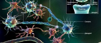
Functioning of axons
Through neural processes, different parts of the cerebral cortex are connected and the body’s vital functions are coordinated. As a result of the creation of connections between neurons through electrical impulses, leading to the formation of centripetal and centrifugal signals, human activity is manifested in great diversity. The furrows and convolutions form four lobes in each hemisphere:
These lobes of the brain are more developed than others and have greater mass. The work of the white matter of the frontal lobes contributes to the formation of voluntary movements, regulates complex forms of behavior, mechanisms for reproducing speech and writing, and thinking processes. The white matter pathways of the brain contribute to absolutely all motor processes. In modern neuropsychology, the nerve centers in the frontal lobes are a software unit that controls and regulates complex forms of life activity.
The following centers are located here: 1) understanding of oral speech, 2) perception of sound signals, 3) vestibular analyzer, 4) center of vision, 5) center of smell and taste, 6) center of music. The functioning of the temporal lobes is asymmetrical. If a person is left-handed, then the right hemisphere will have greater functionality; if you are right-handed, then the left hemisphere will be more active (dominant). The functioning of the white matter of this hemisphere makes it possible to understand speech and learn based on the information heard. By combining olfactory, auditory and visual information, draw conclusions, creating images of a harmonious emotional background and long-term memory. The functions of the non-dominant hemisphere include: recognition of music and rhythm, voice intonations, recognition of faces and their expressions, learning using visual images.
The centers located here give a person general sensitivity: pain, tactile and temperature. There are also centers that carry out complex coordinated movements, brought to the point of automatism, and actions of a purposeful nature, acquired through training and continuous practice throughout life. These are eating, walking, dressing, writing habits, certain work activities and other actions that are unique to humans. The left dominant side provides the ability to write and read; is responsible for actions leading to the desired result; is responsible for feeling the position of your body as a whole and its individual parts; for determining the right and left sides. In the right non-dominant lobe, the process of transforming all information coming from the occipital lobes takes place, a three-dimensional picture of the surrounding world is created, orientation in space is ensured, and distances between landmarks are determined.
Cerebral hemispheres
1. Remember from your zoology course what parts of the brain all vertebrates have. The cerebral hemispheres are derivatives of which division? In which group of animals do they first appear?
All vertebrates are characterized by the presence of 5 parts of the brain: medulla oblongata, hindbrain, midbrain, diencephalon, forebrain.
The cerebral hemispheres are derivatives of the forebrain and first appear in amphibians, but in them they are poorly developed, and the cerebral cortex is practically absent.
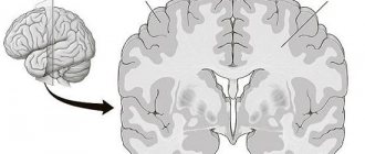
2. Describe the structure of the human cerebral hemispheres. Draw a schematic cross-section of the cerebral hemispheres, indicating the gray matter of the cortex, gray matter of the nuclei, white matter, and ventricles.
In humans, the forebrain is represented by two hemispheres and the corpus callosum, which connects the hemispheres. The cerebral hemispheres: right and left - cover the midbrain and diencephalon and make up up to 80% of the mass of the adult brain. On the surface of each hemisphere there are many grooves and convolutions. There are 4 main sulci (central, lateral and parieto-occipital), which divide each hemisphere into lobes. The superficial layer consists of gray matter (cortex), underneath is white matter, consisting of axons of nerve cells whose bodies lie in the cortex or which carry information to cortical cells. In the thickness of the white matter there are large accumulations of gray matter (subcortical nuclei) and cavities (lateral ventricles). CSF circulates through the ventricles of the brain and the central canal of the spinal cord, which provides nutrition to the subcortical structures.
3. What is the cerebral cortex? Where is it?
The superficial layer of gray matter of the cerebral hemispheres is called the cortex. The cortex consists of several layers of neuron bodies, different in structure and function. It is believed that it consists of about 12-18 billion cells, the thickness is 1.5 - 4.5 mm, and the area is 1.7 - 2.5 thousand cm2.
4. Explain the significance of the grooves and convolutions on the surface of the cerebral hemispheres.
The furrows and convolutions significantly increase the surface of the cerebral cortex, according to some sources up to 10-12 times.
5. What function does the white matter of the cerebral hemispheres perform?
White matter forms pathways that connect areas of the cortex and the cortex with the rest of the nervous system.
6. What lobes are distinguished in the cerebral hemispheres?
The hemispheres are divided into frontal, 2 parietal, 2 temporal and occipital lobes
7. Distinguish between the concepts of “lobes of the cerebral hemispheres” and “zones of the cerebral hemispheres”. Give examples of when they coincide or do not coincide.
The lobes of the cerebral hemispheres are a division of the surface of the cortex according to an anatomical principle: in each hemisphere there are frontal, occipital, parietal, and temporal lobes.
Cortical zones are a section of the cerebral cortex, characterized by uniformity of structure and functions.
In our brain, the lobes and zones do not coincide; one lobe consists of several zones. The occipital lobe consists of the areas of vision and visual recognition. The areas of smell, hearing and taste are located in and adjacent to the temporal lobe.
8. When examining a blind patient, it was discovered that his eyes and optic nerves were not damaged. Why doesn't he still see?
The inability to see can be caused not only by damage to the eyes or optic nerves, but also by damage to the visual areas of the occipital lobes of the cerebral hemispheres.
9. Using additional sources of information, find out whether the functions of the left and right hemispheres of the cerebrum differ.
There is a “functional asymmetry” between the right and left hemispheres of the brain, that is, their functions are different. This was proven during experiments on cutting the communication routes between the hemispheres (subsequently such operations began to be carried out for medical reasons for certain diseases, such as Parkinson's disease). In right-handed people, the leading hemisphere is the left, in left-handed people, the leading hemisphere is the right. The right hemisphere is responsible for imaginative thinking, forms the basis for creativity and making non-standard decisions; The visual area of the right hemisphere is responsible for recognizing faces. The left hemisphere provides logical reasoning and abstract thinking, it contains the centers of oral and written speech, and the formation of decisions; The visual area is responsible for recognizing letters and numbers. Therefore, as a joke, right-handers are called mathematicians, and left-handers are called artists.
10. There are everyday concepts of “male logic” and “female logic”. Is there any basis for such differences?
In the external and internal structure, there is no difference between the brains of men and women, but despite the fact that the structure of our brains is almost the same, we are individual. The main differences in the perception of the world according to the “male” or “female” type are laid down in early childhood, depending on our upbringing and under the influence of gonadal hormones during growing up, but all of them are only psychological in nature.
Brain damage
As a result of a skull injury, damage to the brain and therefore white matter can occur. Another cause is certain diseases that damage the forebrain. The development of pathology, depending on the location, causes paralysis of the muscular system on one side of the body. Such symptoms are typical when a part of the brain is damaged due to a stroke. Paralysis can be mixed, for example, the left half of the face and the right half of the body. White matter damage can impair visual field, swallowing, speech impairment, and many other symptoms. With Alzheimer's disease, the brain areas responsible for memory and recognition are affected, and mental disorders appear. Damage to certain areas of the brain can occur during intrauterine development of the fetus due to an infectious disease of the mother. In severe labor, the child is at risk from birth trauma, and in the first months of life, the threat is infectious diseases that lead to brain damage.
White matter of the brain in the first month after birth
The microstructure of white matter required for the efficient and coordinated transmission of neural impulses undergoes particularly pronounced development in the first years of life, while deviations from this trajectory of brain development are likely to lead to changes in brain connections relevant to the behavior of the child and subsequently adult. Assessing white matter microstructure during normal brain development is critical to neurological approaches to both typical and pathological early development.
Research results reveal changes in the microstructure of white matter in the earliest periods of development and demonstrate different periods of regional development and regional asymmetries.
The neural architecture that forms bundles of myelinated nerve fibers, known as white matter microstructure, is fundamental to brain connectivity and contributes to higher-level cognitive functioning. The development of white matter structure (circuitry) occurs due to a cascade of complex processes such as axon formation, dendritic sprouting and myelination, which begins in late stages of pregnancy and continues to develop through childhood, adolescence and adulthood with the most pronounced maturation during the first two years life. During this developmental period, neural tissue substrates that govern individual differences in vulnerability or resistance to adversity are likely to begin to emerge early in brain development. Despite the importance of white matter microstructure for healthy brain function and connectivity, a significant gap remains in our knowledge regarding the normative characteristics of infant white matter microstructure, such as sexual dimorphism and asymmetry, especially in the weeks immediately following birth.
Magnetic resonance imaging (MRI) provides detailed images of the brain and can non-invasively monitor changes associated with white matter development. Diffusion MRI (dMRI) is very sensitive to changes in tissue microstructure and is widely used to study the fiber architecture of white matter. Diffusion tensor imaging (DTI) provides measurable measures of diffusion properties, including fractional anisotropy (FA), mean diffusivity (MD), radial diffusivity (RD), and axial diffusivity (AD), each of which provides quantitative measures of diffusion in brain tissue. Given the degree of anisotropic diffusion in white matter—due to tissue barriers such as axonal fibers and the myelin sheath—FA, MD, RD, and AD provide indirect markers of white matter microstructure.
More recent dMRI techniques, such as neurite orientation dispersion and density imaging (NODDI), use biophysical modeling to increase the level of detection of microstructural specificity available to neuroimaging. NODDI measures quantitative parameters of three specific diffusion processes: intraneurite diffusion (ν IC, diffusion within axons and dendrites), extraneurite diffusion, and isotropic (free) water diffusion (ν ISO). A similar model also quantifies the degree of neurite angular change through the orientation dispersion index (ODI). NODDI provides important information about brain development in infants and young children, with changes in parameters consistent with neurodevelopmental mechanisms of myelination and axonal fiber development during the first few years of life. For example, a nonlinear increase in the volume fraction of NODDI intraneurites (ν IC) occurs around the first 3 years of childhood, and ν IC continues to increase nonlinearly until 7.5 years. In clearly preterm infants, NODDI parameters indicate increased axonal dispersion and lower axonal density compared with timely born infants, and these parameters are also associated with poorer functional outcomes in very preterm infants. These studies provide valuable insight into brain development profiles and also demonstrate the utility of dMRI methodology.
Even within a narrow age range, measures of white matter microstructure vary by gestational age and exhibit broad left-sided differences. Sex differences were minimal, and no significant associations emerged between white matter parameters and neonatal growth markers. Finally, a comparison of DTI and NODDI values shows that although these methods measure similar aspects of white matter microstructure, they provide differential microstructural information early in child development. DTI diffusion indices are strongly associated with age adjusted for length of gestation, reflecting a decrease in total brain water content and an increase in membrane density. RD decreases and FA increases in central white matter regions, which may indicate early myelination or increased organization of white matter fibers and bundles. The advantage of NODDI is that it uses biophysical modeling to determine specific microstructural characteristics. ν IC is interpreted to provide an index of neurite density (i.e., axons and dendrites), whereas ODI is an index of the degree of fiber coherence.
Central white matter regions, such as the corpus callosum, internal capsules, and radials, develop in front of the more peripheral white matter, and the posterior regions develop in front of the more anterior ones, with most of the white matter microstructure being established after one month. Qualitatively, the FA and ODI maps at one month appear adult-like, suggesting that fiber architecture and organization are developing in the uterus. On the other hand, developmental processes such as myelination mainly occur postnatally and are likely to influence subsequent changes in RD and ν IC parameters
Preventative measures for brain health
The speed of nerve impulses directly depends on the integrity of the white matter. Its healthy state determines its normal functioning. It has been scientifically proven that with increasing age, the quality of white matter and its functionality decline. Therefore, you need to comply with some simple conditions:
- Exercise regularly at any age - from simple morning exercises to serious sports.
- Monitor your health and consult a doctor on time.
- If diseases that can cause brain damage occur, treat under the supervision of a doctor.
- Remove bad habits from your life that can worsen your health.
- Increase immunity using hardening procedures.
- Keep your emotional state under control.
- Give food for brain activity: read, write, solve crosswords and other puzzles.
- During pregnancy, be under constant supervision of a specialist.
An active physical life and intellectual pursuits in both work and leisure will prolong normal performance and clarity of mind, and maintain strong memory. Teach children to take their health seriously as early as possible. Play sports and games that develop intelligence. It’s good to work together, proving its usefulness by example.
Only humans have higher nervous activity, and this is their direct difference from other species of mammals. The conditioned reflex actions that he masters in the process of life put him at the highest level of development.