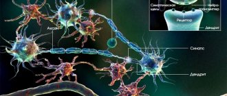
All structures of the nervous system are composed of neurons, which form the gray and white matter of brain tissue.
The distribution of these structures depends on the functionality of the region to which they belong: for example, the gray matter of the brain covers the white substance, while in the spinal region the nuclei, consisting of gray neurons, are located inside the medullary canal formed by the white component.
Functions of gray matter
Extraembryonic organs, which develop during embryogenesis outside the body of the embryo, perform diverse functions that ensure the growth and development of the embryo itself. Some of these organs surrounding the embryo are also called embryonic membranes. These organs include the amnion, yolk sac, allantois, chorion, placenta.
The amnion is a temporary organ that provides an aqueous environment; it appears at the second stage of gastrulation. The wall of the vesicle forms the extraembryonic ectoderm and is connected to the extraembryonic mesoderm.
The main function of the amniotic membrane is the production of amniotic fluid. The amnion also performs a protective function, preventing harmful agents from entering the fetus.
The amnion epithelium is formed by large polygonal cells closely adjacent to each other. At the 3rd month, the epithelium transforms into prismatic. The epithelium of the amnion in the area of the placental disc performs a secretory function and resorption of amniotic fluid.
In the connective tissue stroma of the amniotic membrane, there is a basement membrane, a layer of dense fibrous connective tissue and a spongy layer of loose fibrous connective tissue.
The yolk sac stores nutrients necessary for the development of the embryo. It is formed by the extraembryonic endoderm mesoderm. Appearing in the 2nd week. Blood islands develop in the wall of the yolk sac, forming the first blood cells and the first blood vessels.
Above the yolk sac, the intestinal tube is formed. The connection between the embryo and the yolk sac remains in the form of a hollow cord called the yolk stalk.
The umbilical cord connects the embryo (fetus) to the placenta. It is covered with the amniotic membrane and rudiments of the yolk sac and allantois.
Mucous connective tissue ensures the elasticity of the cord, protects the umbilical vessels from compression, providing the embryo with nutrients and oxygen.
At the beginning of the 3rd week, blood capillaries grow into the chorionic villi and tertiary villi form.
The work of the central nervous system provides a large number of connections in the body that perform two main functions: control of muscle activity (motor reflex) and provision of sensory perception (sensory reflexes) and higher mental functions: memory, speech, emotions.
The functions of the substantia grisea are determined by its location, for example:
- In the cerebral cortex, the substance is responsible for connecting the body with the outside world, and also carries information and regulates the activity of internal organs, is responsible for ensuring higher nervous activity, thanks to which a person is able to think, remember, perceive, etc.
- In the medulla oblongata, the nuclei of the substance regulate motor processes, balance, ensure coordination of movements, and also regulate metabolism, respiratory processes and blood supply.
- In the cerebellar cortex, the gray nuclei are responsible for coordination of movements and orientation in space.
- In the diencephalon, the nuclei are responsible for controlling the activity of internal organs, regulating reflexes and body temperature.
- In the telencephalon, the nuclei provide motor, reflex control and regulation of higher mental functions: coherent speech, vision, smell, taste, hearing, touch.
The spinal cord is a complex structure that has the following functions: reflex, motor, sensory and conductive. The first three functions are assigned to the gray matter, and the third - to the white matter.
- Reflex function - regulation of unconditioned reflexes: sucking reflex, knee reflex, instant reaction to painful stimuli, etc.
- Motor function – control of muscle reflexes associated with the motor system. The corresponding cells of the spinal cord send signals to a specific group of muscles, prompting one or another action, thanks to which we can purposefully turn our head, move our neck, raise and lower our arms, and walk.
- Sensory function is the transmission of an impulse coming from the afferent fibers of the torso to the parts of the brain, from where the command comes, containing a reaction to the stimulus.
- The conductor function is to ensure the passage of an impulse to the brain, and from there, the passage of an action command going to the corresponding organ. Regulated by the white substance.
The gray substance ensures the normal functioning of a person, his interaction with the outside world, types of human activity, is the basis of cognitive and sensory perception, as well as the basis of motor, reflex, regulatory and all mental functions.
Where is the white matter located?
The white matter of the brain begins to form by 6 months of intrauterine development of a person, while its formation does not stop throughout the subsequent years of life. This feature allows the body to train and accumulate experience.
White matter itself is the opposite of gray matter and is a dense network of neuron branches that transmit information from the cerebral cortex to the underlying nerve centers of the spinal cord and brain. At the same time, the functioning of the connection is influenced by the quantity and quality of the formed nerve pathways: the denser and stronger the connection between the structures, the more developed and talented the individual turns out to be.
The largest accumulation of white matter is located in the cranium and is represented by large lobes. This is understandable: all the control centers of the body are located in the brain, and also in its structures the formation and fulfillment of higher mental tasks occurs, the presence of which distinguishes humans from the rest of the animal world. Moreover, in addition to the main one, the white matter also performs a protective function: in appearance and physical characteristics, it is a gelatinous, fat-like mass that plays the role of a shock absorber for the underlying structures.
Also, the white matter forms the peripheral meninges for the gray matter of the spinal cord - like the head of the central nervous system, it contains all types of fibers (commissural, associative and projection), with a characteristic myelin color, which are collected in special bundles that provide communication between the spinal cord and other parts peripheral and central NS.
Chemical composition of the cell. Chemical organization of the cell
Organogens are chemical elements that are part of all organic compounds and make up about 98% of the cell mass.
| Oxygen | 65—75 | It is part of most organic cell substances. Formed during photosynthesis during photolysis of water. For aerobic organisms, it serves as an oxidizing agent during cellular respiration, providing cells with energy. It is found in the largest quantities in living cells in water. |
| Carbon | 15—18 | Included in all organic substances; a skeleton of carbon atoms forms their basis. In addition, in the form of CO2 it is fixed during photosynthesis and released during respiration, in the form of CO (in low concentrations) it participates in the regulation of cellular functions, and in the form of CaCO3 it is part of mineral skeletons. |
| Hydrogen | 8—10 | It is part of all organic substances of the cell. It is found in the largest quantities in water. Some bacteria oxidize molecular hydrogen to produce energy. |
| Nitrogen | 2—3 | It is part of amino acids, proteins (including enzymes and hemoglobin), nucleic acids, chlorophyll, and some vitamins. |
Important Tetanus vaccination: side effects, reactions and complications
Elements present in a cell in smaller quantities - tenths and hundredths of a percent.
| Calcium | 0,04—2,00 | Contained in the cell membrane, intercellular substance and bones. Participates in the regulation of intracellular processes, maintaining membrane potential, transmission of nerve impulses, and is necessary for muscle contraction and exocytosis. Insoluble calcium salts are involved in the formation of bones and teeth of vertebrates and mineral skeletons of invertebrates. |
| Phosphorus | 0,2—1,0 | It is part of ATP in the form of a phosphoric acid residue (PO43-). Contained in bone tissue and tooth enamel (in the form of mineral salts), and is also present in the cytoplasm and intercellular fluids (in the form of phosphate ions). |
| Potassium | 0,15—0,4 | Participates in maintaining membrane potential, generating nerve impulses, and regulating cardiac muscle contraction. Contained in intercellular substances. Participates in photosynthesis. |
| Sulfur | 0,15—0,2 | Contained in some amino acids, enzymes, thiamine. It is present in small quantities as sulfate ion in the cytoplasm of cells and intercellular fluids. |
| Chlorine | 0,05—0,1 | Participates in the formation of the osmotic potential of blood plasma and other liquids in the form of an anion. Contained in gastric juice. |
| Sodium | 0,02—0,03 | Participates in the maintenance of membrane potential, the generation of nerve impulses, osmoregulation processes (including the functioning of the human kidneys) and the creation of a blood buffer system. |
| Magnesium | 0,02—0,03 | Cofactor for many enzymes involved in energy metabolism and DNA synthesis; maintains the integrity of ribosomes and mitochondria, is part of chlorophyll. In animal cells it is necessary for the functioning of muscle and bone systems. |
Microelements, constituting from 0.001% to 0.000001% of the body weight of living beings, include vanadium, germanium, iodine (part of thyroxine, the thyroid hormone), cobalt (vitamin B12), manganese, nickel, ruthenium, selenium, fluorine ( tooth enamel), copper, chromium, zinc, molybdenum (participates in the binding of atmospheric nitrogen), boron (affects growth processes in plants).
Ultramicroelements make up less than 0.000001% in the organisms of living beings, these include gold, silver, which have a bactericidal effect, mercury, which suppresses the reabsorption of water in the renal tubules, affecting enzymes. Ultramicroelements also include platinum and cesium, beryllium, selenium, radium and uranium. The functions of ultramicroelements are still poorly understood.
How is gray and white matter distributed in the cerebral hemispheres?
To visually study the structure of the central nervous system, there are several methods that allow you to see the brain in cross-section. The most informative is the sagittal section, with the help of which the brain tissue is divided into 2 equal parts along the central line. At the same time, to study the location of gray and white matter in the thickness, a frontal section of the anterior section, and accordingly the cerebral hemispheres, is ideal, allowing one to isolate the hypothalamus, corpus callosum and fornix.
The white matter of the anterior section is located in the thickness of the large lobes, which are the springboard for the gray matter that makes up the cortex. It covers the entire surface of the hemispheres with a kind of cloak and belongs to the structures of higher nervous activity in humans.
Moreover, the thickness of the gray matter of the cortex is not the same throughout and varies from 1.5 to 4.5 mm, reaching its greatest development in the central gyrus. Despite this, it occupies about 44% of the volume of the forebrain, as it is located in the form of convolutions and grooves, which make it possible to increase the total area of this structure.
At the base of the white matter of the cerebral hemispheres, there are also separate accumulations of gray matter, which make up the basal ganglia. These formations are subcortical structures or central nodes of the base of the terminal section. Experts distinguish 4 types of such functional centers, which differ in form and purpose:
- caudate nucleus;
- lenticular nucleus;
- fence;
- amygdala.
All these structures are separated from each other by layers of white matter, which transmits information from them to the underlying parts of the brain through the black substance located in the middle section, and also connects the nuclei with the cortex and ensures their coordinated work.
Metals in the periodic table
In the Mendeleev system, alloys have a predominant number and the list of them is very large - they start with Boron (B) and end with polonium (Po) (the exceptions are germanium (Ge) and antimony (Sb)). This group has characteristic features; they are divided into groups, but their properties are heterogeneous. Their characteristic features:
- plastic;
- electrical conductivity;
- shine;
- easy release of electrons;
- ductility;
- thermal conductivity;
- hardness (except mercury).
Due to the different chemical and physical essence, the properties may differ significantly between two representatives of this group; not all of them are similar to typical natural alloys, for example, mercury is a liquid substance, but it belongs to this group.
In its normal state, it is liquid and without a crystal lattice, which plays a key role in alloys. Only chemical characteristics make mercury similar to this group of elements, despite the conventionality of the properties of these organic compounds. The same applies to cesium, the softest alloy, but it cannot exist in nature in its pure form.
Some elements of this type can exist only for a fraction of a second, and some do not occur in nature at all - they were created in artificial laboratory conditions. Each of the groups of metals in the system has its own name and characteristics that distinguish them from other groups.
However, their differences are quite significant. In the periodic table, all metals are arranged according to the number of electrons in the nucleus, i.e. by increasing atomic mass. Moreover, they are characterized by periodic changes in their characteristic properties. Because of this, they are not placed neatly in the table and may not be placed correctly.
In the first group of alkalis there are no substances that would be found in pure form in nature - they can only exist as part of various compounds.
How to distinguish a metal from a non-metal?
How to determine the metal in a compound? There is a simple way to determine it, but for this you need to have a ruler and a periodic table. To determine you need:
- Draw a conditional line along the junctions of the elements from Bor to Polonium (possibly to Astat).
- All materials that will be on the left of the line and in the side subgroups are metal.
- The substances on the right are of a different type.
However, the method has a flaw - it does not include Germanium and Antimony in the group and only works in a long table. The method can be used as a cheat sheet, but in order to accurately determine the substance, you should remember the list of all non-metals. How many are there in total? Few - only 22 substances.
In any case, to determine the nature of a substance it is necessary to consider it separately. Elements will be easy if you know their properties
It is important to remember that all metals:
- At room temperature they are solid, with the exception of mercury. At the same time, they shine and conduct electricity well.
- They have fewer atoms at the outer level of the nucleus.
- They consist of a crystal lattice (except mercury), and all other elements have a molecular or ionic structure.
- In the periodic table, all nonmetals are red, metals are black and green.
- If you move from left to right in a period, the charge of the nucleus of the substance will increase.
- Some substances have weakly expressed properties, but they still have characteristic features. Such elements are classified as semimetals, such as Polonium or Antimony, and are usually located at the boundary of the two groups.
It is important to remember that when moving in the table from top to bottom, the non-metallic properties of substances become stronger, since elements that have distant outer shells are located there. Their nucleus is separated from the electrons and therefore they attract weaker
What does white matter consist of?
White matter is a special component of the central nervous system, represented by bundles of nerve fibers covered with a special myelin sheath, thanks to which the main purpose of this brain structure is fulfilled, which is to transmit information from the main functional centers of the nervous system to the underlying parts of the nervous system.
The myelin sheath allows electrical impulses to be transmitted over long distances at high speed without loss. It is a derivative of glial cells and, due to its special structure (the sheath is formed from a flat outgrowth of the glial body devoid of cytoplasm), wraps the nerve fiber along the periphery several times, interrupting only in the area of interceptions.
This characteristic feature allows you to increase the strength of the impulse sent by the gray matter several times. In addition, it performs an isolating function, allowing the signal strength to be maintained throughout the entire axon.
Regarding the chemical composition of white matter, myelin is mainly formed by lipids (organic compounds including fats and fat-like substances) and proteins, so white matter, at first glance, is a fat-like mass with corresponding characteristics.
The distribution of white matter in different parts of the central nervous system is heterogeneous in chemical composition: the spinal cord is “fatter” than the brain section of the nervous system. This is due to the fact that from the gray matter of this section, a greater amount of efferent information comes out to the peripheral nervous system.
Where is gray matter located?
The gray matter of the brain is represented mainly by an accumulation of a large number of neurons with unmyelinated axons interwoven into glial tissues, their dendrites and blood capillaries that ensure their metabolism.
The largest accumulation of gray neurons forms the cerebral cortex, which covers the surface of the terminal section. The thickness of this structure is no more than 0.5 cm throughout, but it occupies more than 40% of the volume of the telencephalon, and its surface is many times greater than the plane of the cerebral hemispheres. This characteristic is determined by the presence of wrinkles and convolutions, which contain up to 2/3 of the area of the entire cortex.
Also, accumulations of gray matter in the brain form special nerve centers or nuclei, which have a characteristic shape and their functional purpose. The peculiarity of the structure of this structure is that the term “nucleus” means a paired or dispersed formation of neuron cells that do not have a myelin sheath.
Important Treatment of insomnia with folk remedies in the elderly
There are a large number of nuclei of the nervous system, which, for the sake of a general concept and ease of perception, are usually identified according to the operation they perform, as well as their appearance. This distribution does not always correctly reflect reality, since the brain is a poorly understood structure of the central nervous system and sometimes scientists make mistakes.
The main cluster of nuclei is located within the brainstem, for example in the thalamus or hypothalamus. At the same time, the basal ganglia are located in the anterior section, which to some extent influence a person’s emotional behavior and are involved in maintaining muscle tone.
The gray matter of the cerebellum, like the terminal cortex, covers the hemispheres and the vermis at the periphery. Also, its individual ones form paired nuclei deep in the body of this rudiment.
Anatomically, the following types of nuclei are distinguished:
- Serrated. Located in the lower part of the white matter of the cerebellum, its pathways are responsible for the motor function of skeletal muscles, as well as for a person’s visual-spatial orientation in space.
- Spherical and cork-shaped. They process information received from the worm, and also receive afferent signals from parts of the brain responsible for somatosensory, auditory and visual data.
- Tent core. It is located in the tent of the cerebellar vermis and receives information about the position of the human body in space according to data received from the sense organs and the vestibular apparatus.
A characteristic feature of the structure of the spinal cord is that the gray substance in the form of nuclei is located inside the white component, but at the same time is an integral part of it. This arrangement can be seen in most detail when studying the spinal part of the central nervous system in a cross section, where a clear transition of gray matter into white matter from the center to the periphery will be clearly visible.
How the nervous system works, what is white matter, gray matter
The human nervous system has a complex structure. Conventionally, experts distinguish between the peripheral and central nervous systems of humans.
The human central nervous system includes all parts of the brain (terminal, middle, medulla, intermediate, cerebellum), as well as the spinal cord. These components control the functioning of all body systems, connect them with each other and ensure their coordinated operation in response to external influences.
Functional features of the central nervous system:
- The human brain is located in the cranium and plays a controlling role: it participates in the processing of information received from the environment and regulates the vital functions of all systems of the human body, and is a kind of steering wheel.
- The main function of the spinal region of the central nervous system is to transmit information from nerve centers located in other parts of the body to the brain. Also, with its support, motor reactions to external stimuli are performed (using reflexes).
The peripheral nervous system includes all branches of the spinal cord and brain that are located outside the central nervous system or, in other words, on the periphery. It includes cranial and spinal nerves, as well as autonomic nerve fibers that connect the structures of the central nervous system with other parts of the human body. With its help, unconscious (at the level of reflexes) control of the vital functions of certain organs occurs, be it heartbeat or automatic muscle contraction in response to external stimuli (for example, blinking).
This part of the nervous system is especially vulnerable to various toxins or mechanical damage, since it does not have protection in the form of bone tissue or a special barrier separating the blood and its components.
Peripheral NS includes:
- Vegetative or autonomous NS. It is controlled by the human subconscious and controls the performance of vital functions of the body. The main task of this part of the NS is to regulate the internal environment of the body, through the circulatory, endocrine system, as well as various endocrine and exocrine glands. Anatomically, it is divided into sympathetic, parasympathetic and metasympathetic NS. In this case, centers or vegetative nuclei, consisting of a gray cerebral component, are located in the spinal and head sections of the central nervous system, and the latter is represented by clusters of neurons located in the walls of the bladder, gastric tract and other organs.
- Somatic NS. Responsible for human motor function - with its help, afferent (incoming) signals are transmitted to the neurons of the central nervous system, from where, after processing, information is received through efferent (descending motor) fibers to the limbs and organs of the human body to reproduce the corresponding movement. Its neurons have a special structure that allows them to transmit data over long distances. Thus, most often the body of a neuron is located in close proximity to parts of the central nervous system or enters it, but at the same time its axon extends further, eventually reaching the surface of the skin or muscles. Through this part of the NS, various protective reflexes are carried out, which are carried out at the subconscious level. This feature is achieved by the presence of reflex arcs, which allow the action to be performed without the participation of the main center, since in this case the nerve fibers connect the dorsal part of the central nervous system with the area of the body directly. In this case, the final point of information perception is the cerebral cortex, where memories of all actions performed remain. Thus, the somatic nervous system is involved in learning, protection and the ability to process information received from the environment.
- Some experts classify the human sensory nervous system as the peripheral nervous system. It includes several groups of neurons located on the periphery of the central nervous system, which are responsible for the perception of information from the environment through the organs of hearing, vision, touch, taste and smell. Responsible for the physical perception of concepts such as temperature, pressure, sound.
As mentioned earlier, the structures of the human nervous system are represented by a white and gray substance, each of which has its own structure and contains different types of nerve cells that differ in appearance and functionality.
Thus, the white matter mainly performs a conductive function and transmits nerve impulses from one part of the brain matter to another. This feature is due to the structure of the neurons of this structure, the bulk of which consists of long processes or axons covered with myelin, which has a high conductivity of electrical impulses (about 100 m/s).
Neuron axons can be divided into 2 main groups:
- Long (intracortical), connect distant areas, located in the depths of the medulla.
- Short processes that connect the gray cells of the cortex and nearby structures of the white matter have a second name - subcortical.
Also, depending on the location and functionality of the nerve cell fibers of the white matter, it is customary to distinguish the following groups:
- Associative. They differ in size: they can be either long or short and perform various tasks, but at the same time they are concentrated in one of the hemispheres. Long axons are responsible for connecting distant convolutions, while short axons connect nearby structures.
- Commissural. They connect the 2 hemispheres and ensure their coordinated work, located in opposite parts. Such axons can be considered during the anatomical study of this organ, since they consist of the anterior commissure, the corpus callosum, and the fornix commissure. Projection axons connect the cortex with other centers of the central nervous system, including the spinal cord. There are several types of such fibers: some connect the thalamus with the cortex, the second - the cortex with the nuclei of the bridge, and the third conduct impulses, thanks to which the command and control of certain limbs is carried out.
There are 2 types of similar fibers, which differ in the direction of the transmitted information:
- Afferent. Through them, information comes from the underlying structures of the brain, organ systems and tissues to the cortex and subcortical structures that process the incoming information.
- Efferent. They carry out a response impulse from the centers of higher mental activity to controlled structures.
The opposite of the white medulla is the gray component, which, like its predecessor, consists of a cluster of neurons - with their help, all functions of higher nervous activity of a person are performed.
Its main part is located on the surface of the white medullary component located in the head and makes up the cortex, which has a conventionally gray color. It also lies deep in the brain and along the entire length of the spinal cord in the form of nuclei. The gray matter includes several groups of nerve cells, their dendrites and axons, as well as glial tissues that perform an auxiliary function.
Branched processes of neurons or dendrites, through synapses, receive and transmit information from the axons of neighboring cells to their own. The quality of the impulse depends on the density of their branching - the more developed the branches of the main fiber and the more extensive the network of synapses, the more data will flow to the cell nucleus from neighboring ones.
Since neurons and, accordingly, the nuclei of gray matter cells are located close to each other, they do not require long axons, while the main flow of information is transmitted through the dendritic synaptic connection of nearby cells. For the same reason, their axons do not require a myelin sheath.
Individual accumulations of gray matter are called nuclei, each of which controls the performance of a specific vital function of the body, and they can be divided into 2 large groups: those related to the central nervous system and those responsible for the peripheral nervous system.
The anatomical structure of gray matter neurons in all parts of the central nervous system has a similar structure and approximately the same composition. Therefore, the pattern of arrangement of neurons in the terminal section is no different from the totality of these elements in other structures.
Notes
- ↑
- Khudayberdiev, Kh. Kh. Neurosurgical anatomy of the substantia nigra of the brain: abstract. diss. ...cand. medical sciences / Kh. Khudaiberdiev. - Leningrad, 1970. - 15 pages
- . Retrieved August 15, 2013.
- (English). www.sciencedirect.com
. Retrieved June 12, 2021. (inaccessible link) - . Retrieved March 17, 2013.
- . Retrieved March 19, 2013. (inaccessible link)
- Template:Better source
- Markov A.
Human evolution. Book 2. Monkeys, neurons and the soul. - Corpus, 2011. - T. 2. - 512 p. - (Dynasty). — 5000 copies. — ISBN 978-5-271-36294-1, 978-5-17-078089-1, 978-5-17-078089-1. - (unavailable link). Retrieved March 19, 2013.
- ↑ Yakhno N.N., Shtulman D.R. Diseases of the nervous system. - M.: Medicine, 2001. - T. 2. - P. 76-95. — 744 p. — ISBN 5-225-04540-5
- Yakhno N. N., Shtulman D. R. Diseases of the nervous system. - M.: Medicine, 2001. - T. 2. - P. 76-95. — 744 p.
- Reference Guide to Psychopharmacological and Antiepileptic Drugs Approved for Use in Russia / Ed. S. N. Mosolova. — 2nd, revised. - M.: BINOM Publishing House, 2004. - P. 17. - 304 p. — 7000 copies. — ISBN 5-9518-0093-5.
- ↑ (inaccessible link). Retrieved March 18, 2013.
- .
- .
- ↑ (inaccessible link). Retrieved March 18, 2013.
- (unavailable link). Retrieved March 18, 2013.
- (unavailable link). Retrieved March 18, 2013.
- (unavailable link). Retrieved March 18, 2013.
- (unavailable link). Retrieved March 18, 2013.
- (unavailable link). Retrieved March 18, 2013.
- (unavailable link). Retrieved March 18, 2013.
- (unavailable link). Retrieved March 18, 2013.
- (unavailable link). Retrieved March 18, 2013.
- ↑ (inaccessible link). Retrieved March 18, 2013.
- (unavailable link). Retrieved March 18, 2013.
- (unavailable link). Retrieved March 18, 2013.
- (unavailable link). Retrieved March 18, 2013.
- (unavailable link). Retrieved March 18, 2013.
- (unavailable link). Retrieved March 18, 2013.
- (unavailable link). Retrieved March 27, 2013.
- (unavailable link). Retrieved March 27, 2013.
- A.P. Ashmarin.
Neurochemistry: a textbook for biological and medical universities / Ed. acad. RAMS A.P. Ashmarin and Prof. P.V. Stukalov. - M.: Publishing house of the Institute of Biomedical Chemistry of the Russian Academy of Medical Sciences, 1996. - 470 p. — ISBN 5-900760-02-2. - ↑
- . DrugBank
. University of Alberta (February 8, 2013). Retrieved October 13, 2013.
How gray matter influences some human abilities
The gray tissue of the brain, regulating the processing of signals from the outside and generating effector impulses, is not only responsible for the functioning of the entire human nervous system, but also affects his abilities: mental, cognitive, physical, etc.
Various experiments by scientists have shown that a person’s abilities depend on the volume of the gray substance, while changes in the amount of white substance did not show any noticeable changes.
Experiments by British scientists have shown that the thinner the cerebral cortex, therefore, the smaller the volume of gray substance, the worse a person copes with solving logical problems, the less various abilities he has, and also with a low volume of substance, subjects often had problems with reaction speed , speech dysfunctions, memory problems and poor intellectual abilities.
At the same time, studies have shown that learning foreign languages, memorizing poetry, scientific or artistic works, and playing music affect the enlargement of the cerebral cortex. The longer and more intense the learning process, the greater the volume of gray substance becomes, therefore, the more abilities, including mental ones, a person displays.
A decrease in the amount of gray matter is affected by:
- a person’s lifestyle is a sedentary, inert, inactive, from a physical and mental point of view, way of life;
- abuse of bad habits - alcohol, drug addiction and smoking reduce the volume of gray substance.
For example: those suffering from alcoholism experience a significant decrease in the amount of brain tissue, which is reflected in behavior and mental functions: incoherent speech, problems with memory and perception, inhibition of thought processes.
Phosphorus and its compounds
Orthophosphoric acid is used as a reagent in inorganic and organic synthesis, an intermediate for the production of mineral fertilizers, as a component of anti-corrosion coatings, in the food industry, etc.
Phosphorus-containing mineral fertilizers are of particular importance. Phosphorus is a necessary element for plant life and improving soil quality.
Phosphoric acid salts are included in nitrogen-phosphorus and nitrogen-phosphorus-potassium fertilizers. Of these, we note ammophos, which is a mixture of mono- and diaphosphates of ammonium NH4H2PO4 and (NH4)2HPO4, as well as nitrophoska, a mixture of NH4H2PO4, (NH4)2HPO4, CaHPO4, NH4NO3, KNO3, KCl.
Phosphorus is an organogen. Its compounds form the basis of the skeleton and teeth of animals and humans. Phosphorus is part of proteins and nucleic acids. Sugars and fatty acids can be used by cells as an energy source only if they are first phosphorylated.
Organophosphorus compounds are of great interest. Among them, effective medicines and chemical plant protection products were found. The most toxic and effective chemical warfare agents (sarin, soman, VX) are also organophosphorus compounds. The chemistry of organophosphorus compounds is a huge independent branch of organoelement chemistry.
Among the inorganic phosphorus compounds used in medical practice are aluminum phosphates AlPO4 and zinc phosphates Zn3(PO4)2, which are part of phosphate cements used in dentistry as a filling material.
External structure
The midbrain is located between the pons and the diencephalon. Of all the structures we have analyzed, the midbrain is the shortest - its size barely reaches two centimeters. In this illustration, the myelencephalon is tinted blue, the pons is yellow, and our mesencephalon, that is, the midbrain, is red.
The midbrain has two surfaces - ventral and dorsal. The ventral surface is directed towards the abdomen, and the dorsal surface is directed towards the back. If we look at the ventral surface of the midbrain, we see two massive projections that are located horizontally. These projections are called the cerebral peduncles (pedunculi cerebri). You've probably heard that brains can leak, according to the term "brain drain"? So, they may rather not flow away, but run away - after all, they have brain stems.
I would like to immediately warn you against a popular misconception. By association with the name, the cerebral peduncles are often mistakenly classified as part of the cerebral hemispheres. Of course, the cerebral peduncles are connected to the cerebral hemispheres by many pathways, however, they are a direct part of the midbrain, and not the cerebral hemispheres. Also, do not confuse the cerebral peduncles (1 pair) and the cerebellar peduncles (like the cockroach, 3 pairs) - these are completely different anatomical formations.
On our sagittal section we can see not only the cerebral peduncle, but also a fairly large trunk of the oculomotor nerve (nervus oculomotorius). To avoid confusion, I have outlined the oculomotor nerve in green and the cerebral peduncle itself in black.
The space located between the cerebral peduncles is called the interpeduncular fossa (fossa interpeduncularis mesencephali). The interpeduncular fossa is an area of white matter through which many vessels pass. If these vessels are removed, we will see a white substance with many holes. That is why the bottom of the interpeduncular fossa is called the posterior perforated substance (substantia perforata posterior).
We can only see these structures in a horizontal section of the bridge, so I'll have to jump ahead a little and use a horizontal section. The arrow points to the interpeduncular fossa, and the space with small circles is the perforated substance:
The dorsal surface of the midbrain looks like four small elevations, two of which are located above and are called the superior colliculi (colliculi superioris), and two below (colliculi inferioris). If we remove the cerebellum and the thin plate called the superior medullary velum (velum medullare superius), and then expand it to the frontal plane, we will see the lamina quadrigemina in all its glory:
Just above the superior colliculi hangs a small, round structure called the epiphysis. This is a very important endocrine gland, which belongs to the diencephalon (diencephalon). I talked a little about the pineal gland here, but undoubtedly, this gland’s finest hour on my blog is yet to come. In this illustration (unfortunately, I don’t know who the author is - it’s very coolly done) we see the epiphysis (which the arrow points to) and the quadrigeminal.
Posterior brain tissue
Directly above the medulla oblongata is the pons, and to the right is the cerebellum. The first section is presented in the form of a light-colored roller. It is associated with the cerebral peduncles and myelencephalon.
Transverse fibers divide the bridge into the following parts:
- Ventral (gastric). In this area, the substantia alba is represented predominantly by conductive fibers, and the substantia grisea has its nuclei here;
- Dorsal (dorsal). It consists of the following elements: Switching cores;
- Network formation;
- Sensory systems;
- Nerve pathways.
The cerebellum is located just below the occipital part of the brain. It consists of 2 hemispheres and a middle part. Gray matter is presented in the form of nuclei (dentate, cork-shaped, spherical, tent-shaped) and cortex. The white substance is under the dark shell. It is located in all convolutions and mainly consists of fibers that perform the following purposes:
- Connect the cerebral lobes and gyri;
- They follow the nuclei localized inside;
- Link departments.
Important Tenoten
What is white matter responsible for?
As mentioned earlier, the white matter of the brain performs several tasks: first of all, it is a link between the gray matter of the cortex and other functional clusters of neurons located in deep structures.
Other functions of the white matter of the brain are also known - it acts as a link between the cerebral hemispheres through the corpus callosum, and also ensures the interaction of remote areas of the cortex with other parts of the nervous system, including the spinal cord, using specific fibers.
Its main feature and distinctive feature is that the white matter is formed by a cluster of long nerve processes or fibers covered with a myelin sheath, which ensures rapid transmission of electrical impulses and relevant information to functional centers.
The white matter of the telencephalon forms the cerebral hemispheres, which are the most developed and massive structure of the central nervous system. This feature is due to the presence of a large number of projection fields in the cortex, which require a developed network of connecting fibers for their normal functioning. Otherwise, the connection and parallel execution of higher mental functions of the brain are disrupted: for example, speech becomes slow and inarticulate.
In the middle part of the brain, the white matter is located mainly over its entire surface, as well as ventral to the gray matter of the quadrigeminal colliculi. It also consists of the upper legs, which connect the midbrain with the cerebellum and transmit efferent information from this motor center to other parts of the central nervous system.
The white matter of the oblong section includes all types of fibers: both long and short. The long ones perform a transient function and connect the descending pyramidal tracts with the spinal nerve cords, and also carry out the coordinated work of the medulla oblongata with the thalamic structures, while the short ones form a connection between the nuclei of this department and send information to the overlying structures of the central nervous system.
Location in the cerebral cortex and basal ganglia
The large hemispheres of the telencephalon are separated by a fissure, and they are connected by the corpus callosum and commissures. Most of them are a cloak, the surface of which is called the neocortex - the new cortex, this is truly the phylogenetically newest structure, where the centers of all HMFs are localized, including those that allowed the formation of the second signaling system, which, according to Darwin's theory, makes humans the highest link in evolution. It is covered with well-known convolutions, which form a complex pattern of grooves and ridges. The thickness of the gray matter here is only 1.3 - 4.5 mm, or six neural layers.
Each hemisphere is divided into large sections - lobes. There are five lobes that form lobules - smaller sections, and convolutions. The pattern that the convolutions form varies in shape for each person, even in two halves of the same brain it looks different.
Useful to know: White matter of the brain: structure, functions
This substance is also located in the basal ganglia, also called the subcortical formations, or the old brain. It is believed that animal instincts are localized here, since the old brain allows you to automatically make decisions in a difficult situation.
The olfactory brain, the most ancient formation, is also located here. The olfactory organ is the main sense organ in fish and animals. When the distant ancestors of humans came out of the water onto land, those species survived that based their behavior on the basis of smell: it helped to find food, a sexual partner, and run away from the enemy in time. Then, gradually, other structures developed from this part of the brain, in particular, the limbic system, which is responsible for emotions. Now the sense of smell no longer plays a vital role; a person can survive without it, however, his emotions will be significantly impoverished.
Location in other parts of the brain
The medulla oblongata regulates all automatic functions, protective and pro-survival reflexes (for example, heartbeat and sneezing). There is also a center that distributes tension between certain muscles responsible for holding the body in any position. The gray matter here forms the nuclei of the olive, several pairs of the lower head and vagus nerves, as well as the reticular formation, the sphenoid and thin bodies.
The hindbrain consists of the pons and the cerebellum. The cores of the bridge are dispersed between fibers directed in different directions. The cerebellar cortex has three sections: external (molecular), ganglionic and granular (granular). The outer layer contains several scattered cell nuclei. The ganglion contains a number of Purkinje cells, their processes extend deep into the cerebellum. The white matter contains the subcortical nuclei: spherical, mushroom, dentate, tent nucleus.
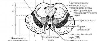
The midbrain has a cavity in its center filled with central gray matter. The so-called aqueduct is located here; at its bottom there are several nuclei, the processes of which mainly innervate the organs of vision, as well as the nuclei of the III and IX cranial nerves. In front of it are the cerebral peduncles, divided by the substantia nigra into a base and a tegmentum, where the red nucleus stands out. Together with the substantia nigra, they belong to the extra pyramidal system and control unconscious movements and muscle tone with the help of the hormone dopamine. The visual and auditory centers are located in the colliculi of the midbrain roof.
Useful to know: How the diencephalon functions and why it is needed
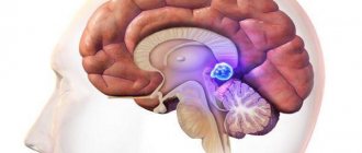
The nervous tissue of the diencephalon forms its four parts. The first of them is the Thalamus, where the nuclei of the thalamus lie. The epithalamus - the second part - includes the leash, its commissure and triangle, as well as the epiphysis, otherwise called the pineal gland. It is interesting for its mystical reputation - esotericists believe that it is this gland that causes clairvoyance and other supernatural abilities. In fact, the pineal gland produces the hormone melatonin, which is responsible for regulating sleep and wake cycles. The third part, the Metathalamus, contains the geniculate bodies. The latter, the Hypothalamus, has two sections. In the anterior one there is the so-called gray tubercle, and in the posterior one there are the mastoid bodies, containing two nuclei.
to contents ^
The structure of protein molecules
Currently, protein molecules have been studied quite well. Based on the available data, a modern definition of proteins has been formulated.
Proteins are high-molecular organic compounds built from amino acids connected by peptide bonds and having a large molecular weight and complex structural organization.
Based on methodological considerations, several levels of organization are distinguished in the structure of protein molecules: primary, secondary, tertiary and quaternary.
Primary structure
The formation of protein molecules begins with the joining of amino acids with each other. This is the first level or primary structure of the protein.
The primary structure of a protein is a nolinentide chain in which amino acids are connected by peptide bonds.
Establishing the primary structure of a protein requires performing several operations in a certain sequence, which are listed in Table. 4.2.
Algorithm for determining the primary structure of a protein
| Operation to be performed | The purpose and essence of transformation |
| Breaking SS bridges (if any) | Unfolding of a polypeptide chain. Carry out the oxidation of S-S bridges with adformic acid to SO:jH-rpynn, which are not destroyed during further analysis |
| Partial hydrolysis of nolineitide chains | Shortening of amino acid sequences, which facilitates their further decoding. Carry out by selective enzymatic hydrolysis (trypsin or chymotrypsin) or chemical agents (bromocyanium, 2,4-dinitrofluorobenzene, etc.) |
| Fractionation of the resulting peptides | Separation of the resulting polypeptide chains from each other. Performed by electrophoresis |
| Decoding the amino acid sequence in short peptides | Determination of the primary structure in individual polypeptide chains. Carry out by mass spectrometric method or using a sequencer |
Why is damage to white and gray matter dangerous?
As a result of any pathological processes occurring in the structures of the white and gray matter, pronounced symptoms of the disease can manifest themselves in different ways and depend on the location of the destroyed area and the extent of focal brain damage.
Particularly dangerous diseases are characterized by the presence of several or multiple hard-to-reach lesions, which are aggravated by blurred symptoms, consisting of a large number of signs of pathological changes.
Diseases of the central nervous system accompanied by changes in the structure of white matter:
- Leukoatherosis. Refers to many focal changes in the structure of the brain. As a result of this disease, there is a gradual decrease in the density of the white matter located in the hemispheres of the cerebellum and the trunk of this organ. It leads to degenerative changes in human behavior and is not an independent disease, since it most often develops against the background of an insufficient supply of nutrients to the nervous tissue.
- The most common cause of a disease such as multiple sclerosis is demyelination of white matter or destruction of the myelin sheath of nerve fibers. Just like with the first disease, the process is focal in nature and affects all structures of the central nervous system, which is why it has an extensive clinical picture, which can combine many signs and symptoms of the disease. Typically, patients with multiple sclerosis are easily excitable and have problems with memory and fine motor skills. In especially severe cases, paralysis and other motor dysfunction develop.
- A pathological condition such as heterotopia of the gray matter of the brain is characterized by an atypical arrangement of neurons of the gray component in the structures of this part of the central nervous system. It occurs in children with epilepsy and other mental pathologies, such as mental retardation. It is the result of a genetic and chromosomal abnormality in human development.
Advances in modern medicine make it possible to diagnose pathological changes in the brain matter at an early stage of development, which is extremely important for subsequent therapeutic actions, since it is known that any progressive changes in the structure of both the white and gray matter of the brain ultimately lead to degenerative changes and other severe neurological problems.
Diagnosis of the disease includes an in-person examination of the patient by a neurologist, during which, using special tests, almost all pathological changes in the gray and white matter are detected, without the use of special equipment.
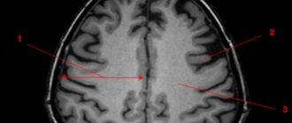
The most informative methods for studying both white and gray matter are MRI and CT, which make it possible to obtain a number of images of the internal state of brain structures. Using these research methods, it became possible to study in detail the general anatomical picture of both single and multiple foci of changes in these functional units of the NS.
What does the bear have to do with it?
The bear face is an excellent association for remembering the basic structures of the midbrain. If you turn our drawing over, you will see this:
You can also use the picture from Wikipedia:
Here we see:
- Eyes (red cores);
- Ears (brain legs);
- The line separating the head and ears (substantia nigra);
- Nose (central gray matter);
- Nostrils (Sylvius aqueduct).
The triangles inside and next to the gray matter are a schematic representation of the nuclei of the cranial nerves; we will definitely analyze them in more detail in separate lessons. The green plates that are located laterally are the midbrain pathways; now I will publish this article and immediately begin the second part devoted exclusively to them.
Let's mark all the anatomical formations we have analyzed in a separate figure: