The human brain consists of white and gray matter. The first is everything that is filled between the gray matter in the cortex and the basal ganglia. On the surface there is a uniform layer of gray matter with nerve cells, the thickness of which is up to four and a half millimeters.
Let's study in more detail what gray and white matter is in the brain.
Gray and white matter of the spinal cord
The heterogeneous substance of this organ is gray and white. The first is formed by a huge number of neurons, which are concentrated in nuclei and come in three types:
- radicular cells;
- tufted neurons;
- internal cells.
The white matter of the spinal cord surrounds the gray matter. It includes nerve processes that make up three fiber systems:
- intercalary and afferent neurons connecting different parts of the spinal cord;
- sensory afferents, which are long centripetal;
- motor afferent or long centrifugal.
Structure
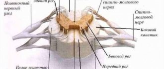
The spinal cord has two thickenings: cervical and lumbar, corresponding to the exit points of the nerves going to the upper and lower extremities. The anterior and posterior longitudinal grooves divide the organ into two symmetrical halves, each in turn has two weakly defined longitudinal grooves, from which the anterior and posterior roots emerge - the spinal nerves. The exit point of the roots does not correspond to the level of the intervertebral foramina and the roots, before leaving the canal, are directed to the sides and down. In the lumbar region they run parallel to the filum terminale and form a bundle called the cauda equina.
From the spinal cord, formed from the anterior (motor fibers) and posterior (sensory fibers) roots, 31 pairs of mixed spinal nerves depart. The area corresponding to the origin of a pair of spinal nerves is called a nerve segment, or a segment of the spinal cord. Each segment innervates specific skeletal muscles and skin areas.
The cervical and upper thoracic segments innervate the muscles of the head, girdles of the upper limbs, chest organs, heart and lungs. The lower thoracic segments and part of the lumbar segments are responsible for controlling the muscles of the trunk and intra-abdominal organs. From the lower lumbar segment and the sacral segment, nerves extend to the lower extremities and partially to the abdominal cavity.
Gray matter structure
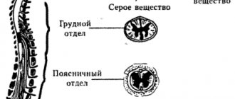
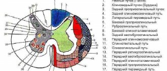
A cross section of the spinal cord has the appearance of a butterfly, which is formed by gray matter surrounded by white matter. The wings of a butterfly are symmetrical sections in which the anterior, posterior and lateral columns (or horns) are distinguished. The front horns are wider than the rear ones. The posterior roots enter the posterior horns, and the anterior roots emerge from the anterior horns. In the center of the gray matter, along its entire length, there is a channel where cerebrospinal fluid circulates, which supplies nerve tissue with nutrients.
Gray matter is formed from more than 13 million nerve cells. Among them, three types are distinguished: radicular, bundle, and intercalary. The anterior roots contain the axons of the root cells. The processes of tuft cells connect the parts of the spinal cord with each other, and the intercalary ones end at synapses within the gray matter.
Neurons with a similar structure are united into the nuclei of the spinal cord. In the anterior horns, ventromedial, ventrolateral, dorsomedial and central pairs of nuclei are distinguished, in the posterior horns - proper and thoracic. In the lateral horns there is a lateral intermediate nucleus formed by association cells.
Structure of the spinal cord
White matter structure
The white matter consists of processes and bundles of nerve cells that form the organ's conduction system. Constant and unhindered transmission of impulses is ensured by two groups of fibers:
- Short bundles of nerve endings that occupy different levels of the spinal column are association fibers.
- Long fibers (projection) are divided into ascending, which go towards the cerebral hemispheres, and descending, which go from the hemispheres to the spinal cord.
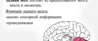
hindbrain
This brain consists of the pons and the cerebellum. Let's look at the gray and white matter in them. The bridge is a large white ridge on the back side of the base. On the one hand, its border with the cerebral peduncles is pronounced, and on the other, with the medulla oblongata. If you make a cross section, the white matter of the brain and the gray nucleus will be visible very clearly. Transverse fibers divide the bridge into ventral and dorsal sections. In the ventral part, the white matter of the pathways is mainly present, and the gray matter forms its nuclei here.
The dorsal part is represented by nuclei: switching, reticular formation, sensory systems and cranial nerves.
The cerebellum is located under the occipital lobes. It includes the hemispheres and the middle part called the “worm”. Gray matter makes up the cerebellar cortex and nuclei, which are tent-shaped, spherical, corky and dentate. The white matter of the brain in this part is located under the cerebellar cortex. It penetrates into all gyri as white plates and consists of different fibers that either connect the lobules and gyri, or are directed to the internal nuclei, or connect sections of the brain.
Pathways
Long ascending and descending pathways connect the periphery to the brain using two-way communication. Afferent impulses along the spinal cord pathways are carried to the brain, transmitting to it information about all changes in the external and internal environment of the body. Along descending pathways, impulses from the brain are transmitted to effector neurons of the spinal cord and cause or regulate their activity.
Ascending paths:
- Posterior funiculi (sensory tracts), which carry signals from skin receptors to the medulla oblongata.
- Spinothalamic, send impulses to the thalamus.
- Dorsal and ventral (spinocerebellar) are responsible for conducting excitation from proprioceptors to the cerebellum.
Descending Paths
- Pyramidal - runs in the anterior and lateral columns of the spinal cord and is responsible for performing movements.
- The extrapyramidal tract starts from the structures of the brain (red nucleus, basal ganglia, substantia nigra) and goes to the anterior horns and is responsible for involuntary (unconscious) movements.
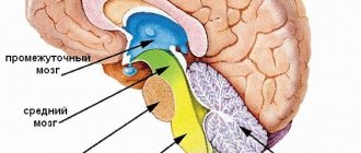
Midbrain
It starts from the mesencephalon. On the one hand, it corresponds to the surface of the brain stem between the pineal gland and the superior medullary velum, and on the other, to the area between the mammillary bodies and the anterior part of the pons.
It includes the cerebral aqueduct, on one side of which the boundary is provided by the roof, and on the other by the covering of the cerebral peduncles. In the ventral region, the posterior perforated substance and the peduncles of the cerebrum are distinguished, and in the dorsal region, the roof plate and the handles of the inferior and superior colliculi are distinguished.
If we look at the white and gray matter of the brain in the cerebral aqueduct, we will see that the white surrounds the central gray matter, which consists of small cells and has a thickness of 2 to 5 millimeters. It consists of the trochlear, trigeminal and oculomotor nerves, together with the accessory nucleus of the latter and the intermediate nucleus.
Diencephalon
It is located between the corpus callosum and the fornix, and on the sides it fuses with the telencephalon. The dorsal section consists of the visual tuberosities, on the upper part of which there is the epitubercle, and in the ventral part there is the inferior tuberosity region.
The gray matter here consists of nuclei that are associated with centers of sensitivity. White matter is represented by conducting pathways in different directions, guaranteeing the connection of formations with the cerebral cortex and nuclei. The diencephalon also includes the pituitary gland and pineal gland.
Spinal cord membranes
The organ is protected by three membranes: hard, arachnoid and soft.
- The dura mater is located on the outside of the spinal cord and does not fit tightly to the walls of the spinal canal. The resulting space is called the epidural; connective tissue is located here. Below is the subdural space at the border with the arachnoid membrane.
- The arachnoid membrane consists of loose connective tissue and is separated from the soft membrane by the subarachnoid space.
- The pia mater directly covers the spinal cord, limited from it only by a thin glial membrane.
Finite brain
It is represented by two hemispheres, which are separated by a gap running along them. It is connected in depth by the corpus callosum and commissures.
The cavity is represented by the lateral ventricles located in one and the second hemisphere. These hemispheres consist of:
- a cloak of neocortex or six-layer cortex, distinguished by nerve cells;
- striatum from the basal ganglia - ancient, old and new;
- partitions.
But sometimes there is another classification:
- olfactory brain;
- subcortex;
- gray matter of the cortex.
Without touching on the gray matter, let's focus immediately on the white matter.
On the characteristics of the white matter of the hemispheres
The white matter of the brain occupies all the space between the gray and basal ganglia. There is a huge number of nerve fibers here. The white matter contains the following areas:
- central substance of the internal capsule, corpus callosum and long fibers;
- radiant crown of radiating fibers;
- semi-oval center in outer parts;
- a substance found in the convolutions between the furrows.
Nerve fibers are:
- commissural;
- associative;
- projection.
The white matter includes nerve fibers that are connected by the convolutions of one and the other cerebral cortex and other formations.
Nerve fibers
Mostly commissural fibers are found in the corpus callosum. They are located in the cerebral commissures, which connect the cortex on different hemispheres and symmetrical points.
Association fibers group areas on one hemisphere. In this case, short ones connect neighboring convolutions, and long ones connect those located at a far distance from each other.
Projection fibers connect the cortex with those formations located below, and then with the periphery.
If the internal capsule is viewed in section from the front, the lenticular nucleus and the posterior limb will be visible. Projection fibers are divided into:
- fibers located from the thalamus to the cortex and in the opposite direction, they excite the cortex and are centrifugal;
- fibers directed to the motor nuclei of the nerves;
- fibers that conduct impulses to the muscles of the whole body;
- fibers directed from the cortex to the pontine nuclei, providing a regulatory and inhibitory effect on the work of the cerebellum.
Those projection fibers that are located closest to the cortex create the corona radiata. Then their main part passes into the internal capsule, where the white matter is located between the caudate and lenticular nuclei, as well as the thalamus.
There is an extremely complex pattern on the surface, with alternating grooves and ridges between them. They are called convolutions. Deep grooves divide the hemispheres into large areas called lobes. In general, the grooves of the brain are deeply individual; they can vary greatly from person to person.
The hemispheres have five lobes:
- frontal;
- parietal;
- temporal;
- occipital;
- island.
The central sulcus originates at the top of the hemisphere and moves down and forward to the frontal lobe. The area posterior to the central sulcus is the parietal lobe, which ends in the parieto-occipital sulcus.
The frontal lobe is divided into four convolutions, vertical and horizontal. In the temporal lobe, the lateral surface is represented by three gyri, which are delimited from each other.
The furrows of the occipital lobe are variable. But everyone, as a rule, has a transverse one, which is connected to the end of the interparietal groove.
On the parietal lobe there is a groove that runs horizontally parallel to the central one and merges with another groove. Depending on their location, this lobe is divided into three convolutions.
The island has a triangular shape. It is covered with short convolutions.
Brain lesions
Thanks to the achievements of modern science, it has become possible to conduct high-tech brain diagnostics. Thus, if there is a pathological focus in the white matter, it can be detected at an early stage and therapy can be prescribed in a timely manner.
Among the diseases that are caused by damage to this substance are its disorders in the hemispheres, pathologies of the capsule, corpus callosum and syndromes of a mixed nature. For example, if the hind leg is damaged, one half of the human body can be paralyzed. This problem may develop with sensory disturbances or visual field defects. Malfunctions of the corpus callosum lead to mental disorders. In this case, the person ceases to recognize surrounding objects, phenomena, etc., or does not perform purposeful actions. If the lesion is bilateral, swallowing and speech disorders may occur.
The importance of both gray and white matter in the brain cannot be overstated. Therefore, the earlier the presence of pathology is detected, the greater the chance that treatment will be successful.
Functions
Functions of the spinal cord
The spinal cord has two functions: reflex and conduction.
As a reflex center , it carries out complex motor and autonomic reflexes, and is also the site of closure of reflex arcs, which consist of three links: afferent, intercalary and efferent.
It is connected by afferent (sensitive) pathways to receptors, and by efferent (motor) pathways to muscles and internal organs.
An example is the congenital and acquired reflexes of a person; they are closed at different levels of the spinal cord: the knee at the level of the 3-4 lumbar segment, the Achilles at the 1-2 sacral segment.
The conduction function is based on the transmission of impulses from the periphery (from skin receptors, mucous membranes, internal organs) to the brain along the ascending pathways and back through the descending pathways.
Similarities and Differences in Brainstem and Spinal Cord Functions
The brainstem is the structure into which the spinal cord passes through the foramen magnum and has a structure similar to it. The similarity lies in their performance of reflex and conduction functions.
They differ in the location of the gray matter: the brain stem is characterized by accumulations of gray matter in the form of nuclei, which are responsible for vital functions: breathing, blood circulation, etc., and in the spinal cord it is in the form of columns. Also, the trunk is an autonomous substance in the regulation of sleep, vascular tone, consciousness, and the spinal cord carries out all actions under the control of the brain.
Please rate the article. We tried our best:)