Norms of rhythm indicators on EEG
Based on the EEG results, the doctor makes a conclusion, based on which the patient will be diagnosed and a treatment strategy will be determined. In this case, the individual characteristics of the body are taken into account - age, presence of chronic diseases, etc. Deviations in brain activity indicators may indicate a disease.
Alpha rhythm norms and disorders
These are oscillations, the frequency of which normally varies from 8 Hz to 14 Hz, and the maximum amplitude is limited to 100 μV. Signs of pathological changes in the alpha rhythm include:
- its stable fixation in the frontal areas of the skull;
- too large a difference in oscillations between the hemispheres (over a third);
- disrupted sinusoidality of the signal, distortion of the wave structure;
- high degree of frequency dispersion;
- a drop in amplitude below 25 μV or an increase above 95 μV.
The listed disorders indicate asymmetry of the hemispheres, which may be a symptom of the presence of a tumor, hemorrhage, stroke or other brain pathology localized in one hemisphere. Exceeding the frequency norm is a sign of injury to the skull or brain tissue.
Beta rhythm norms and disorders
Today, normal indicators are considered to be fluctuations from 3 μV to 5 μV, which is recorded in both hemispheres of the brain. Excessively high amplitude of beta rhythms may indicate a concussion. The so-called short spindles on the EEG are a sign of encephalitis. If the duration and frequency of spindles increase, this is a sign of inflammation of the brain tissue.
For children, beta rhythms, the frequency of which is stabilized within 15-16 Hz, and the amplitude lies between 40 μV and 50 μV, are considered a sign of pathology. The doctor is especially concerned about the localization of vibrations in the anterior or central zone of the brain. In this case, we can talk about the possibility of delays in the mental development of the baby.
Norms and disorders of delta and theta rhythms
Doctors may suspect a functional brain disorder if the amplitude of delta and theta rhythms increases to more than 45 microvolts and the increase is persistent. If such a picture is observed for all lobes of the brain, with a high degree of probability, we can talk about severe damage to the nervous system.
Excessively high amplitude of delta oscillations is often a symptom of tumor development. An increase in theta and delta indices, localized for the occipital part of the brain, is an alarming sign when recorded in a child: this may indicate a delay in his development, a retarded psyche, and even circulatory disorders in the brain.
EEG for patients
Electroencephalography (EEG) is an excellent method for diagnosing epilepsy and various brain injuries. Unfortunately, EEG is often prescribed to everyone, including patients who do not need it at all.
The essence of the method
EEG is a method that records electrical signals from neurons (nerve cells in the brain). Indeed, some diseases can manifest as severe disturbances in the electrical activity of the brain.
Most often this is epilepsy, in which a group of neurons exhibits excessive activity, and structural changes in the brain (tumor, cyst, consequences of stroke and hemorrhage). Almost always, using an EEG, a doctor (neurophysiologist) can determine where this focus of excitation is located.
Indications for EEG
In our country there are diagnostic standards for all diseases. Unfortunately, in accordance with Russian standards, such an excellent method as EEG is often used to diagnose not only epilepsy and brain tumors, but also any neurological disorders.
For example, a patient complains of fainting in a stuffy room, with a crowd of many people, in a confined space. Or for a paroxysmal headache. Here are the readings for the EEG according to the standards.
Moreover, in most cases, a routine EEG is used with a recording of up to 20 minutes. Unfortunately, such a short recording often does not record even some types of epilepsy, in which changes in activity are quite pronounced. For a detailed assessment of electrical activity in epilepsy, a longer EEG recording is needed, and preferably overnight monitoring or recording after a sleepless night (sleep deprivation). And if we are talking about “vegetative-vascular dystonia” or headaches, then the EEG will most likely only confuse both the doctor and the patient.
Problems decoding results
The doctor receives the EEG report and the patient waits hopefully for the verdict. If a stroke or tumor has already been established, then usually there is no intrigue. Even such a short recording will show that yes, indeed, there is a focus of pathological activity. The recording, in particular, will help evaluate the effectiveness of treatment for excessive neuronal activity in the affected area.
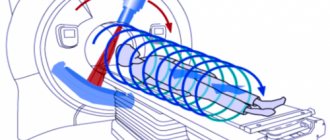
How does electroencephalography work?
Signal transmission in the human nervous system is carried out both chemically (using neurotransmitters) and electrically (action potentials). A single action potential or membrane voltage of a single neuron is too weak to be detected by non-invasive diagnostic methods. However, electrodes can detect the summation of synchronously occurring action potentials and make fluctuations in electrical activity visible.
There is a certain connection between a person’s mental state and EEG waves. Abnormalities or unusual brain waves may indicate pathology. A neurologist analyzes and describes such waves.
The electrodes measure activity in parts of the cerebral cortex that have a high density of nerve cells. However, an EEG measures not only the electrical potential of nerve cells in the brain, but also the muscles of the head and skin. Accordingly, fundamental EEG rhythms do not reflect precise neuronal activity. EEG rhythms and their relationship with the functional state of the brain are the subject of controversy in the scientific community.
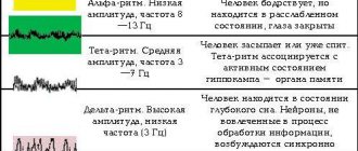
Delta rhythms
EEG delta rhythms have a low frequency ranging from 0.1 to <4 Hz. Delta waves are typical functional waves of the deep, dreamless sleep phases. In infants, the delta rhythm is also present upon awakening.
Theta waves
Theta wave is a slow rhythm in the frequency range from 4 to <8 Hz. They are more common during drowsiness and dozing. EEG rhythms and their characteristics depend on the patient’s age. They are present in the waking state in children, but presence in adults may indicate brain dysfunction or damage.
Alpha waves
The normal alpha rhythm on the EEG has the following features:
- frequency 8-12 Hz: the lower limit of normal alpha rhythm in adults and children over 8 years of age is 8 Hz;
- location: occipital regions;
- morphology: rhythmic and regular;
- amplitude: usually 20-100 mV;
- reactivity: appears when the eyes are closed and disappears when they open.
Beta waves
The normal EEG beta rhythm has the following characteristics:
- Frequency (by definition) greater than 13 Hz.
- Location: diffuse distribution.
- Morphology: usually rhythmic and symmetrical.
- Amplitude: range 5-20 mV.
Reactivity: Beta activity increases during the first and second stages of sleep, and decreases in the deep phases. Central beta activity may be reactive to voluntary movements and proprioceptive stimuli.
Gamma waves
A gamma wave is a signal in the frequency range above 30 Hz. This rhythm occurs during strong concentration, during study or meditation. Recent studies have shown that the occurrence of gamma rhythms is necessary for the integration of various stimuli.
It should be noted that gamma rhythms are not visible on the EEG strip with the naked eye.
Alpha - EEG activity
The main type of bioelectrical activity of the adult brain is considered to be alpha activity (8-13 Hz) or alpha rhythm. He is recorded in a state of quiet wakefulness with his eyes closed in a dark room. Usually the alpha rhythm is zonally differentiated, better expressed in the occipital regions. From the occipital to the frontal regions, the index and the regularity of the alpha rhythm decrease. The alpha activity index is considered low when its value is up to 25%, medium - up to 50% and high - more than 70%. Alpha activity is considered primary if it is expressed in the occipital-parietal leads with an index of at least 60% or dominates in all areas of the brain with an index of at least 50%. Modulation of the amplitudes of alpha rhythm - the correct increase and decrease in the amplitudes of individual waves forming horizontal spindles. Alpha activity is considered regular if the periods of adjacent waves differ by no more than 0.5 Hz. Signs of alpha rhythm disorganization are recorded when the periods of adjacent waves differ by 1-2 Hz or more; amplitude modulations are unclear or erratic; The shape of the waves is not smooth, but pointed or jagged. By the age of three, most children have an alpha rhythm of 8 Hz, and over 10 years, it is 10 Hz. The frequency may transiently increase after opening the closed eyes, quickly moving to the frequency band characteristic of the beta rhythm (13-30 Hz). This phenomenon, independent of the frequency of the beta rhythm, is sometimes referred to as “alpha squeak”. Alpha rhythm manifests itself better when the patient is in a state of relaxation, peace, and during periods of relative physical and mental passivity. The frequency and amplitude of the alpha rhythm typically change with age, reflecting changes in the physiological activity of the brain as we age. Only 1% of healthy adults have alpha activity at a frequency of 8 Hz, but as noted above, this rhythm is typical for adolescents. With increasing age, the frequency of the alpha rhythm gradually and steadily slows down (decreases). In people over 80 years of age, the frequency of alpha activity reaches 7.5 Hz.
The normal level of alpha activity in the occipital region in adults is from 8 to 13 Hz. The alpha rhythm frequency is associated with cerebral blood flow and decreases when blood supply to a particular area of the brain decreases. There are numerous attempts in the literature to clarify the relationship between alpha rhythm and cognitive and mental functions.
Reactivity is a characteristic feature of alpha rhythm. weakening of alpha activity occurs as a response to opening the eyes. When the eyes are closed, alpha activity returns to its amplitude and frequency. in the occipital region. Apparently, the reactivity of the alpha rhythm can also change from various stimuli, for example, with increased cognitive activity. In approximately 25% of normal adults, the alpha rhythm is poorly visualized. or may only be noticeable intermittently. The amplitude is also variable among different individuals and even fluctuates throughout his life. Low alpha rhythm amplitude is observed in less than 10% of patients. (voltage less than 15 millivolts (mu V). Alpha rhythm is maximally expressed in the occipital region and moves to the anterior parts of the brain at the moment of falling asleep, when drowsiness appears.
Alpha rhythm asymmetry is best assessed by comparing the two posterior electrode leads. (parietal and occipital) with bipolar recording of reference quality. Higher amplitude is usually characteristic of the right hemisphere and ranges from 20 to 60 microvolts (peak to peak). Using P4-O2 derivation, normal amplitude is 15-45 microvolts. High amplitude is more characteristic of a slow alpha rhythm. If the asymmetry of alpha activity voltage reaches 50%, one should think about the presence of a pathological process. If the voltage of alpha waves in the left hemisphere is higher by more than 35% compared to the right, it is also worth looking for pathological disorders.
From the point of view of morphology, the alpha rhythm is sinusoidal, however, especially in young patients, it can be temporarily interrupted by sharp superficial negative components. It can either intensify or weaken, provoking the so-called “beating effect”. Such a spindle-shaped ri) may be present in some people or absent in others. The fusiform rhythm is especially noticeable when studying the structure of sleep. The appearance of a high-amplitude alpha rhythm in the temporal regions may indicate the presence of epileptiform changes. Sometimes such an alpha rhythm (temporal areas) is designated by some authors with the term (“third rhythm”), in contrast to the “posterior alpha rhythm phenomemon”. Paradoxical alpha activity occurs when there is a presentation of alpha activity in the anterior leads, but there is no drowsiness.
Sex differences in alpha rhythm have not been identified. The dependence of the amplitude and frequency of the alpha rhythm depending on the menstrual cycle was noted. An increase in the frequency of alpha activity and a decrease in its amplitude are observed in the premenstrual phase, and a slowdown in alpha frequency and an increase in amplitude are recorded during menstruation (Chang B., et.al., 2011). As body temperature rises, the frequency of the alpha rhythm increases. The cardiac pacemaker (“pacemaker”) also increases the frequency of alpha activity by more than 1-2 Hz. , possibly due to increased cardiac output and therefore increased cerebral blood flow (Kellaway P., 2003). Medicines can slow down the alpha rhythm. In oncological diseases accompanied by hyperthermia, depression of the alpha rhythm and a slowdown in its frequency are also observed. Alpha harmonics are typical of a normal electroencephalogram.