Many specialists, including neurologists and cardiologists at MedicCity, are confident that MRI scanning of cerebral vessels should certainly be included in the program of mandatory medical examination of the population.
MRI diagnostics allows you to detect pathological changes in blood vessels at the earliest stages. This means that we are able to prevent the development of the most dangerous conditions, such as atherosclerosis, stroke, heart attack, and tumor processes. MRI angiography of cerebral vessels can save your life and the lives of your loved ones!
Diagnostics based on the phenomenon of magnetic nuclear resonance are not only extremely informative and accurate, but also do not have a harmful effect on the body and can be carried out many times.
The study of the condition of cerebral vessels makes it possible today to identify and treat diseases that for many years have robbed people of their mental and physical health at an unacceptably early age.
1 MRI of cerebral vessels
2 MRI of cerebral vessels
3 MRI of cerebral vessels
Why is MR angiography needed?
The study of the circulatory system is used in surgery, oncology, neurology and other areas of medicine. MRI of a given area with MR angiography allows you to assess the functionality, fullness, lumen, and condition of the walls of veins and arteries.
Based on the scan results, the nature of the blood supply to the area under consideration is determined, and foci of ischemia and necrosis are identified. In case of injuries, tomograms visualize hematomas, a violation of the integrity of the vascular wall.
Indications for prescribing MRI scanning in angiography mode are:
- dizziness, fainting;
- lack of coordination;
- severe headaches (cephalgia) that cannot be relieved with medication;
- decreased visual acuity, flickering of spots before the eyes;
- hearing loss, tinnitus;
- paresis and paralysis of the facial muscles;
- numbness, weakness of the upper and lower extremities;
- speech disorder;
- memory impairment;
- “shaky” gait;
- convulsions;
- leg pain;
- swelling of the lower extremities;
- dryness, peeling, ulceration of the skin of the legs, etc.
Decreased tone, decreased lumen, and ruptures of vessel walls lead to the development of pathological changes in surrounding tissues and organs. Impaired patency of the coronary arteries causes destructive, dystrophic processes in the heart muscle, which can cause a stroke or heart attack.
MRI angiography of cerebral vessels (sagittal section)
Pathologies of the aorta, superior and inferior vena cava are accompanied by dysfunction of the abdominal and pelvic organs and contribute to the development of diseases of the lower extremities.
In the diagnosis of cerebral vascular lesions, MRI using the MR angiography mode visualizes the slightest changes in the functioning of the circulatory system.
The procedure allows you to diagnose:
- stenosis, occlusion - narrowing and blockage of the vascular bed;
- acute disturbance of blood circulation in tissues;
- aneurysm - protrusion of the arterial wall;
- thrombosis - the formation of a clot in the lumen of a vein;
- vascular wall ruptures;
- atherosclerosis - cholesterol deposits in the lumen of the artery;
- vasculitis - inflammatory changes, thickening and stratification of the walls;
- embolism - blocking the lumen of the artery with foreign particles (gas, parasitic, etc.);
- extravasal compression syndrome - external compression of the vascular bed;
- arteriovenous malformations - pathological anastomoses (connections) of blood channels;
- congenital anomalies of the vascular system - kinks, bifurcations, etc.;
- dissection (dissection) of the aorta;
- thrombophlebitis - inflammation of the walls of the veins with the formation of bloody clots;
- vascular tumors.
The results of magnetic resonance angiography are analyzed by an MRI physician. The study protocol indicates:
- diameter, patency of the vascular bed;
- the condition of the walls, the fullness of the vessel;
- the presence of foreign particles (emboli) in the lumen;
- localization of the affected area;
- condition of surrounding tissues;
- functionality of veins and arteries;
- degree of circulatory disturbance in tissues;
- causes of hemorrhagic and ischemic phenomena.
MRI angiography examination is prescribed before surgery. Based on the scan results, the location and extent of the affected area are determined, the nature of the pathology and the scope of the upcoming operation are clarified.
During the rehabilitation period, recovery processes are monitored using MR angiography; MRI helps to timely diagnose the development of complications.
Scanning allows you to track the course of chronic diseases. In this case, vascular MRI angiography is prescribed at a frequency of 2 times a year. Early detection of relapses facilitates timely correction of treatment and helps achieve stable remission.
There are two types of MR angiography in MRI. The methods are identical, the difference lies in the purpose of the study.
MRI of the neck with MR angiography
Venography is aimed at studying the work of the vessels of the same name. Scanning allows you to diagnose inflammatory, degenerative processes, signs of reverse flow, and determine the causes of pathological phenomena.
Arteriography is indicated in case of symptoms of vascular insufficiency. If necessary, a comprehensive study of the vascular system is performed.
Methodology for conducting SCA
SCA refers to an invasive test where a radiopaque contrast agent is injected into an artery.
General scheme of conducting SCA
- The arterial puncture site is treated with an antiseptic and numbed.
- A catheter is inserted through the puncture, which is passed into the target vessel, and a contrast agent is supplied into it.
- A series of x-rays are taken.
- The catheter is removed, the puncture site is treated with an antiseptic, and a pressure bandage is applied.
Scheme of selective cerebral angiography
Carotid artery catheterization
Direct carotid angiography involves catheterization of the carotid artery.
The patient is laid down, the head is turned in the opposite direction and thrown back. The puncture point is determined by the pulsation of the carotid artery - it is located between the upper level of the thyroid cartilage and the inner side of the sternocleidomastoid muscle.
Currently practically not used.
Catheterization of the vertebral artery
With direct vertebral angiography, catheterization of the vertebral artery can be performed in different ways.
- Direct puncture of the vertebral artery. An angiographic needle is inserted at the level of the IV–V cervical vertebrae, advanced to their transverse processes and the vertebral artery is punctured between them.
- Puncture of the subclavian artery. In the area of the clavicle, the pulsation of the subclavian artery is palpated, it is punctured with a needle, and a catheter is inserted to the site of the branch of the vertebral artery.
- Puncture of the brachial artery is performed in the axillary fossa. A catheter with a side hole and an internal plug is inserted into the lumen of the needle and advanced to the mouth of the vertebral artery. Then a catheter of smaller diameter is inserted through it on a flexible metal string.
Currently, these accesses are practically not used.
It is important! Since the lumen of the vertebral artery is smaller than that of the femoral and carotid arteries, the contrast agent must be supplied more slowly.
Femoral or radial artery catheterization
More often, selective angiography is performed using an indirect method. In this case, catheterization of the femoral or radial artery is performed. This method makes it possible to diagnose four arteries at once: two paired carotid and two paired vertebral arteries.
For this:
- Prepare the puncture site.
- The femoral artery in the medial third of the inguinal ligament or the radial artery in its distal segment is palpated.
- After inserting the main catheter, it is advanced along the vessel to the aortic arch.
Arterial catheterization technique
When performing SCA, arterial catheterization is performed using the following technique:
- An angiographic needle punctures the skin at an acute angle.
- Move the needle forward until the artery pulsates.
- They pierce the artery with a sharp push, trying not to damage its opposite wall.
- After a stream of blood appears, a conductor is inserted into the needle, secured, and the needle is removed.
- Using a guidewire, an introducer-dilator is inserted into the vein. It is necessary for atraumatic installation of the catheter and is equipped with a three-way stopcock to prevent blood loss.
- The guidewire is removed and the catheter is inserted into the introducer lumen.
On a note! Advancement of the catheter along the vascular bed is absolutely painless, since the inner walls of the vessels are devoid of pain receptors.
Use of contrast agents
Contrasting brain vessels with 3D reconstruction
Radiocontrast agents are used in angiography to visualize blood vessels.
These are iodine-containing drugs that are used to fill the bloodstream using a catheter, thereby increasing the clarity of the X-ray image of vascular formations.
Iodine-containing preparations are ionic and non-ionic.
- Ionic ones can provoke an allergic reaction, causing an increase in the osmotic concentration of blood plasma.
- Non-ionic ones are much safer, but their cost is higher.
Table 1. Iodine-containing contrasts.
| Compound | Drug name | Active substance | Osmolarity level |
| Ionic | Vizotrust Urografin Verografin Trazograph Diatrizoate | Sodium amidotrizoate | High |
| Ionic | Hexabrix 320 Ioxaglat | Ioxagloic acid | Short |
| Nonionic | Iopamidol Yopamiro Scanlux Tomoscan | Iopamidol | Short |
| Nonionic | Yomeron | Yomeprol | Short |
| Nonionic | Omnipack Iobriz Introvise Iohexol | Iohexol | Short |
| Nonionic | Oxylan Telebrix | Yoxitalamic acid | Short |
| Nonionic | Yopromide Ultravist | Yopromide | Short |
| Nonionic | Iodixanol Visipak | Iodixanol | Short |
| Nonionic | Optiraeus | Ioversol | Short |
One of the mandatory points in preparation for angiography is an iodine sensitivity test . It consists of slowly injecting 2 ml of contrast intravenously and observing the body’s reaction for three to four hours. At the moment, with the use of modern non-ionic contrasts, this procedure is not required.
The examination is canceled if the following symptoms appear:
- skin redness;
- rash;
- itching;
- swelling;
- nausea and vomiting;
- headache;
- cough;
- suffocation.
Before introducing an iodine-containing drug into the catheter, it is heated to body temperature. The rate of administration should be commensurate with the speed of blood flow.
Possible unpleasant sensations include a metallic taste in the mouth, a rush of blood to the face, a feeling of heat throughout the body, especially in the pelvic area.
Remember! All radiopaque agents are nephrotoxic. If renal function is impaired, strict monitoring of creatinine and urea levels is necessary.
Tools needed to conduct SCA
The following set of instruments is used to perform angiography:
- puncture angiographic needle;
- standard conductor with Teflon coating;
- introducer-dilator;
- catheter, sometimes of slightly different diameters.
Angiographic installation
The angiography procedure is performed in the department of X-ray surgical methods of diagnosis and treatment.
The angiographic installation consists of the following complex:
- A table that transmits x-rays and moves in a horizontal plane.
- X-ray tube with electron-optical converter.
- Image recording and playback system.
- Monitor.
A series of x-rays is taken in frontal and lateral projections, at a speed of one or more images per second. Simultaneous recording allows, at the end of the study, to examine each image in detail and decipher it.
MR angiography with contrast
Magnetic resonance imaging of the circulatory system involves various scanning modes:
- time-of-flight;
- phase contrast;
- MRI angiography of vessels with an intensifier.
Time-of-flight tomography provides T1-weighted images. The liquid medium under study is brightly colored against the background of the surrounding soft tissue.
Phase contrast MR angiography involves receiving a signal from blood moving through the vessels. The method allows you to determine the characteristics of fast arterial and slow venous flow with equal accuracy. The simultaneous use of time-of-flight and phase-contrast MR angiography helps to differentiate between hematomas and other static areas.
Magnetic resonance imaging with enhancement visualizes the bloodstream in the area of interest. A solution of gadolinium salts is used as a contrast agent. The drug has paramagnetic properties and provides a hyperintense signal.
Contrast for vascular MRI angiography is administered intravenously. The injection is given using a catheter connected to an automatic device. This system allows the “coloring” solution to be supplied at a constant speed.
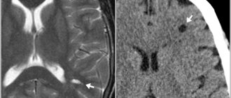
Layer-by-layer images during MRI angiography of cerebral vessels
Data recording begins after the lumen of the bloodstream is filled with contrast. 1-2 ml of solution is pre-injected, which helps to identify the initial moments of the arterial and venous phases. In this way, the signal registration time during vascular MRI angiography is determined. For further measurements, the peak arterial concentration of the gadolinium solution is used.
Angiograms make it possible to study the nature of the blood supply to a certain area. A broken line with a hyperintense signal indicates stenosis and occlusion of the vascular bed. The consequence of the pathology is ischemia and necrosis of the underlying tissues.
MRI angiography of vessels with contrast does not show calcifications, blood clots, or cholesterol plaques. In case of atherosclerosis, embolism, the images show changes in the lumen, fullness, blood channel, and parietal foci with a hypointense signal.
Contrast MR angiography for MRI diagnostics of neoplasms visualizes arteries and veins in the area of interest, shows the condition of surrounding tissues, and helps determine the nature of the disease.
The hemodynamics of the tumor circulatory network depends on the malignancy of the process. The vessels of a malignant tumor have a large number of anastomoses and bends, so the tumor tissue accumulates and releases the contrast agent more slowly.
Subarachnoid hemorrhage on MRI with MR angiography
The final diagnosis is made by the attending physician. The results of MRI angiography of veins and arteries for vascular neoplasms are supplemented with data from clinical tests, biopsy and other research methods.
Indications
- Presence of headaches of unknown origin;
- Detection of atherosclerosis, in order to identify the risk of stroke;
- Presence of head injuries;
- If other diagnostic methods do not provide convincing information regarding the diagnosis;
- Systemic and cerebral vasculitis;
- Risk of hemorrhage;
- To assess the patient’s condition after surgery;
- The presence of malignant and benign neoplasms in blood vessels;
- Any abnormalities in vascular development.
What types are there
The area of interest in MRI determines the type of MR angiography. Main areas:
- head;
- neck;
- lower limbs;
- abdomen;
- pericardium;
- upper limbs;
- small pelvis
MRI angiography of the vessels of the brain, neck, and lower extremities is most often prescribed.
A study of the blood supply system of the central nervous system can prevent the development of organic damage to the cerebral substance. MRI of the brain with MR angiography involves studying the lumen of the arteries and determining the functionality of the veins.
A neck scan is performed to assess the patency and fullness of the great vessels.
MRI of the lower extremities with MR angiography is prescribed for damage to the venous network, inflammatory processes, and thrombosis. Early diagnosis of diseases contributes to the effectiveness of treatment measures.
Angiography of cerebral vessels
Acute and chronic insufficiency of cerebral blood supply is manifested by decreased performance, inhibition of certain functions, and deterioration of well-being. In the absence of treatment, an increase in symptoms is observed:
- cephalgia;
- loss of memory, intelligence;
- paralysis;
- seizures, etc.
Severe disease can lead to disability and death.
MRI angiography of cerebral vessels with contrast is prescribed for diagnostic and preventive purposes. Patients at risk for cerebrovascular pathologies are recommended to undergo regular tomographic examinations.
MRI of the head with vascular angiography allows you to diagnose:
- arteriovenous malformations - there is no intermediate capillary network in the junction zone, the surrounding tissues are atrophic, signs of gliosis are observed;
- carotid-cavernous anastomosis - due to the intensive discharge of blood from the internal carotid artery into the sinus cavity, the pressure in the sinus increases, dilation of the ophthalmic vein occurs on the affected side, and pulsating exophthalmos develops (displacement of the organ of vision forward);
- aneurysms of cerebral vessels - there is a uniform increase in the diameter of the artery in a limited area or protrusion of the wall in the form of a bag (diverticulum);
- intracranial atherosclerosis - against the background of degenerative processes, a narrowing of the lumen of the blood channel occurs with subsequent expansion;
- vasculitis of cerebral vessels - the formation of multiple foci of cerebral infarction is possible;
- stenosis, deformation of the main arteries - pathological tortuosity, coiling (loop formation), kinking (curvature at an acute angle).
Brain MRI with vascular MR angiography is used in the diagnosis of strokes. Magnetic resonance imaging is informative already in the first 6 hours after an attack.
MRI angiography of head vessels
Acute disturbance of cerebral circulation can be caused by ischemic and hemorrhagic phenomena. In the first case, changes in the hemodynamics of the arteries are observed on MRI images of the brain with MR angiography. Intracranial hemorrhages occur when the integrity of the vascular wall is disrupted. Venography images reveal single or multiple hematomas.
Angiography of the lower extremities
The venous system of the legs experiences high loads, which is due to the structural features of the human body. Blood rises through the vessels, overcoming the force of gravity. The veins of the lower extremities have a system of valves that prevent reverse flow.
Vascular diseases of the legs are accompanied by swelling, a feeling of heaviness, and impaired motor function. When blood clots form, there is a risk of the latter breaking off and being transported through the arteriovenous network.
MRI angiography of leg vessels
A serious complication is blockage of the pulmonary circulation by an embolus (high risk of death).
MRI angiography of the veins and arteries of the lower extremities allows you to diagnose:
- thrombosis;
- atherosclerosis;
- thrombophlebitis;
- traumatic vascular lesions;
- stenosis, occlusion of the arteries of the lower extremities;
- diabetic foot syndrome.
Timely detection and treatment of thrombosis during venography reduces the risk of life-threatening complications.
Angiography of the neck
The study of the great vessels makes it possible to assess the blood supply to cerebral structures, soft tissues, the thyroid gland, etc. In the diagnosis of diseases of the central nervous system, complex MRI angiography of the neck with examination of the arteries and veins of the brain is often used.
Scanning reveals:
- thrombosis of intracranial venous sinuses;
- causes of cerebrovascular accident;
- stenosis, occlusion, thromboembolism of the main and brachiocephalic vessels;
- aneurysms;
- vasculitis of neck vessels;
- malformations;
- disruption of venous outflow from the cranial cavity, etc.
MRI of the neck with MR angiography (series of layered photos)
The scan results in detailed images of the vasculature and surrounding tissues. MRI of the brain and neck with MR angiography allows you to assess the patency, fullness of the vertebral arteries, the quality of cerebral blood supply, and the functionality of the internal jugular veins with the main tributaries.
Alternative Research
If it is not possible to use CT angiography to diagnose pathologies of the neck vessels or there are contraindications, the following methods are used:
- MR angiography with contrast.
- Ultrasound with duplex scanning: replaces CT for vascular spasms, injuries, developmental anomalies, and determination of neoplasms.
- Triplex scanning: it is used to obtain a color image illustrating blood movements, which allows you to accurately calculate blood flow parameters and assess the condition of the vascular wall.
Preparation for vascular angiography
MRI examination of the arteriovenous system with contrast is performed by appointment. You need to bring to the procedure:
- passport;
- Compulsory medical insurance policy, VHI policy;
- results of previous examinations;
- referral from the attending physician.
You should arrive 10-15 minutes before the scan starts.
Before MRI angiography of cerebral vessels, it is recommended to rest well and avoid stressful situations. It is advisable to monitor your blood pressure several days before the procedure.
Alcohol and smoking should be avoided 2-3 days before MRI with MR angiography. Avoid taking tonic substances, including strong tea, coffee, and energy drinks.
Before MRI angiography of veins and arteries, you need to warn your doctor about possible contraindications, diseases, and use of medications.
In case of suspected pregnancy, the woman needs to undergo additional examination. To clarify the condition, an analysis is performed to determine the level of the hCG hormone, and an ultrasound is performed. MRI angiography of blood vessels is a contrast procedure, which is a contraindication for diagnostic purposes.
During lactation, women are advised to prepare milk or formula to feed the baby. After administration of the contrast solution, the breasts must be expressed for 6-12 hours.
Before MRI angiography of veins and arteries, the patient removes jewelry and metal accessories. You can change into a comfortable home set and pajamas.
The patient's condition after the procedure and possible complications
The patient is prescribed bed rest for 24 hours. The patient’s well-being is monitored by the attending doctor, who measures body temperature and examines the area of invasive intervention. The next day, the bandage is removed and if the person’s condition is satisfactory and there is no bleeding in the puncture area, he is sent home.
For the vast majority of people, angiography does not pose any risk. According to available data, the risk of complications during angiography does not exceed 5% and may consist of the following:
- Allergic reactions to contrast agent, antiseptic or anesthesia used during the procedure;
- Hemorrhages or bleeding from the puncture site of the vessel;
- In rare cases, in the presence of severe concomitant diseases, serious pathologies such as myocardial infarction, acute renal failure, etc. may develop.
How is MR angiography done?
The procedure takes 40-45 minutes.
Scanning steps:
- The patient is placed on the tomograph conveyor, the limbs and body are fixed with special rollers. Random movements during an MRI session lead to distortion of MR angiography results. The device may produce noise when operating; use headphones for protection.
- After briefing, medical personnel are located in an adjacent room. Communication with the patient is maintained using an intercom. For emergency communication, the patient can use the panic button.
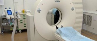
MRI of the neck with MR angiography on a closed tomograph
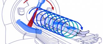
- For more accurate scanning, a gradient coil is placed above the area of interest, creating an alternating electromagnetic pulse. The table with the subject is moved into the tube of the apparatus, where the constant force field generator is located.
- After a series of native images, the patient is injected with a gadolinium solution. Scanning continues after filling the vascular bed with a contrast agent.
- During an MRI session, angiography of veins and arteries may require holding your breath for 10-20 seconds.
The shooting is carried out in axial, sagittal and frontal projections. Based on the results of MR angiography, a three-dimensional image of the arteriovenous network can be reconstructed. The 3D model helps to clarify the localization of the pathologically changed area. The thickness of the scanned section is from 1 mm.
MRI angiography of vessels is performed using open and closed type devices. The first ones do not have a tunnel module, the power of the tomograph is a maximum of 0.8 Tesla. Such devices are recommended for MRI in patients with claustrophobia and overweight patients.
Closed tomographs provide a magnetic field strength of 1.5 Tesla.
MRI of the neck with contrast MR angiography
High power allows you to make high-quality images of the studied area.
Aneurysm
An aneurysm is a specific section of a blood vessel that is intensively supplied with blood and grows in size. The convex wall of the vessel often compresses neighboring tissues and nerve endings. Damage to the aneurysm, which results in hemorrhage, poses a particular threat.
An aneurysm is diagnosed in any part of the brain, but is more often localized in the area where branches originate from arteries. Aneurysms develop asymptomatically and only when they reach large sizes do they manifest clear symptoms. Main signs of pathology:
- deterioration in the quality of vision;
- severe headaches;
- nausea;
- sudden loss of consciousness.