Computed tomography and magnetic resonance imaging are modern informative diagnostic procedures that allow us to visualize the internal structures of the body with a high degree of accuracy and detail. Both methods involve obtaining layer-by-layer images of the area of interest in different planes. The main difference between CT and MRI is the scanning principle. The methods are based on different technologies and are used in different clinical situations. Based on the diagnostic tasks, the attending physician determines which is better - CT or MRI, and then prescribes the most rational diagnostic method for a particular case.
All advanced diagnostic methods are available to medical patients. The clinic offers computed tomography and magnetic resonance imaging using modern equipment. The results are interpreted by experienced radiologists who regularly improve their skills in the best scientific centers in the Russian Federation and abroad.
Types of CT
Computed tomography is a relatively new diagnostic method, however, despite its relative youth, the technology of tomographic examination, the method of interpreting results, and most importantly the diagnostic equipment itself have undergone significant improvements.
According to the decision of the attending physician, based on objective indications, the patient may be offered one of four types of tomographic examination. Despite the fact that all diagnostic techniques have significant differences, they are united by the operating principle of the tomograph.
Spiral computed tomography
The first safe method of tomographic examination was the method of spiral computed tomography. This technique has been tested in practice many times and is considered the most informative. This method received its name because of a specific examination procedure, during which the radiation source and the table with the patient rotate in a spiral relative to each other. During the entire procedure, the patient, located on a special table, is scanned using a source of directed x-ray radiation, obtaining an accurate layer-by-layer picture of the area of interest. During scanning, the coverage area continuously increases, and the duration of the examination decreases.
Multislice computed tomography
The next computed tomography method is a logical development of the previous one. Due to the specificity of the effects of X-ray radiation, it was called multislice computed tomography; in addition, it is also called multislice and multilayer. In this type of tomography, X-rays passing through the area under study are captured by an ultrasensitive detector, thereby forming an image of the object of interest. The radiation is directed not in a fan-shaped manner (as with the spiral method), but in a volumetric manner, which allows not only to study a large area, but also to significantly reduce the received radiation dose. The advantage of this method is the possibility of simultaneous use of several radiation tubes, which significantly reduces the procedure time, without significantly increasing the radiation dose to the body. It is logical to conclude that this type of computed tomography is potentially less dangerous than all others.
Cone beam computed tomography
The next method is used exclusively in maxillofacial surgery and is called cone beam computed tomography. Its main difference from the previous ones is the design of the tomograph, the ring of which differs from other versions in size (lower than in previous versions) and location (the ring is located strictly vertically, presumably at head level). The patient's head is placed inside the ring part of the tomograph, around which the beam tube rotates. This diagnostic method allows you to obtain three-dimensional contrast images of teeth and skull bones, making it possible to detect pathology even at the initial stages of its occurrence.
Emission computed tomography
The next method is emission computed tomography. It is used in exceptional cases and is carried out only in those diagnostic centers that have special equipment and appropriate permission to work with radioactive pharmacological drugs. The main feature of this method is that to carry out diagnostics, a drug containing a special solution of radionuclides that accumulate in tissues and organs is injected into the patient’s body, making them clearly visible in the photographs. Most often, emission tomography is used to identify severe systemic pathologies that cannot be detected by other diagnostic methods. Emission tomography is only in its infancy, however, its scope of application is growing every year. Modern medicine uses emission CT in diagnosing cancer, pathologies of the brain and the cardiovascular system.
In modern medical practice, multilayer or spiral types of computed tomography are most often used.
CT scan of the chest: what will it show and how will it help the patient?
Computed tomography of the chest (CT CT) is one of the modern diagnostic methods. When and for what purpose may you need to do a CT scan of the chest? What does it include?
These and other questions are answered by Yulia Aleksandrovna Rutskaya, Deputy Chief Physician for Radiation Diagnostics, radiologist of the highest qualification category “Clinic Expert” Kursk.
— Yulia Aleksandrovna, how often do patients come to you for a CT scan of the chest?
“People come to our clinic for this service every day. Computed tomography of the chest organs accounts for on average more than a third of all examinations per day. Now, during the coronavirus epidemic, these numbers are, of course, much higher.
— What does a chest CT scan show?
- Anything that goes into the chest. First of all, the study allows us to study the condition of the lungs. We can assess the location of the vessels and bronchi, their patency, and find out whether there are neoplasms, inflammatory processes, or congenital pathologies in the lungs. Also, with the help of this study, you can see the esophagus, dilatation (expansion) of the chambers of the heart and large vessels, and the condition of the mediastinal lymph nodes.
In addition, we see a frame formed by the ribs, the thoracic spine, and the sternum. These bony structures are assessed for destructive changes.
— What diseases can be identified or excluded during this study?
— The main pathologies that can be detected are pneumonia (pneumonia) of various natures, including viral, tuberculosis, the detection of which is of social importance, and lung cancer. Interstitial lung diseases are also detected, diffusely affecting the lung tissue; accumulation of fluid in the pleural cavity; tumors and metastases in the lungs, bones and mediastinum (for example, thymoma, lymphoma, dermoid tumors, cysts, esophageal tumors); abnormalities of bone structure.
Read materials on the topic:
Tuberculosis: a disease that knows no boundaries What is a dermoid cyst? COVID-19 and more: what are the features of viral pneumonia?
— Yulia Aleksandrovna, please tell us how a CT scan of the chest organs is done
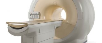
— First of all, the patient undergoes a registration procedure in our clinic. This takes 5-10 minutes. Then the laboratory assistant accompanies him to the locker room, where the patient undresses to the waist. A mandatory requirement is to remove all metal jewelry. The patient lies on a special tomograph table on his back. The tomograph itself is a ring-shaped device that rotates smoothly around the patient and takes pictures.
At the command of the radiologist, the patient inhales and holds his breath 2-3 times for a certain time (from 10 to 30 seconds). Next comes the processing of information received by the radiologist from the tomograph computer. The doctor previews the images over the next 10-15 minutes. If no changes are detected, the result is reported to the patient immediately, but the specialist needs time to describe the images. The conclusion is sent to the patient via email within a few hours, or he can pick it up from the clinic himself the next day.
Read material on the topic:
Electronic media for recording MRI studies: marketing ploy or benefit to patients?
— What is the purpose and how is a CT scan of the chest organs with contrast performed? What is special about this type of research?
— Such a study is carried out, for example, if there is a suspicion of a blood clot in the pulmonary artery. This condition is life-threatening; contrasting is required to clarify the diagnosis. In addition, we perform CT of the chest with contrast when there are space-occupying formations in the mediastinum or roots of the lungs. Contrasting allows you to study in detail the localization and spread of the tumor, assess its nature, and the location of the vessels in it.
The contrast agent is an iodine-containing substance that is injected using a special syringe-flask at the beginning of the procedure.
— Is preparation required for this study? If so, how to prepare for a chest CT scan?
— No special preparation is required for traditional CT examination of the OGK. If a contrast study is necessary, the patient must first undergo blood tests to assess the kidney's glomerular filtration rate and creatinine level. The doctor also finds out whether the patient has a history of severe allergic reactions, bronchial asthma, or individual iodine intolerance.
— In what cases is CT scanning of the chest organs contraindicated?
— Absolute contraindications are pregnancy and excess weight (more than 150 kg). A contrast study is not performed in cases of severe renal failure or individual iodine intolerance; it is used with caution in cases of diabetes mellitus and thyroid pathologies.
— Is it possible to do a CT scan of the chest organs for children?
— It is possible, at any age, only if there are direct indications and no contraindications. I would like to note that this requires a referral from a specialist with justification for conducting this particular study.
— And if an adult wants to sign up for a CT scan of the chest organs, does he need to take a referral from a doctor?
— It is desirable because from the referral the radiologist receives preliminary information about the expected diagnosis and, based on it, decides how best to conduct the study, and also determines further tactics for managing the patient.
Interviewed by Sevilya Ibraimova
If you need a CT scan of the chest, you can sign up for the study here ATTENTION: the service is not available in all cities
The editors recommend:
What does MRI of the mediastinum show? How to live with chronic obstructive pulmonary disease without losing quality of life? Formula of Love by Alexander Abdulov
For reference:
Rutskaya Yulia Alexandrovna
Graduate of the medical faculty of Kursk State Medical University in 2002. In 2004, she completed an internship in radiology. Currently, she is the deputy chief physician for radiation diagnostics, a radiologist of the highest qualification category at the Expert Clinic Kursk. Receives at the address: st. Karl Liebknechta, 7
Why do a CT scan?
The list of indications for tomography is quite extensive, however, in addition to suspicion of the presence of diseases, it makes sense to conduct a computed tomography scan if the attending physician has doubts about the correctness of the diagnosis.
Recently, it has become fashionable to undergo a CT scan to assess the condition of the whole body. This is reasonable if the patient has sufficient financial resources and some free time. However, some experts are very critical of this approach, arguing with the following statements:
Tomography is not always able to detect existing diseases, since there are some individual characteristics that limit visualization.
CT scan of the whole body (regardless of the chosen diagnostic technique) is a very expensive undertaking.
This type of study makes sense to prescribe if there is an immediate threat to the health and life of the patient.
From the standpoint of safety and economy, it makes sense to conduct research only on those organs whose normal functioning is in doubt. Also, if they are at risk for any chronic diseases.
Several types of diagnostics can be used simultaneously (ultrasound, MRI, laboratory tests).
Most often, computed tomography of the following organs is performed:
- bones, muscles, ligaments and joints;
- internal organs;
- heart and blood vessels.
Indications for CT and MRI
CT and MRI are in demand in pulmonology, cardiology, neurology and neurosurgery, traumatology and orthopedics, urology, gynecology, gastroenterology, oncology and other areas of medicine. Both methods are considered expensive, so they are rarely used in routine diagnostics. More often they are used when it is necessary to clarify the clinical situation.
Computed tomography is mainly used to diagnose diseases of bone tissue and organs with a heterogeneous structure (for example, lungs). The ability to visualize loose elements of the body using a contrast agent allows the method to be used in the diagnosis of neoplasms and vascular pathologies. CT is also prescribed when cerebrovascular accidents are suspected (for example, when diagnosing hemorrhagic strokes).
Magnetic resonance imaging is used to study soft tissues and internal organs of various locations. The reason for prescribing the procedure is the suspicion of pathology of any parenchymal organ, muscles, nerves, blood vessels, soft elements of joints (cartilage, capsule and ligaments), subcutaneous fat, skin, etc. MRI is recommended for questionable ultrasound results, as well as inconsistencies clinical picture and established diagnosis. The study is indispensable in cancer research. The method makes it possible to visualize primary and secondary tumors at early stages of development and accurately assess their effect on surrounding tissues.
What to do to the patient, CT or MRI, is always determined by the attending physician. The clinician takes into account the medical history, complaints, results of other diagnostic tests and a preliminary diagnosis. The studies are not mutually exclusive and do not replace each other. In controversial situations, the doctor may prescribe both diagnostic methods. The results of CT and MRI qualitatively complement each other, allowing you to collect maximum information about pathological changes and make the correct diagnosis.
How does CT differ from MRI?
Many people, especially those encountering computed tomography for the first time, are trying to understand: what is better than CT or MRI?; What is the difference between CT and MRI? And which method will be more effective in their case.
To answer the question: what is the difference between MRI and CT? — one should understand the mechanisms of their influence and areas of use. For example, if in computed tomography a directed beam of X-ray radiation is used to obtain an image, then in MRI the principle of nuclear magnetic resonance is used for the same purposes.
It is worth noting that although in some cases (especially when it is necessary to obtain images of blood vessels or the spinal cord), MRI is superior in efficiency to CT, it can also cause more serious harm to the patient’s body. Therefore, the decision regarding its use can only be made by a qualified physician, after a thorough assessment of all risks.
The principle of operation of CT and MRI: what is the difference
Computed tomography and magnetic resonance imaging are radiation diagnostic methods. The main difference between CT and MRI is the technology used to influence the body to obtain an image.
Computed tomography uses ionizing radiation (X-ray scanning). The method is based on the ability of body tissues to retain X-rays to varying degrees. When passing through the area of interest, the rays are maximally absorbed by dense tissues (for example, bones) and easily penetrate soft tissue elements. As a result, the images clearly visualize bones and other dense structures - stones, foreign bodies.
The difference between computed tomography and standard radiography is the number of images obtained. During a CT scan, a tube rotates around the patient, sending out rays, and special sensors capture them. As a result of the procedure, step-by-step X-ray images of the study area are obtained. The more often the tube rotates, the smaller the scanning step, and, accordingly, the higher the resolution.
MR imaging is based on the phenomenon of nuclear magnetic resonance, characteristic of hydrogen atoms contained in water molecules. Most water is present in soft tissues. Tissues that contain little water (for example, bones) are poorly visualized. The tomograph sensor records changes in the state of hydrogen atoms under the influence of radio radiation in a constant magnetic field.
As a result, step-by-step images are obtained in three planes, in which the soft tissue structures of the body are visible.
Special MR scanning modes allow improved visualization of various internal structures. Angiography involves scanning a specific part of the body and subtracting areas that do not give a signal (such as moving blood in the vessels). There are special modes for visualizing the conduction tracts in the brain - required for complex operations, a mode for studying the heart, which allows assessing the contractility of the myocardium, and other modes that provide unique opportunities for diagnosing diseases of various organs and systems.
Understanding the difference between MRI and MRI, we can draw a logical conclusion that the procedure is more informative regarding certain types of tissue. However, the diagnostic capabilities of the methods can be expanded with the help of contrast agents. CT scans use iodine-containing contrast, which is distributed throughout the body through the bloodstream and makes blood vessels and some soft tissue visible. This allows, in some cases, the use of CT to study internal organs, tumors and blood vessels.
Enhanced MRI involves injecting gadolinium into the body. The reasons for prescribing contrast studies for MRI are similar to those for CT—the need to visualize structures that are impossible or difficult to examine in native mode. The accumulation of contrast in areas of inflammation and tumors simplifies the diagnosis of certain diseases. The amplifier does not affect the quality of bone tissue visualization during MRI.
Is the computer examination procedure safe?
There is an opinion based on the assumption of the potential harm that X-rays used in the research process can cause. Note that although these fears are completely unfounded, they encourage many potential patients to abandon CT in favor of MRI. A use that is completely unjustified.
Thanks to the use of the latest technologies and radioactive materials in modern tomographs, the radiation dose is much lower than when using an X-ray machine. Of course, one should not think that minimal radiation is absolutely safe, but it can be argued that the use of CT will not cause much damage (even if it is used repeatedly).
Only a qualified radiologist can determine the number of procedures that will not harm the patient’s health, based on the need for the study and the radiation dose previously received by the patient.
Diagnostic capabilities of SM-Clinic
CT and MRI are performed using modern high-tech equipment
The clinic uses European equipment. MRI is performed on the new generation Siemens MAGNETOM ESSENZA 1.5 Tesla and Siemens SOMATOM AERA 1.5 Tesla devices. The center has modern computed tomographs, including Siemens SOMATOM Perspective (128 slices).
Specialists in the field of radiology diagnostics with many years of experience
The effectiveness of modern diagnostic methods largely depends on the skills and experience of the specialists who interpret the images. SM-Clinic radiologists regularly improve their skills and join forces in difficult clinical situations.
Contraindications for computer diagnostics
The following contraindications exist for patients:
- Pregnancy and lactation – despite the fact that the area of exposure to directed X-ray radiation is relatively small and the dose received by the patient is minimal, the procedure can damage the health of the fetus and significantly reduce the quality of breast milk.
- Claustrophobia - due to the specifics of the study (the patient is placed in a confined space for the period of the study), patients with claustrophobia may develop nervousness and a panic attack. If no alternative is available, patients must be sedated prior to the procedure.
- Renal pathologies - the introduction of a contrast agent required for CT scanning can provoke an exacerbation of existing kidney diseases.
Contraindications to examinations: CT and MRI
CT and MRI have contraindications, including absolute (the procedure is impossible or dangerous for the patient) and relative (diagnosis can be carried out under certain conditions).
Absolute contraindications to CT:
- established pregnancy;
- the patient's condition does not allow him to be in the tomograph.
With intravenous contrast, allergic reactions to iodine-containing drugs and food products.
Relative contraindications:
- when using an amplifier: creatinine and urea levels are higher than normal;
- bronchial asthma (severe)
- thyrotoxicosis.
- claustrophobia (fear of closed spaces).
Absolute contraindications to MRI:
- the presence in the body of ferromagnetic metal fragments, implants, staples (for example, on blood vessels), prostheses, etc.;
- the presence of implanted systems in the body (for example, an inner ear implant, an insulin pump);
- serious condition of the subject (need for mechanical ventilation, etc.);
- pregnancy (first 12 weeks).
Relative contraindications:
- inappropriate behavior of the patient;
- acute mental illness;
- claustrophobia (fear of closed spaces).
With intravenous contrast:
- allergic reactions to contrast agent;
- creatinine and urea levels are higher than normal.
Why is contrast needed?
In some cases, if there are indications, the attending physician decides on the need to use specialized contrast agents. Contrast agents are a class of chemicals (iodinated) that accumulate in certain tissues.
Depending on the purpose of the procedure, the contrast agent is administered intravenously or through the oral cavity, and also in some (very rare cases) through the rectum.
In most cases, the contrast agent is cleared from the body within 3-7 days.
How is the procedure done?
The procedure is as follows:
- Before starting, the patient is placed on a special table, which slides inside the tomograph, in which the scanning itself is carried out.
- During the examination, the tomograph ring rotates around the table with the patient. At this stage, it is critical that the patient remains completely calm.
- At the time of the procedure, the diagnostician is in a closed room, observing the progress of the procedure through transparent glass.
What equipment and are there any restrictions?
The type of CT scanner used is important for the procedure to be successful. The tomograph can be:
- Open.
- Closed - looks like a sealed tube into which the patient is placed using a retractable table. This type of design allows you to study only the necessary area of the body, which minimizes the impact of a directed flow of ionizing radiation on the entire body.
A closed-type device cannot be used for CT examinations of patients with fear of closed spaces and obesity. To examine such patients, only an open type apparatus is used.
How to prepare for a CT scan, what to take with you?
Basically, computed tomography does not require any preparatory measures. However, in some cases, preparation will still be needed.
Preparation is carried out in the case of CT examination of the gastrointestinal tract. In this case, preparation is carried out according to the following algorithm:
- A few days before the procedure, fatty, fried and salty foods are excluded from the diet.
- On the day of the procedure, a cleansing enema is performed.
- On the day of the procedure, it is prohibited to eat food of any kind.
To undergo a CT scan, you must provide the attending physician with the following documents:
- Referral from a specialist providing treatment.
- Results of past diagnostic studies.
What will I get after a computer examination, where to go with the results?
After completing the diagnosis, the patient is given the resulting images, as well as their interpretation and a medical report. By prior agreement, the results of the study can be sent to the attending physician or patient by e-mail or recorded on an electronic storage medium.
With the results obtained, you must contact your doctor, who, after reviewing and interpreting the results, will give the necessary recommendations.
If the tomography was carried out without a medical prescription (at personal request), contact a specialized specialist with the results.
Computed tomography in St. Petersburg
If you need to have a CT scan in St. Petersburg and the choice of a medical institution is difficult due to lack of time, on the website mrt-v-spb.ru you can find all the necessary information regarding CT scanning in St. Petersburg (addresses and prices).
The patient can also learn about:
- where to get a CT scan in St. Petersburg (in total, information about more than 100 medical institutions is available);
- where to get a brain tomography in St. Petersburg;
- what equipment is used when performing CT scans in St. Petersburg.
The price for CT scans in St. Petersburg ranges from 700 to 20,000 rubles. You can make an appointment by calling the phone number located on the main page of the site.
MSCT of the head: what is it, indications, preparation - MEDSI
Table of contents
- What is MSCT for brain research?
- MSCT of the head allows
- How is MSCT performed?
- Differences between MSCT and MRI
- Contraindications and risks
- Advantages of carrying out the procedure at MEDSI
MSCT (or CT)
– multislice computed tomography. A radiation diagnostic method that allows you to achieve highly detailed images of an organ.
What is MSCT for brain research?
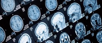
MSCT of the brain is a multi-slice scan of brain structures. Depending on the settings of the tomograph, it can record both soft tissues of different densities (vessels, glands, brain tissue and meninges) and bone structures of the skull. The procedure lasts up to 20 minutes, depending on the purpose and scope of the study.
MSCT of the brain is prescribed:
- If a stroke is suspected - in order to clarify the focus and extent of the lesion (ischemia, hemorrhage, hematoma)
- For the diagnosis of neoplasms (cysts, tumors, infiltrates), their location and size
- For obsessive headaches, dizziness, double vision and other meningeal symptoms of unknown origin - in order to clarify the diagnosis
- If you have symptoms of increased intracranial pressure
- When performing a biopsy of neoplasms of brain structures - for the purpose of better visualization, for greater accuracy of the procedure
- After a head injury to assess the degree of damage to the brain and membranes, the presence of bleeding, foreign bodies
- To assess the condition of blood vessels and the level of blood supply to the brain (for atherosclerosis, ischemia, aneurysm)
To study the condition of the vessels, a radiopaque substance is used, which makes it possible to trace the vessel along its entire length: to assess its integrity, filling, and patency. With the blood flow, it passes through the vessels, highlighting them in the picture. CT uses iodine-based drugs, which are then eliminated from the body by the kidneys.
Contraindications for the use of contrast:
- Kidney failure
- Continuous use of Metformin (for diabetics)
- Intolerance to iodine-containing drugs
- History of hypersensitivity reactions (anaphylactic shock, angioedema), asthma
MSCT devices administer a contrast agent as a bolus - automatically, in a strictly dosed mode. This method allows you to reduce the dose of the drug.
MSCT of the head allows:
- examine the bone structures of the skull for microdamages, structural changes, metastases
- examine the temporal bones in the presence of pathologies of the auditory or vestibular system
- conduct an oncological search: determine the location of the tumor in the organ (if its presence is suspected) and diagnose small focal neoplasms up to 1 mm in size
- monitor antitumor therapy
- establish the causes of impairment or lack of consciousness
- diagnose stroke, cerebral edema, intracranial bleeding, hematoma or hemorrhage, vascular disorders
- assess the condition of the hypothalamic-pituitary system
How is MSCT performed?
- The device is adjusted to the desired structure depending on its density
- The patient changes into loose clothing without metal parts (usually a light disposable gown)
- All jewelry, watches, and dentures are removed during the procedure.
- The subject lies comfortably on a flat tomograph table
Further movements will be performed only by the device:
- The table progressively moves into the device as the desired area of the body is scanned, and the sensors rotate around it, illuminating the selected structures
- The patient does not experience any sensations during the procedure
- When a contrast agent is injected, a slight metallic taste in the mouth and a feeling of spreading warmth may occur, but this quickly passes
Differences between MSCT and MRI
- Imaging method: MSCT is x-rays, MRI is magnetic waves that are reflected from tissues
- Oncological diagnostics: MSCT is preferable
- Indications: MSCT is the method of choice for visualizing bone structures, MRI – soft organs and tissues
- Contrast agent: in MSCT – iodine preparation, in MRI – gadolinium-based (less allergenic than iodine-based)
- Frequency: MSCT – according to indications, MRI – without restrictions
- The presence of metal and electronic implants in the body, tattoos with metal-based paint: MSCT - no contraindications, MRI - contraindicated
- Children's age, pregnancy: MSCT is contraindicated, MRI is allowed from the 12th week of pregnancy (children under 7 years old are performed under anesthesia)
- Duration of the procedure: MSCT – 5–15 minutes, MRI – up to 30 minutes
Contraindications and risks
The main limitations to CT scanning relate to the use of contrast media.
But there are some risks that the patient should be aware of:
- Frequent CT scans at a young age increases the risk of development, worsening or recurrence of cancer
- Research may lead to malfunctions of electronic implants
Advantages of carrying out the procedure at MEDSI
- Modern MSCT devices allow you to carry out the procedure as quickly as possible and minimize the radiation dose
- High accuracy and detail of images allow diagnosing pathology at an early stage, which improves the prognosis of the disease
- Tests are carried out by qualified and experienced radiologists
- More than 25 types of research
Make an appointment by calling 24/7 8 (495) 7-800-500.