We usually don’t hear about epilepsy as often as, say, heart attacks and strokes. However, almost each of us, one way or another, is familiar with this disease, if not personally, then from the stories of others whose relatives or friends suffer from this disease. And this is no coincidence, because epilepsy is observed in almost 5% of the population of our planet, and in children this disease is registered three times more often than in adults. And the general term “epilepsy,” according to the conclusion of modern practicing neurologists, combines more than fifty diseases that differ in clinical manifestations and treatment, including the most common generalized epilepsy.
Definition of idiopathic epilepsy
Idiopathic generalized epilepsy is a form of epilepsy in which seizure activity is recorded from all areas of the brain. Forms in which secondary generalization of seizures occurs cannot be included in this group. If a local component is nevertheless observed, then the likelihood of the disease belonging to the group of idiopathic generalized epilepsy is low.
The IGE group includes:
- Childhood epilepsy with absence seizures;
- Juvenile absence epilepsy;
- Juvenile myoclonus epilepsy;
- Epilepsy with isolated generalized seizures.
BIBLIOGRAPHY:
- Diagnosis and treatment of epilepsy in children. Ed. Temina P.A., Nikanorova M.Yu. // M.: Mozhaisk-Terra. – 1997. – 656 p.
- Evtushenko S.K., Omelyanenko A.A. Clinical electroencephalography in children // Donetsk. – 2005. – p. 539-546, 585-594
- Martinyuk V.Yu. Protocol for the treatment of epilepsy and epileptic syndromes. Protocol for the treatment of status epilepticus in children. – Kiev, 2005.
- Mukhin K.Yu., Petrukhin A.S. Idiopathic forms of epilepsy: systematics, diagnosis, therapy - M.: Art-Business Center, 2000 - 319 p.
- Epileptology of childhood: A guide for doctors / Ed. A.S. Petrukhina. - M.: "Medicine", 2000. - 620 p.
Signs and symptoms of idiopathic epilepsy
Idiopathic generalized epilepsy manifests itself in different ways, but there are common signs:
- The first symptoms of idiopathic epilepsy begin to appear in childhood and adolescence. With a history of close relatives, in 50% of cases it is possible to identify the presence of a genetic predisposition;
- Seizures are recorded at a certain time of day;
- Psychoneurological symptoms specific to this pathology are extremely rarely detected during routine examination;
- Higher cortical functions (thinking, memory) are rarely affected;
- The treatment prognosis is favorable. Children outgrow this disease, but relapses sometimes occur.
The name of the nosological group of the disease in question indicates the nature of the epileptic attacks - generalized. The attacks themselves can take various clinical forms: absences, tonic-clonic, or mixed as a combination of the first two types.
results
The results of the study showed that SHSPs can be part of the structure of many epileptic syndromes (Table 1).
Table 1. Main characteristics of forms of epilepsy associated with SHSP Note. IFE-PHP is idiopathic focal epilepsy with pseudogeneralized seizures.
Table 1. Main characteristics of forms of epilepsy associated with SHSP (end) Note. IFE-PHP is idiopathic focal epilepsy with pseudogeneralized seizures. Most often, in patients with SHSP, symptomatic focal epilepsy was diagnosed (33.8% of cases), cryptogenic focal epilepsy (23.8%), rolandic epilepsy (12.6%), and FEDSIM-DEPD - 12.3% of cases. Other forms of epilepsy were detected much less frequently.
The onset of epilepsy in patients with SHSP varied over a wide age range from the first month of life to 18 years. The average age of onset was 5.7±4.96 years. Most often, epilepsy associated with SHSP debuted in the following age intervals: from the first month of life to 1 year - 16.8%, from 1 year to 3 years - 25.3% of cases, from 4 to 6 years - 20.8% . As they grew older, the likelihood of developing an epileptic syndrome in patients with SHSP gradually decreased: from 7 to 9 years of age - 15.7%, from 10 to 12 years - 9.3%, from 13 to 15 years - 5.7%, from 16 to 18 years old - 6.4%. It is noteworthy that in most cases (62.9% of patients), the onset of the disease was noted in children of preschool age. The age of onset of various syndromes associated with SHSP could differ significantly (see Table 1).
At the onset of epilepsy, SHSPs were noted in 207 cases, which accounted for 43.9% of patients. SHSP as the only type of paroxysms was observed in 28.3% of cases during the entire period of the disease. In other cases, SHSPs were combined with other types of seizures. Two types of attacks or more were observed in 71.7% of cases, three types or more - in 39.3%.
Along with SHSP, 19 different types of seizures could also be observed in the clinical picture of patients in our group. The following paroxysms were most often observed in epilepsy associated with SHSP: focal automotor - 22% of cases, focal hemiclonic - 20.4%, focal sensory - 20.4%, febrile seizures - 14.6%, focal motor (including versive) - 14.2%, asymmetrical tonic seizures - 8.9%. Other types of seizures were less common: focal atonic (so-called “temporal syncopation”) - 6.8% of cases, atypical absences - 6.2%, autonomic paroxysms - 6.2%, seizures originating from the occipital cortex - 4.7%, focal myoclonus - 3.6%, epileptic spasms - 3.4%, negative myoclonus - 2.9%, myoclonic seizures - 2.5%, focal hyperkinetic - 2.1%, focal adversive and gelastic - 1.3%, atonic - 1.1%, epileptic myoclonus of the eyelids - 0.8% of cases.
It should be noted that for different epileptic syndromes associated with SHSP, the frequency of occurrence of different types of seizures differed (see Table 1).
During continued VEM, various types of pathological activity were detected in most cases (91.3%).
The absence of epileptiform activity was noted only in 8.7% of cases (41 patients). Moreover, all these patients were observed with diagnoses: symptomatic or cryptogenic focal epilepsy. In other groups of epileptic syndromes - idiopathic focal forms of epilepsy and epileptic encephalopathies - epileptiform changes were recorded in 100% of patients during prolonged VEM with sleep included.
Regional/multiregional epileptiform activity was recorded in 91.3% of cases of all forms of epilepsy associated with SHSP.
Interictal diffuse epileptiform activity was detected in 30.1% of cases. Moreover, in 100% of cases, diffuse discharges were detected in epileptic encephalopathies, progressive myoclonus epilepsy and idiopathic focal epilepsy with pseudogeneralized seizures (IFE-PHP syndrome).
The study revealed that 175 (37.2%) patients had DEPD. These graphelements were found in all patients with idiopathic focal forms of epilepsy, pseudo-Lennox syndrome, Landau-Kleffner syndrome, electrical status epilepticus syndrome of slow-wave sleep, cognitive epileptiform disintegration, FEDSIM-DEPD syndrome.
During continued VEM with sleep included, HFSPs were recorded in 39 patients. In all cases, a similar EEG pattern of the attack was revealed - at the beginning of SHSP, the appearance of regional rhythmic epileptiform activity on the EEG is noted. The tonic phase corresponds to the appearance of diffuse rhythmic pointed theta activity in combination with rhythmic fast peak-wave and polypeak-wave complexes. As the tonic phase continues, an increase in the amplitude of the acute-slow wave complexes and their slowdown to 1.5-3.5 Hz are observed, which corresponds to the transition to the clonic phase. Clonic seizures were characterized by the appearance of groups of diffuse polypeak-wave complexes of a low degree of synchronization in combination with rhythmic motor and myographic artifacts. As the attack ends, diffuse polypik-wave discharges become less frequent. The stage of post-ictal confusion corresponds to the appearance of diffuse slowing. In all cases, pronounced myographic and motor artifacts were superimposed on the ictal EEG recording, making it difficult to assess the bioelectrical activity of the brain; in 12 cases, an objective assessment of the EEG pattern of the attack was impossible due to the large number of artifacts.
Analysis of the use of therapy demonstrated that patients were prescribed the following groups of drugs, both in mono- and polytherapy in various combinations: carbamazepine, oxcarbazepine, valproate, levetiracetam (tablets and in the form of an oral solution - Keppra), topiramate, perampanel, lamotrigine, ethosuximide.
Prescription of PET led to the achievement of complete remission in 57.1% of cases of epilepsy associated with SHSP. A reduction in the frequency of attacks by 50% or more on the background of PET was observed in 33.6% of patients. No effect on seizures was noted in 9.3% of cases.
The study showed different effectiveness of PET in the treatment of certain groups of epileptic syndromes associated with SHSP (Table 2).
Table 2. Efficacy of PET in various forms of epilepsy associated with SHSP (%) The highest percentage of remission was observed in idiopathic focal forms of epilepsy (94.7% of cases) and in FEDSIM-DEPD syndrome (77.6%). In the largest group of patients with symptomatic/cryptogenic focal epilepsy, remission was achieved in only 42.0% of cases. In epileptic encephalopathies, remission was observed in 45.2% of patients.
Separately, the effectiveness of PET was analyzed in forms of epilepsy associated with DEPD, which were recorded in patients with idiopathic focal forms of epilepsy, pseudo-Lennox syndrome, Landau-Kleffner syndrome, electrical status epilepticus syndrome of slow-wave sleep, cognitive epileptiform disintegration, FEDSIM-DEPD syndrome (37 .2% of all patients with SHSP). High effectiveness against attacks was obtained, which was significantly higher than in the general group of patients with SHSP. Remission of attacks was observed in 84.6% of cases, a reduction in the frequency of attacks by 50% or more in 14.3%, and no effect on paroxysms in only 1.1% (see Table 2).
Causes of idiopathic epilepsy
It is very difficult to identify an objective etiological factor contributing to the occurrence and development of this disease. In a number of experimental studies, it was found that one of the reasons is a genetic factor, which determines the hereditary nature of the pathology. However, at the same time it was revealed that different forms of the disease correspond to mutations in different parts of the human genome.
Factors that can trigger an epileptic attack:
- Excessive physical or mental stress;
- Sudden interruption of sleep;
- Alcohol consumption;
- Mental and emotional stress.
Ketogenic diet for the treatment of epilepsy in children
At our center, we advocate the use of a ketogenic diet in patients with epilepsy. The course format (2-3 weeks) does not allow us to put the child on a diet. However, we strongly advise parents to consult a doctor upon arrival home and begin the process of treating epilepsy with diet. In 10-15% of cases, children with pharmacological resistance achieve remission within 2 years.
The ketogenic diet is not just a diet for epilepsy, it is a treatment. Any slight deviation from the diet, even the use of toothpaste that contains sugar, affects its effectiveness. The diet is prescribed by a doctor, accompanied throughout the entire treatment of epilepsy (usually 2 years) by a nutritionist, and begins in the hospital with a 24-hour fast.
The diet was developed in 1920 to alleviate epileptic seizures in children. It almost excludes carbohydrates, the basis of the diet is fats and a moderate amount of protein foods. The ratio of fat to protein + carbohydrates is 4 to 1.
The ketogenic diet in the treatment of epilepsy works in 50% of all cases, 34% of children have good, lasting results after its use. Almost all children (more than 90%) show a marked improvement in their condition within 6 to 24 months of using the diet. (3, 4)
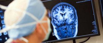
Diagnosis of idiopathic epilepsy
Diagnostics includes analysis of electroencephalography data and techniques that allow one to see structural changes in the brain substance. When conducting an EEG examination, seizure activity is recorded in symmetrical parts of the brain. A characteristic feature of IGE is the absence of tomographic criteria for brain changes. However, if studies are carried out using high-resolution expert-class devices, a specialist can identify areas of degenerative changes: microcysts and foci of pathological architectonics of the brain matter. However, in this case we should talk about other forms of epilepsy.
Material and methods
The study included 471 patients with identified SHSP. Among the patients we examined, there were no significant quantitative differences by gender: 244 (51.8%) were male patients, 227 (48.2%) were female patients.
Diagnosis of epileptic syndromes associated with SHSP was based on the criteria of the international classification of epilepsies, epileptic syndromes and similar diseases (1989), as well as on the basis of the report of the ILAE commission on classification and terminology (2001).
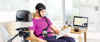
In all cases, continued video-EEG monitoring (VEM) was performed with sleep included (electroencephalograph-analyzer device EEGA-21/26 ENCEPHALAN-131−03, modification 11, Medicom MTD, Russia; VEM Neuroscope 6.1.508, Biola ", Russia). The VEM program included: research in a state of wakefulness (before and after sleep) with functional tests, as well as natural sleep (without the use of sedatives). Functional tests included hyperventilation for 3 minutes and rhythmic photostimulation in the frequency range from 3 to 24 Hz. All patients underwent magnetic resonance imaging (MRI) (Sigma Infinity GE magnetic resonance system with a magnetic field voltage of 1.5 Tesla).
Vitamins in the treatment of epilepsy
Taking anticonvulsant drugs for the treatment of epilepsy leads to a decrease in the concentration of useful substances in the blood (E, D, B1, B6, B9, Omega-3, Selenium, Zinc, Magnezium).
Drug treatment of epilepsy is always associated with a deficiency of vitamins, to replenish which it is necessary to eat the most nutritious healthy foods and take vitamin complexes. For more effective treatment of epilepsy in children, we help you choose a course of vitamins. We provide brief information on why vitamins and minerals are required for the comprehensive treatment of epilepsy in a child.
B9 (Folic acid)
AEDs inevitably reduce the level of vitamin B9 in the blood. According to some data, taking folic acid reduced epileptic seizures in children, other studies do not indicate the benefit of taking vitamin B9 for epilepsy. However, after an attack, the level of folic acid in the child’s blood drops sharply; the growing body constantly spends reserves to make up for this deficiency. Folic acid deficiency in a child with epilepsy can be compensated for with a synthetic complex, or with increased consumption of foods rich in folic acid: spinach, oranges, cherries, etc.
Omega-3 (fish oil) Professor at the University of California, K. DiGiorgio, experimentally proved that with the systematic consumption of omega-3 fatty acids, the number of epileptic seizures is reduced by a third. Omega-3 fatty acids were taken for 10 weeks; they are found in large marine fish, and can also be sourced from dietary supplements and vitamin complexes.
The professor hypothesized that omega-3s have a calming effect on the nervous system and neuronal excitability, so the number of epileptic seizures decreases. Simply taking fish oil once a day is effective in treating epilepsy in children and adults in a third of cases.
Magnesium and zinc and epilepsy in children
The frequency of epileptic seizures and benign myoclonic seizures in children may be associated with magnesium and zinc deficiency. When the temperature rises, a slight deficiency of magnesium in the blood occurs, which can lead to a seizure. Insufficient zinc content affects the conductivity of nerve cells in the brain, but its excess is also dangerous for children with epilepsy, since it increases the excitability of the nervous system. Before taking any vitamin complexes, you must undergo appropriate tests.
Craniocerebral stimulation
Doctor L.I. Levit has mastered various osteopathic techniques for the rehabilitation of children, and has been working with children with mental and psychophysical development disorders, epilepsy and other neurological diseases since 1996.
Dr. Levit's original method - craniocerebral stimulation - is a painless and very gentle method of treating epilepsy. Outwardly, it represents a barely perceptible touch on the head, spine, sacrum - very light and gentle manipulations with the child’s body. The technique has a beneficial effect on a wide range of neurological disorders, including epilepsy.
Over the past decades, osteopathy and cranial therapy have become part of official medicine. Every year the position of this science is strengthening: more and more hospitals and medical centers in the USA, England, France, and Israel are using it in their practice. There is not a single advanced country in the world in which osteopaths and cranial therapists do not work, relying on the developments of doctors Still, Maqun, W. Sutheiland, M. Littlejohn and others. Dr. Lev Levit works within the framework of the American school of John E. Upledger.
There are several rhythmic cycles in the body: cardiac, respiratory, gastrointestinal, etc. The human nervous system has cyclicity and periodicity.
There is also a so-called craniosacral rhythm, which determines the amplitude and frequency of the craniosacral system, which primarily affects the state of the central nervous system. Therefore, restoration of the craniosacral rhythm has a very beneficial effect on the condition of a child suffering from epilepsy.
Our main method of rehabilitation is craniocerebral stimulation (cranio - skull, cerebrum - brain). This is an original technique based on osteopathy. In all forms of neurological disorders, the dynamics of the outflow of fluid that circulates in the brain and spinal cord is disrupted. It helps remove old cells and toxins, and nourishes our brain with new microelements.
This process is called liquor dynamics. If the system fails, as a result, the functioning of the brain and cranial system is disrupted. When carrying out procedures in patients with epilepsy, the circulation of cerebrospinal fluid, metabolic processes in the brain are improved, and the conductivity of nerve impulses is improved. Treatment with cranial therapy and craniocerebral stimulation in children with epilepsy reduces the intensity and frequency of seizures. In the practice of the center, there are also children who have achieved complete remission; epilepsy attacks have not recurred after the course.