General information
Inside both hemispheres, in addition to the cortex, there are separate nuclei of gray matter.
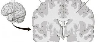
They, in turn, form the upper layers of the telencephalon, are located next to the base, inside the body of the white area, due to which they are called the basal (subcortical) nuclei. They also include the amygdala and striatum. The gray-white area is essentially the subcortical part of the teleencephalon and the main input base of the basal ganglia system. In sections taken horizontally or frontally, the striatum appears as an alternation of gray and white stripes, which is why it was called the striatum.
Damage to the striatum and consequences
When the striatum stops functioning, a person experiences the following disorders:
- Athetosis. Banal alternating movements of the limbs.
- Chorea. Incorrect movements that are performed without any sequence or order, involving the entire musculature of the body.
- Strengthening unconditioned reflexes (defensive, indicative, etc.).
- Hyperkinesis. Significant strengthening of auxiliary movements that accompany each main movement.
- Hypotonicity. Upset muscle tone, its decrease.
- The appearance of Tourette's syndrome.
- The onset of Parkinson's disease contributes to the death of neurons in the body, which is why domafin, which is responsible for the motor system of the human body, is not produced.
- The appearance of Huntington's disease.
In addition, damage to the striatum and caudal nucleus in particular:
- Completely or partially prevents the perception of painful, visual, auditory and other types of stimulation.
- Reduces or increases salivation.
- Makes it difficult to orient in space.
- Impairs memory.
- Slows down the growth of the body.
- Promotes the disappearance of conditioned reflexes for a long time. Human behavior can be inert and stagnant.
History of terms
In the seventeenth and eighteenth centuries, the term "corpus striatum" was used to designate the various, deep, intercortical elements of the hemisphere. In 1941, the nomenclature was simplified by proposing the term "striatum" for all elements constructed with striped segments.
The concept "neostriatum" was also used, but as a collective designation for comparative anatomists comparing subcortical structures between vertebrates, because it was thought to be a phylogenetically newer segment of the striatum. The term is still used by some sources, including medical subject headings.
Anatomy of the structure
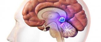
Striatum consists of two parts:
- Dorsal (posterior) region. Placed externally, it includes both the caudate and lenticular segments, as well as the shell.
- The ventral (anterior) region, which consists only of the nucleus accumbens. Layers of white matter divide it into segments: the medial globus pallidus, the putamen of the lenticular area, and the lateral globus pallidus.
The kernel was called lentiform because of its appearance resembling a lentil grain. The fence itself looks like a thin plate consisting of a gray substance located on top of a lenticular segment.
The ancient striatum (paleostriatum) is a formation that is a pale ball. It stands out both macroscopically and histologically against the background of other parts of the striatum.
The formation of a new structure (neostriatum) in mammals occurs taking into account the development of sensorimotor centers of the neocortex. The neostriatum consists of: the caudate body, the shell itself and the fence.
The trunk of the striatum is:
- A critical component of the motor mechanism system.
- Includes glutamatergic and dopaminergic inputs from various sources.
- Serves as a major input to the rest of the basal ganglia.
In primates, the striatum is divided into the ventral striatum and the dorsal striatum, subdivisions based on function and connections. The ventral striatum includes accumbens cells and the olfactory tubercle. The dorsal striatum includes only the caudate nucleus and putamen. White matter, a nerve tract (internal capsule) in the dorsal striatum separates the caudate region from the putamen. Anatomically, the term striatum describes the striped appearance of the grey-white matter.
The ventral striatum consists of accumbens cells and the olfactory tubercle. The accumbens nucleus is composed of the accumbens nucleus itself and the accumbens nucleus sheath, which have distinct neuronal populations. The olfactory tubercle receives input from the olfactory bulb but is not involved in odor processing. The ventral striatum is connected to the limbic system and is involved as the main region of the circuitry responsible for decision making and coordination of behavior, reward motivation.
The dorsal striatum consists of two sections: the caudate and the putamen. Staining can differentiate the dorsal striatum into compartments of striosomes and surrounding matrix. The striped material is spatially organized according to several levels.
Anatomical divisions and territories
The dorsal striatum forms a continuous large mass, topographically separated by the internal capsule in the caudal segment medially, the putamen on the side and the floor below, connecting the two preceding ones ventrally into a single whole. The striatum is homogeneous in its neuronal components.
Structure
The striatum consists of:
- Caudate nucleus.
- Lenticular nucleus.
- Fences.
If we examine the body under a microscope, it consists of large neurons with long tails extending beyond the boundaries of the striopallidal system.
The parts of the caudate are the head, body and tail. The head forms the lateral wall of the anterior horn of the lateral ventricle; the body of the nucleus is extended along the central part of the ventricle; the tail is located on the upper wall of the inferior horn of the ventricle and ends at the level of the lateral geniculate body.
The posterior wall of the nuclear head is located on the border with the thalamus, separated by a strip of white matter.
The lenticular kernel, as the name implies, is shaped like a lentil.
It is located sideways to the caudate nucleus and thalamus. If you cut the kernel in half, it has a wedge-shaped shape, with the top facing the middle and the base facing the side.
And small layers of white matter divide the nucleus into several parts:
- The shell.
- Lateral globus pallidus.
- Medial globus pallidus.
The globus pallidus is a specific ancient formation (ancient body), which differs from other parts of the striatum both in macroscopic and histological appearance.
The fence is located outside the lenticular core. Externally, it is a thin, up to two millimeters, plate of a gray substance. The middle of the plate is smooth, and on the lateral edge there are small bulges of gray matter.
Organized interaction
The dorsal striatum is a single entity, closed and continuous with a toric topology. The observed anatomical subdivisions of the dorsal striatum (caudate nucleus and putamen), essentially induced by the internal capsule, do not completely overlap with the currently accepted anatomical and functional subdivisions. The selective distribution of axon terminal arborizations of cortical sources differentiates the sensorimotor striatum, mainly putaminal, but located in its dorsal part and in the lower part of the caudate. Most of the remaining volume (mostly caudate), derived from axon terminals from the frontal, parietal, and temporal cortex, forms the associative striatum. The division between these two territories is quite clear and observable using calbindin immunochemistry. The third entity, the most sterile, is more problematic because there is no general agreement regarding its boundary with the associative stripe.
The ventral striatum is clearly delineated, tracing the subic territory. This corresponds to the olfactory tubercle and the nucleus, which is not actually an olfactory tubercle, being a striped part consisting of gray-white elements.
Functions of education
Functionally, the striatum coordinates multiple aspects of cognition, including both motor behavior and action planning, decision making, motivation, reinforcement, and reward perception.
The striatum performs a number of functions:
- planning the entire complex of actions and establishing control over movements;
- participates in cognitive processes in order to implement multidirectional moves;
- involved in learning together with midbrain neurons containing dopamine;
- participates in making and finding decisions together with the prefrontal cortex;
- used in the mechanism of the reward system (a person receives pleasure from what is happening both in the past and present, including from the very sensation while waiting for them);
- the limbic system is involved in short-term memory, emotions, motivation and reinforcement; responsible for pleasure, satiety, guilt, punishment, inhibition, wakefulness, alertness, excitement and autonomic activity; involved in cortical speech and language.
Metabotropic dopamine receptors are present on both spiny neurons and cortical axon terminals. Second messenger cascades caused by activation of these dopamine receptors can modulate pre- and postsynaptic functions, both in the short and long term. The striatum is best known for its role in planning and modulating movement pathways, but is also involved in a variety of other cognitive processes including executive function.
In humans, the striatum is activated by reward-related stimuli, as well as by aversive, novel, unexpected, or intense stimuli and cues associated with such events.
Recent functional magnetic resonance imaging data indicate that the common stimuli to which the striatum responds are relevant for understanding all mechanisms. A number of other brain regions and circuits are also associated with reward. For example, frontal areas.
For sources regarding the importance of the reward pathway (thought to be related to dopamine), one can look at the work of Dr. John Salmone (from the early to late 1990s) and Wolfram Schultz. Ventral tegmental dopaminergic neurons, which innervate parts of the striatum, have long been recognized as the site of the sense of reward. Intracranial stimulation studies conducted in the 1960s indicate that implants in this area of the brain will produce bar pressure in rats for many hours. However, collective work by researchers in the 1990s shows that blocking dopamine receptors does not eliminate rewarding sensations, but rather affects how willing the animal is to work, making it more motivated to seek rewards rather than reward itself.
Pathologies and symptoms
- Problems and damage to the nucleus accumbens. Addiction occurs, which is the behavior of a person with a characteristic obsessive need for some specific activity (for example, a person, when he gets used to medications, may experience an irresistible desire to take them again).
- Lack of the hormone dopamine. This is dangerous because the hormone is necessary for correct movements. The number of dopamine receptors decreases with age, and as a result, the concentration of the neurotransmitter decreases. Excessively high production of dopamine system genes can also disrupt motivation itself, while other manifestations of behavior are not affected.
- Disorders due to genetic factors lead to the death of striatal cells, which ultimately affects the functions of the basal ganglia as a whole.