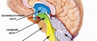
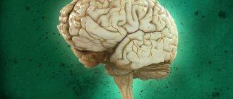
Structure and functions of the hindbrain
The metencephalon (posterior part) develops at the stage of embryonic ontogenesis, originating from the anterior part of the rhomboid region. During development, the structure of the hindbrain is supplemented and gives rise to the pons and cerebellum. Thus, the rhomboid section becomes the myelencephalon (secondary medullary vesicle) from which the medulla oblongata of the CNS organ originates.
The anatomy of this structure is quite well studied and in evolutionary development it belongs to the most ancient area. The physiology of the hindbrain was studied using the ablative method (removing part of the organ and then observing changes in the body), which helped to deeply study the functioning.
Having studied the back of the brain in section, scientists were able to describe which cavity communicates it with the medulla oblongata of the central nervous system - the fourth ventricle. The cranial nerves pass through it; the medullary striae serve as the border.
The posterior (diamond-shaped) section of the central nervous system consists of:
- Oblong section;
- The hindbrain itself.
The second structure, in turn, is divided into the pons and the cerebellum. The rhomboid part of the central nervous system is involved in reflex activity, since it contains nerve bundles and cranial nerves that perform various functions.
Hindbrain - structure and functions of reflex activity:
- Gaulle's bundle - represented by axons, regulates muscle and joint sensitivity (from the lower extremities);
- Burdach's bundle - includes axons, regulates muscle and joint sensitivity of the entire upper body (from the neck).
These bundles form the path of proprioceptive sensitivity and allow you to recognize the position of body parts in space, perceive postures and feel both active and passive movements. If there are disturbances in the cortical direction, the coordination of movements is lost and they become disproportionate.
Reflex activity is also carried out with the help of innervation by cranial nerves (nuclei from 5 to 12 pairs), which form the classification of various reflexes in the corresponding structures of the central nervous system.
The hindbrain is involved in the implementation of the following reflexes:
- Responsible for tactile sensitivity - the function is provided by the trigeminal nerve (5th pair), located between the pons and the middle cerebellar peduncle. Supports unconscious reflexes in response to pain or touching a hot object. At the exit from the bridge it connects with the mandibular nerve and innervates the muscles of mastication. Damage to the fibers is accompanied by sharp pain and hyperemia of the skin on the face. If disturbances affect the motor nuclei, then atony of the masticatory and temporal muscles occurs.
- Responsible for the work of the oculomotor muscle (rectus) - the function is supported by the abducens nerve (6th pair), lies in the thickness of the bridge, exiting in the oblong part of the central nervous system and penetrates into the orbital area. Damage to the fibers leads to blurred vision (double vision) and the inability to direct gaze in any direction.
- Provides facial expressions - innervation of the facial nerve (7th pair). The roots extend from the bridge tire. After which a loop is formed and then the fibers run through the thickness of the bridge, exiting through the gap between the oblong structure of the central nervous system organ. Damage to the fiber (its branches) leads to a complete absence of facial expressions, the face becomes like a mask (all folds are smoothed out, blinking movements are impossible, eyelids do not droop).
- Responsible for hearing and vestibular regulation - the vestibular nerve (8th pair), is divided into two parts. The first (cochlea) conducts auditory impulses to the central nervous system, the second (vestibular) is located at the bottom of the auditory canal and regulates balance. From the first part, the nerve fibers end in the tegmentum of the bridge, from the second, in the cavity of the diamond-shaped structure - the diamond-shaped fossa. Damage to the nuclei leads to hearing loss and impaired balance (staggering gait).
- Responsible for swallowing movements - muscle contraction is provided by the glossopharyngeal nerve (9th pair), and in addition to the pharynx, it innervates the middle ear and tongue (posterior third). It comes out of the hole in the skull and runs to the cavity of the rhomboid region (4th ventricle). Damage to the fibers causes difficulty swallowing (or pain) and impaired taste sensitivity of the tongue.
- Responsible for the functioning of organs in the abdominal and thoracic cavities - the function is provided by the vagus nerve (10 pair), starting from the oblong part of the central nervous system organ, extending down through the neck to the cavities. Damage to the nuclei causes a malfunction of the internal organs, and paresis of the pharynx or larynx is possible.
- Controls contractions of the trapezius and large superficial muscles of the neck - the innervation comes from the accessory nerve (11th pair), the upper part of the fibers starts from the oblong structure, the lower part - from the anterior horns of the spinal part of the central nervous system (upper segments). At the exit from the cranium it intertwines with the vagus nerve. Damage to these fibers causes paresis or paralysis of the innervated muscles.
- Responsible for movements of the tongue - innervation is provided by the hypoglossal nerve (12 pair), located in the rhomboid fossa and has multiple branches, the final branch ends in the tongue. Damage to the fibers leads to impaired motor abilities in the tongue (muscle atrophy).
All parts of the diamond-shaped structure are connected to each other through neural connections that act as conductors.
Brain cells
The brain is made up of two types of cells: nerve cells (neurons) and glial cells.
Nerve cell
Neurons come in many sizes and shapes, but they all consist of a cell body, dendrites, and an axon. A neuron transmits information through electrical and chemical signals. Try to imagine the electrical wiring in your home. An electrical circuit consists of numerous wires connected in such a way that when a light switch is turned on, a light bulb will glow. An excited neuron will transfer its energy to neurons in its immediate vicinity.
Important Causes, symptoms and types of tachylalia
Neurons transmit their energy, or “speak,” to each other through a tiny gap called a synapse (Figure 12). A neuron has many arms called dendrites, which act like antennas that collect messages from other nerve cells. These messages are passed to the cell body, which determines whether the message should be passed along.
Important messages are carried to the end of the axon, where sacs containing neurotransmitters open into the synapse. Neurotransmitter molecules cross the synapse and are placed into special receptors on the receiving nerve cell. This stimulates the cell to transmit the message.
Figure 12. Nerve cells consist of a cell body, dendrites, and an axon. Neurons communicate with each other by exchanging neurotransmitters through a tiny gap called a synapse.
Glial cells
Glia (Greek word meaning glue) are brain cells that provide neurons with nutrition, protection, and structural support. There are approximately 10-50 times more glia than nerve cells and are the most common cell type involved in brain tumors.
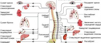
Functions of the pons
The bridge is a small ridge with a depression (basilar groove), which contains a large amount of nerve fiber. The hindbrain is the pons located under the cerebellum. The main purpose of this structure is to transmit information from the back part to the largest part - the forebrain.
The functionality of the bridge is to support unconditioned reflexes (protective) in the body:
- Cough;
- Sneezing;
- Vomit;
- Blinking.
The pons Varoliev forms descending pathways through which impulses pass not only to the cortex of the central nervous system, but also in the opposite direction. Thus, the rhomboid region is connected with the spinal canal and with all the structures in its anatomical structure.
There is a bridge inside the skull
A diagram of the structure of the brain shows that another component, the pons, is located below the peduncles and forms a prominent protrusion (due to transverse fibers directed into the cerebellum). In the anterior zone of the bridge there are predominantly conductive pathways. Namely: the path of the auditory nerve nuclei, the aforementioned pyramidal (motor, cortico-muscular), general sensory (from the spinal cord to the visual thalamus, medial lemniscus), etc.
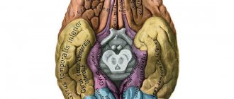
The structure of the human brain includes such an element as the cerebellum. It is located above the medulla oblongata in the posterior cranial fossa. Covered on top by the occipital lobes of the cerebral cortex. The cerebellum has two hemispheres and a central part - the vermis. In a newborn, this organ weighs 20 grams, by five months the weight increases 3 times, by nine months - 4 times.
The final formation of this organ is completed by the age of 15. The surface of the hemispheres is gray matter - the cortex, under which lies the white matter. The organ has three pairs of legs connecting it with other parts: the lower pair - with the medulla oblongata, the middle one - with the pons, the upper one - with the midbrain.
Structure and functions of the cerebellum
The cerebellum is the most posterior part of the rhomboid structure of the brain, as it is located in the cranial fossa.
Anatomical structure:
- Right hemisphere;
- Left hemisphere;
- Worm;
- Brain body.
Visually, the cerebellum is similar to the cerebral hemispheres - lobules, formed on the surface by small grooves and leaves, covered with a cortex on top. The lobules are connected to each other by a worm. The structure is separated by a gap from the central nervous system organ using a tentorium - stretched over the cranial fossa.
Structure functions:
- Regulation of muscle tone;
- Posture coordination;
- Purposefulness of movements.
The connection between the cerebellum and the pons, oblongata and middle section is ensured by bundles of nerve fibers (peduncles). Bundles of axons are located under the layer of gray matter and form a pyramidal pathway for transmitting information.
Tick-borne encephalitis
This disease has a viral etiology and is preceded by an increase in temperature, the development of headaches and malaise, weakening of muscle tissue, which causes rigidity. With an advanced form of tick-borne encephalitis, the symptoms worsen: the patient often exhibits hallucinations and obsessions, he becomes aggressive and agitated, and suffers from seizures.
Measles
With the development of measles encephalitis, damage to the cerebral hemispheres is manifested by a sharp increase in temperature, confusion, as a result of which the patient cannot navigate in time and space, visual hallucinations and nervous excitability. Characteristic symptoms of measles encephalitis are also convulsions that develop in all parts of the body, paresis and weakness of the muscles of the limbs: there is a risk of coma.
Lethargic
If damage to the cerebral cortex is caused by the development of lethargic encephalitis, the patient’s temperature rises, speech deteriorates, and catatonia occurs - a pathological condition in which the body freezes in one position for several hours. Lethargic encephalitis is also characterized by symptoms such as apathy, malaise, and drowsiness caused by disruption of the daily routine.
Important Review and application of parapodiums for the rehabilitation of neurological patients
Functions and significance of the medulla oblongata
Reflexes of the hindbrain are carried out using the medulla oblongata, which acts as a conductor of information. The structure (bulb) is a part of the dorsal part of the central nervous system, but originates from the rhomboid part of the posterior part of the brain.
This structure contains a vital center - the respiratory one, damage to which leads to death. The bulb also regulates balance and coordination of movements.
The bulb contains bundles of axons that provide communication between the spinal cord and other parts of the central nervous system (conducting tracts - long and short). In the oblong part of the central nervous system, autonomic functions are regulated. This structure contains active poles that stimulate the production of various secretions: salivary, lacrimal, and gastric enzymes.
The medulla oblongata of the rhomboid brain has age-related characteristics - at the time of birth, cells are developed only to regulate breathing, blood circulation and control digestive processes. The structure of the bulb begins to function fully (as in an adult) only from the age of 7.
The rhomboid brain belongs to the most ancient part of the central nervous system and regulates vital processes in the body. This section is the main conductor for incoming information, since it connects the centers of the anterior section with the spinal cord.
Pathologies and signs of damage to departments
Extensive damage to the medial areas of the frontal lobe provokes the development of abulia, which is manifested by slow reactions, indifference, and indifference to what is happening. If an area of the prefrontal orbital cortex is damaged, the patient experiences a lack of critical assessment of his own behavior and emotional lability.
Bilateral injury in the frontal region is accompanied by signs: agitation, restless behavior, obsessiveness, verbosity. Abnormal behavior is a sign of dementia, which develops against the background of degenerative processes affecting the frontal lobes. Damage to the motor cortex medulla causes hemiparesis, or muscle weakness.
Disorders develop on the side opposite to the location of the pathological focus in the brain. Damage to the visual area in one hemisphere leads to the development of bilateral blindness in half the visual field. A lesion in area 19 is associated with visual agnosia, a disturbance in visual perception. The patient sees an object, but cannot recognize it.
The information that comes through the visual analyzer is not processed or processed incorrectly, which leads to the inability to distinguish between familiar objects and people's faces. In such patients, color perception is impaired - they cannot distinguish shades.
Damage to field 22 leads to the development of musical deafness (impaired perception of musical works), the appearance of auditory hallucinations, and impaired reactions focused on auditory stimuli. Damage to field 41 is accompanied by the development of cortical deafness (inability to perceive sound signals).
Damage to field 34 is accompanied by impaired perception of smells, including olfactory hallucinations. Pathological structural changes in the nerve tissue of area 39 lead to the inability to read and write. When the field tissue is damaged, 37 people do not remember the names of objects.
Areas of the brain are divided into sensory and motor, as well as associative - and all areas interact with each other. Each department is endowed with certain functions, which together determine higher mental and complex motor activity.