NEXT LEVEL coaching company begins to introduce you to how our brain works. First, let's talk about what the human brain is made of.
The human brain is a complex network of neurons. These neurons serve to build the nervous system. It is with their help that information is transmitted from the brain to the entire body and back.
You probably think that such a complex process requires a huge number of neurons. But how many neurons are there actually in the human brain? Previously, scientists, as one claimed: “100 billion,” but recent studies have shown that there are much fewer neurons in our brain.
How many neurons are there in the human brain?
According to many scientists, the human brain consists of about 100 billion neurons (give or take a couple of billion). It is this figure that has been cited for many years in textbooks on neurobiology and psychology. And all this time, scientists believed that this figure was close to the truth.
That was until Brazilian researcher Suzanne Herculano-Housel discovered that this figure was not entirely accurate. The scientist realized that despite the fact that data on 100 billion neurons are persistently published in many works, it is simply impossible to calculate where this magic figure came from. Then the neuroscientist decided to conduct her own investigation to finally find out how many neurons there actually are in the human brain.
It would seem that the task is elementary. Simply take a sample of the brain, count the number of neurons in that sample, and then purely mathematically calculate the total number of neurons, given the total volume of the brain.
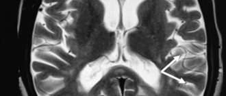
How many neurons were there in the brain studied by scientists?
“We estimate that the brain, on average, consists of 86 billion neurons,” Herculano-Housell said. “No brain that has been studied has even close to 100 billion neurons.” Perhaps someone will consider the difference of 14 billion not so significant, but in fact it is a lot. Judge for yourself: a baboon's brain consists of 14 billion neurons. Half of a gorilla's brain contains the same number of neurons. So, in fact, the difference is quite impressive.
Thus, based on recent research, there are about 86 billion neurons in the human brain.
Electrochemical machine
The human brain weighs only one and a half kilograms, which “fit” about 100 billion cells. Most of them are neurons
.
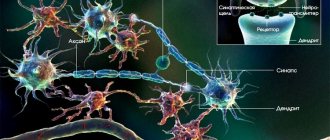
The operating principle of these cells is approximately the same as that of a conventional electrical switch. Neurons have a resting state (off) and an active state (on), in which the electrical impulse is transmitted further along the “wire”. Each neuron consists of a cell body, a “wire” - an axon
, on which there is a kind of “contact” -
a synapse
.
Through it, a neuron connects to another neuron. For this purpose, special chemicals are produced in neurons - neurotransmitters
.
These include, for example, adrenaline, dopamine and others. Different neurons use different chemicals. The release of neurotransmitters to call other neurons occurs at the synapse. By the way, all nerve cells are capable of generating an electrical discharge, the total power of which can reach 60 watts
. Electrical activity of the brain is one of the important indicators of its functioning. It can be measured using a special device - an electroencephalograph (EEG).
How many neurons are there in the brain of animals?
According to Herculano-Housell, the human brain is very similar to the brains of other primates, with one difference: we have many more brain cells, which require enormous amounts of energy to fuel and maintain.
According to experts, approximately 25% of all energy goes specifically to supporting these cells.
If we compare the number of neurons in the human brain and other representatives of the animal world, the difference looks simply huge. So how many neurons are there in the brains of other animals?
Fruit fly – 100 thousand neurons
Cockroach – 1 million neurons
Mouse – 75 million neurons
Cat – 1 billion neurons
Chimpanzee – 7 billion neurons
Elephant – 23 billion neurons
Brain. Neural Factory
We pronounce the phrase “nerve cells do not recover” in dialogues, hinting to the interlocutor that there is no need to worry so much. But what is its origin? For more than 100 years, scientists believed that neurons were not capable of division. And, according to these views, when he died, an empty space remained in his brain forever. Stress is known to be detrimental to nerve cells. So what happens - the more nervous you are, the more “holes” there are in the nervous system?
Nursery for nerve cells
If nerve cells disappeared from the brain forever, then, probably, the Earth would not have seen the rise of civilization. A person would lose his cellular resources before acquiring any skills. Neurons are very “delicate” creatures and are easily destroyed by adverse influences. It is estimated that we lose 200,000 neurons every day. This is not much, but nevertheless, over the years, the shortage can affect health if the losses turn out to be irreparable. However, this does not happen.
Scientists' observation about the impossibility of dividing nerve cells was absolutely correct. But the fact is that nature has found another way to restore losses. Neurons can multiply, but only in three parts of the brain, one of the most active centers is the hippocampus
. And from there the cells slowly migrate to those areas of the brain where they are lacking. The rate of formation and death of neurons is almost the same, so no functions of the nervous system are impaired.
Who has more?
The amount of nerve cell loss varies greatly with age. It would probably be logical to assume that the older a person is, the more irreversible nervous losses he has. However, young children lose the most neurons. We are born with a significant supply of nerve cells, and in the first 3-4 years the brain gets rid of the excess. There are almost 70% fewer neurons. However, children do not become stupid at all, but, on the contrary, gain experience and knowledge. Such loss is a physiological process; the death of nerve cells is compensated by the formation of connections between them.
In older people, the loss of neurons is not fully compensated, even by the formation of new connections between nerve cells.
It's not just about quantity
In addition to restoring cell numbers, the brain has another amazing ability. If a neuron is lost and its place is not occupied for some reason, then neighbors can take over its functions by strengthening connections with each other. This ability of the brain is so developed that even after quite severe brain damage, a person can successfully recover. For example, after a stroke, when neurons in an entire area of the brain die, people begin to walk and talk.
Hit the hippocampus
With many adverse effects and diseases of the nervous system, the restorative function of the hippocampus is reduced, which leads to a decrease in neurons in the brain tissue. For example, regular drinking of alcohol slows down the proliferation of young nerve cells in this part of the brain. With a long “alcoholic experience,” the brain’s recovery abilities decrease, which affects the alcoholic’s state of mind. However, if you stop using it in time, the nervous tissue will recover.
But not all processes are reversible. For Alzheimer's disease
the hippocampus becomes depleted and ceases to perform its functions fully. With this disease, nerve cells not only die faster, but their losses become irreplaceable.
But acute stress is even beneficial because it mobilizes the brain. Another thing is chronic stress.
The nerve cells it kills can still be replaced by the hippocampus, but the recovery process is significantly slower. If stressful circumstances are strong and prolonged, the changes may become irreversible.
Exercises to improve neuroplasticity
All these exercises are related to inviting your brain to do things that are familiar to it in an unusual way and to collect “other teams” of neurons for this. By training the brain to perform unusual actions, we keep it in good shape and improve learning ability.
- Use your non-dominant hand
For starters, you can try brushing your teeth or eating with your other hand. As you get more comfortable with this, switch back to your dominant hand.
- Turn off vision
Try eating, showering, and doing other usual things with your eyes closed (just be careful).
- Change small habits
Find ten new ways to get to work. Have lunch in a different place in your apartment than you usually do.
- See the world upside down
You can do this by looking “under your knees,” as children do, or by turning over familiar objects and looking at them from a different angle.
According to some studies, regular meditation and exercise can also improve neuroplasticity.
What processes are controlled by different hemispheres?
A significant part of the brain belongs to two hemispheres - right and left.
They perform different functions. The right hemisphere is responsible for grouping information, the left hemisphere is responsible for analyzing it. For example, the right hemisphere “sees” a car and recognizes that it really is a car. And the left one “determines” that this is not just a car, but a neighbor’s car. A sober mind until old age
With dementia, a person gradually loses his higher mental functions: memory, speech, attention, intelligence. Find out what dementia is and how to prevent it.
It is widely believed that the right hemisphere is responsible for the perception of abstract things (color and shape), and the left hemisphere is responsible for mathematical abilities, logic and speech. Researchers are finding more and more evidence of such differentiation. For now, scientists can only say with absolute certainty that the right hemisphere controls the left half of the body, and the left hemisphere controls the right.
Human brain
August 25, 2014 Neurology
The human nervous system is represented by the brain, located in the cranial cavity; the spinal cord, located in the spinal cavity, and a branched system of nerves that extend from the brain (cranial nerves) and innervate the organs of the head; a system of nerves that branch from the spinal cord and innervate the arms, legs, torso, and internal organs. The brain and spinal cord represent the central nervous system, and the nerve system represents the peripheral nervous system.
All formations of the nervous system consist of many neurons (cells of the nervous system) and their processes, through which nerve impulses are transmitted in ascending and descending directions due to the diverse connections that exist between neurons.
Despite the fact that different neurons perform different functions and have differences in structure, they all have a body, a receptive structure, and a process, a dendrite, a conducting structure.
According to their functional characteristics, neurons are divided into motor - executive, and sensitive - perceiving, as well as interneurons that interact between them.
The nerve cell performs two main functions: 1) processing of incoming information, transmission of nerve impulses, and 2) biosynthetic, aimed at maintaining its vital functions.
This is the schematic diagram of the structure of a neuron.
This is what the human brain looks like.
This is a complex structure, consisting of many different formations that are in close interaction; carrying out conducting, analyzing, regulating and coordinating functions. All body movements, human feelings, the work of internal organs, his mind, intellect, memory, consciousness, sleep, wakefulness, everything is controlled by the brain. The human brain can be compared to a complex computer with embedded programs that are constantly modified throughout a person’s life.
Schematically, the brain can be divided into lobes: frontal, occipital, temporal, parietal; cerebellum, brain stem. The lobes of the brain are covered with a cortex, which is a collection of highly differentiated neurons that carry out higher integrative activities.
In the frontal lobes there are centers for the regulation of voluntary movements, when damaged, weakness develops in the arms, legs on one side, or only the arms or legs. In the frontal lobes there are also rotations of the eyes and head, when damaged, deviation of the eyes and head occurs towards the pathological focus. The frontal lobes also contain centers for coordination of movements, which, when damaged, cause disturbances in standing and walking. And finally, when the frontal lobe cortex is damaged, behavioral and mental disorders develop.
The parietal lobes are responsible for a person’s ability to recognize objects by touch, the ability to perform complex goal-directed actions, the ability to decipher written characters and the ability to write.
The temporal lobes contain auditory, gustatory and olfactory centers, centers for understanding and reproducing speech, and centers for coordination of movements.
The visual lobes contain the centers for the perception of visual images and visual memory. The cerebellum is one of the main coordinating centers.
In the brain stem there are centers for the regulation of life-supporting organ systems, respiratory, cardiovascular, intermediate centers for the regulation of cranial nerves, the pathways of the motor and sensory systems.
In the brainstem, in its tegmentum, are located the nuclei of the cranial nerves, the bodies of nerve cells responsible for the innervation of the organs of the head and face, providing the functions of the gustatory, auditory, visual, vestibular and olfactory analyzers.
The cranial nerves of the caudal group are distinguished: 1) Accessory nerve, 11th pair, innervates the muscle that turns the head to the side. 2) Hypoglossal nerve, 12th pair, innervating the tongue. 3) Glossopharyngeal nerve, 9th pair, innervating the pharyngeal muscles, tongue, palate, middle ear, salivary glands. 4) Vagus nerve, 10th pair, innervating the muscles of the pharynx, soft palate, larynx, smooth muscles of the bronchi, trachea, esophagus, stomach, intestines.
Next, the cranial nerves of the cerebellopontine angle are distinguished: 1) Facial nerve, 7th pair, innervating the facial muscles. 2) Vestibulocochlear nerve, 8th pair, innervating the inner ear. 3) Trigeminal nerve, 3rd pair, innervating the skin of the face, jaws, and masticatory muscles. Next comes a group of oculomotor nerves: 3, 4, 6 pairs.
And finally, the optic nerve, 2nd pair, innervating the retina, and the olfactory nerve, 1st pair, innervating the nasal mucosa.