Magnetic resonance imaging is a common diagnostic method that allows specialists to visualize the initial stages of the development of many diseases. Thanks to this, patients have a chance for early treatment of the disease and recovery. If ten years ago it was difficult to find a clinic where you could undergo an examination, today MRI can be done in many medical centers at an affordable cost.
Conservative diagnostic methods, unfortunately, do not always reveal pathology. For example, through such a popular and widely used radiography, it is simply impossible to detect abnormalities in the pituitary gland, so patients are referred to undergo an MRI of the pituitary gland. In this article, we will talk about what kind of diagnostic method this is, find out which is better - MRI of the pituitary gland with or without contrast, and also learn how to decide on the choice of clinic.
The essence of diagnosis
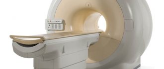
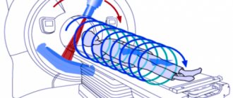
Carrying out an MRI of the pituitary gland is not much different from scanning other organs or parts of the body - it is a non-invasive diagnostic method that does not cause any pain to the person being examined. During the examination, the patient’s body is affected by a magnetic field that is created in the internal part of the tomograph. It is completely harmless, so patients do not have to worry about the negative impact of the device on the body.
During scanning, high-quality images appear on the computer screen. These photographs can be saved, printed, recorded on any digital media or sent by e-mail.
MRI of the pituitary gland shows any, even the most minor changes in both the appendage itself and the tissues adjacent to it, thanks to which the radiologist can accurately establish the correct diagnosis in the shortest possible time.
A special feature of tomography is that the doctor can not only visualize the pituitary gland itself, but also the eye orbits and sinuses. If the patient needs a more in-depth examination, he will definitely be prescribed an MRI of the brain.
Preparing for diagnosis
A significant advantage of this method of examination is that no significant preparatory measures need to be taken before MRI of the pituitary gland without contrast. All that is required from the patient is to comply with a number of simple requirements immediately before starting the scan.
However, if you plan to perform an MRI with contrast, then it is necessary to adhere to some recommendations. So, the patient must come to the tomography on an empty stomach, or observe a 4-hour fasting break. This is done in order to exclude the appearance of any unpleasant sensations in the intestines during the scan. If MRI of the pituitary gland is performed without contrast, then such restrictions do not need to be observed. Before scanning, all patients, without exception, need to:
- remove items of clothing that have metal parts, and ideally give preference to hospital clothing, which is much more comfortable;
- remove jewelry;
- leave your mobile phone, bank cards, and wallet in the locker room.
Each patient must understand that any metal objects can interfere with the examination, which will negatively affect the quality of the resulting images. Therefore, it is so important to take your preparation seriously and listen to all the advice of experts. Moreover, it does not matter significantly whether an MRI of the pituitary gland will be performed without contrast or with its use.
Indications and contraindications for diagnosis
The specialist can refer the patient for a tomography after conducting an initial examination and passing all the necessary tests. In this case, an MRI is prescribed if, based on the results of other examinations, it was not possible to identify the cause of the deterioration in health. Indications for scanning may include:
- metabolic problems;
- decreased visual acuity;
- in women - menstrual irregularities;
- in men - impotence;
- excessively high levels of TSH in the blood;
- headache.
As for contraindications to undergoing MRI of the pituitary gland, they are usually divided into permanent and temporary. If there are permanent contraindications, examination is strictly prohibited:
- People with pacemakers. During operation of the scanner, they can not only fail, but also begin to move, which will cause injuries.
- Patients with metal implants, including dental ones.
- People with tattoos with metal connections.
Temporary contraindications may include:
1. The first 3 months of bearing a child. To date, there has not been enough research into the negative effects on the fetus, so scanning is prohibited in the first 3 months of pregnancy. However, an MRI in early pregnancy can only be performed if the woman's life and health are at risk.
2. The patient's serious condition. If the patient is unconscious after injury or under a ventilator. In this case, it is not possible to carry out the examination.
3. Mental illness. Another contraindication to scanning is the presence of mental illness in the patient. In some cases, for example, if the situation is urgent, MRI of the pituitary gland can be performed in such patients under anesthesia.
4. Uncontrollable tremors of the limbs. It simply does not make sense to conduct examinations of such patients during exacerbations for the reason that the quality of the images will be unsatisfactory.
A separate list of contraindications includes MRI of the pituitary gland with contrast:
- liver pathologies;
- kidney diseases;
- diabetes;
- contrast intolerance.
Before the scan, each patient must inform the radiologist about the presence of dental implants, dentures, metal objects in the body, tattoos, chronic diseases, regardless of whether an MRI of the pituitary gland is planned with or without contrast. This will avoid a large number of complications and make the diagnosis as safe as possible.
Equipment
In order for the images to be highly informative and sufficiently detailed, you need to carefully approach the choice of tomograph. This must be a modern high-field model with a power of at least 1.5 Tesla. Otherwise, the quality of the images will not allow detecting microadenomas and cancerous tumors in the early stages. This is why it is so important to take a serious approach to where to get an MRI of the pituitary gland. People suffering from a fear of closed spaces are better off choosing devices with an open chamber. It is also necessary to take into account the technical characteristics of the device for those patients whose weight exceeds 100 kilograms. To choose a model that has all the necessary qualities, you can consult our service specialists. The website also contains a list of tomograph models that are equipped in leading clinics in St. Petersburg.
MRI of the pituitary gland with contrast
If certain diseases are suspected, the doctor may order an MRI of the pituitary gland with contrast. This does not mean that the doctor suspects the presence of developing cancer or the spread of metastases. With its help, you can get clearer pictures in which even the most minor changes will be noticeable. Contrast is always administered intravenously before scanning. Its main advantages of use include:
- It illuminates the tissue, thanks to which this process is reflected in the resulting images, resulting in the images being of the highest quality.
- It is possible to determine the extent of the pathological process. A specialist can also determine the presence of metastases without any problems.
- The minimum number of restrictions on MRI of the pituitary gland with contrast makes it possible to carry out diagnostics for almost every patient.
In some cases, as soon as the contrast is introduced, the patient may experience a tingling sensation in the limbs, slight dizziness or nausea - this is a normal reaction of the body to the drug and you should not be afraid of this.
As a rule, all unpleasant symptoms disappear within 3-5 minutes after administration. In addition, each patient undergoes a sensitivity test before the examination begins. If it turns out that the patient is intolerant to the drug, then an MRI of the pituitary gland will be performed without contrast.
Interpretation of MRI results
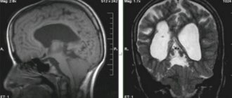
Magnetic resonance imaging of the pituitary gland is interpreted by an experienced, highly qualified doctor. In the absence of pathological processes, the size of the pituitary gland should not exceed 17 mm; it has a rectangular shape, clear and even contours. The pituitary infundibulum is normally located in the midline, but its slight change without the presence of structural abnormalities and pathological processes is normal.
The following indicators may indicate the presence of pathologies:
- Heterogeneous structure of pituitary tissue.
- Fuzzy, uneven contours.
- Shift of the funnel from the midline.
- Deformation of the sella turcica.
Do not diagnose yourself by interpreting a specialist’s report. Some deviations from normal indicators may be a variant of the norm. Therefore, the MRI results must be deciphered for you by your attending physician. Only he can diagnose and prescribe treatment for you!
How is magnetic resonance diagnostics performed?
The method of performing an MRI of the pituitary gland with or without contrast is quite simple. The tomograph is located in a separate room. At the same time, the patient does not remain for a single minute without close attention, because the medical staff monitors the progress of the scan from the next room. Communication with the subject is maintained through special speakers.
Before the scan begins, the doctor conducts an explanatory conversation with the patient, asks about possible contraindications, explains to him the rules of behavior during the examination, and inquires about his well-being. If the patient experiences a feeling of fear, the doctor may suggest taking a sedative.
After this, the patient is escorted to the room with the tomograph, he changes into comfortable clothes. If the MRI is performed with contrast, the substance is administered to the patient intravenously. Then the subject is placed on a special table, the body is fastened with straps, and then placed inside the tomograph. The examination time can vary from 30 to 50 minutes - it all depends on whether an MRI of the pituitary gland is performed without contrast or with its use, as well as on the required number of images.
As soon as the scanning is completed, the patient will be able to go into the corridor to wait for interpretation of the images and a report from the radiologist. There is no need to stay in the clinic or change your usual routine, the only exception is that if the MRI was performed using contrast, a woman in the lactation period will have to stop breastfeeding for 2 days.
If the patient does not have enough free time, he can ask the medical staff to send the images and conclusion to him by email.
What does magnetic resonance imaging show?
MRI of the pituitary gland with or without contrast provides the opportunity to detect both minor and significant changes in the pituitary gland itself and in the tissues surrounding it. In the resulting images you can find:
- malformations of the appendage;
- the presence of any neoplasms;
- cysts;
- vascular pathologies;
- inflammatory processes.
If the subject undergoes a standard MRI of the brain, the radiologist can immediately evaluate nearby tissue. However, to identify pathologies of the examined organ, there is very little such information, so scanning of this appendix is required.
What is the pituitary gland responsible for?
To understand why doctors prescribe an examination of this department, you need to have a general understanding of what the pituitary gland is responsible for.
The pituitary gland is small in size, which usually does not exceed the size of the nail on the little finger. The organ regulates the functioning of the endocrine system, releasing special hormones. For example, growth hormone is responsible for the growth of a child, or TSH is a hormone that helps the thyroid gland produce triiodothyronine. Excess or deficiency of hormones and disruption of functioning lead to serious consequences.
The pituitary gland is located in the brain, so examinations often evaluate the structural features of the entire brain.
Magnetic resonance imaging in children
Children are prescribed an MRI, as a rule, only after 6 years. To obtain high-quality images, the subject must remain completely calm and immobile throughout the entire scan. It is simply not possible for children to fulfill such a condition.
In addition, children are afraid to be in a confined space, so most often they undergo MRI on open-type machines. But even during such an examination, the child is not allowed to make any movements, so doctors may decide to conduct an examination under anesthesia.
During the scan, parents are allowed to be present with the baby, provided that all safety requirements are met. You will have to leave your mobile phone, jewelry, metal objects and other personal items outside the office.
Bibliography:
- Konovalov R.N., Krotenkova M.V., Kremneva E.I. Functional magnetic resonance imaging [Electronic resource] // Journal “Annals of Clinical and Experimental Neurology”, 2011. https://cyberleninka.ru/article/n/funktsionalnaya-magnitno-rezonansnaya-tomografiya
- Kukhtevich I.I., Goryunova T.I., Goryunova V.V. Practice and safety of MRI studies [Electronic resource] // Journal “International Student Scientific Bulletin” - 2021. https://eduherald.ru/ru/article/view?id=18294
- Ternovoy S.K., Sinitsyn V.E., Belichenko O.I., Stukalova O.V. Clinical application of magnetic resonance imaging [Electronic resource] // Regular issues of “RMZh” No. 7, 1996. https://www.rmj.ru/articles/obshchie-stati/KLINIChESKOE_PRIMENENIE_MAGNITNO-REZONANSNOY_TOMOGRAFII/
How to choose a clinic
Any disease always creeps up unnoticed and makes itself felt at the most inopportune moment. Unfortunately, during a visit to a specialist it is not always possible to make a correct diagnosis based on the results of the initial examination and the presence of certain complaints.
In addition, even after an examination using conservative methods, the doctor can not always identify the disease, especially if this pathology concerns the pituitary gland. In this regard, to undergo a more detailed examination, many patients are referred to undergo an MRI of the pituitary gland with or without contrast.
This is where the difficulty arises for patients, namely finding a suitable clinic. Everyone is interested in undergoing a scan in a medical institution with a good reputation, with the latest equipment, at an affordable cost and at the same time under the guidance of highly qualified specialists. But choosing a clinic based on all the necessary parameters can take a lot of time, but still not lead to the desired result.
In such a situation, we advise you to contact our website, where medical centers are presented, in each of which you can undergo an MRI of the pituitary gland with or without contrast. Thanks to the well-thought-out functionality of the site, a convenient filtering system based on the necessary parameters, as well as operator assistance, you can easily make the right choice and make a pre-registration for any day. Here you can find a clinic where you can perform both an MRI of the pituitary gland without contrast and with it.
In addition, you can read reviews that were written by real patients who have undergone MRI in the past. To ensure that contacting us guarantees not only a quick search, but also the lowest cost, we offer each applicant an additional discount on the examination.