Published: 07/07/2021 11:15:00 Updated: 07/07/2021
Thrombosis is a complete or partial blockage of the lumen of a vessel by a parietal or mobile thrombus. A thrombus is a dense blood clot that appears as a result of a change in its fluidity. Normally, thrombus formation is a protective mechanism. Damage to the vascular wall leads to a slowdown in blood flow and accumulation of platelets around the damage. The thrombus literally “darns” the wall of the vessel.
The classic causes of thrombosis are described by Vikhrov’s triad: damage to the vascular wall, slowing of blood flow and changes in blood properties [3]. Some blood clots (they are called emboli) are able to move to narrower areas of the vessel, which are completely or partially blocked. Every year, about 25 million people die from thrombosis, and even more face trophic disorders caused by blood clots [3].
Types of vascular thrombosis
The most common thrombosis of the lower extremities, however, the greatest danger is pulmonary embolism - PE - and disseminated intravascular coagulation syndrome - DIC syndrome.
Arterial thrombosis develops when its lumen is blocked by a thrombus or embolus. Clinical signs are determined by the location where the blockage occurs, an organ or tissue that has little or no blood supply. If blockage with impaired vessel patency occurs slowly, “spare” collateral vessels open, which mitigates the clinical symptoms of arterial thrombosis [3]. Arterial thrombosis occurs more often in middle-aged and elderly men [7].
Vein thrombosis varies depending on the location of the lesion into deep or superficial vein thrombosis and pulmonary embolism. Among all cardiovascular pathologies, venous thrombosis ranks third in frequency of occurrence, second only to ischemic heart disease and atherosclerosis. The third place in the structure of causes of mortality is occupied by pulmonary embolism. Starting at age 40, the risk of developing venous thrombosis doubles every 10 years [5].
Two variants of damage to the veins of the lower extremities are described: phlebothrombosis (primary thrombosis, the thrombus is not firmly fixed) and thrombophlebitis (secondary thrombosis due to inflammation of the vessel wall, the thrombus is firmly fixed) [6]. Thrombophlebitis is more often associated with superficial vein thrombosis [2]. The larger the vein affected by thrombosis, the more pronounced its clinical manifestations. The surrounding tissues are compressed by stagnation of blood, since the blood stays at the site of occlusion, but does not move towards the heart. Venous blood clots tend to break off and travel through the bloodstream (thromboemboli). When they enter vital organs, life-threatening conditions develop [3].
How do blood clots occur?
If a blood clot has formed on the wall of an artery, its appearance can be described in the following stages:
- Some process damages the artery wall.
- The body notices the violation and begins to build protection against blood loss, forming a large number of special blood cells - platelets, which, attaching to the damaged area, form a kind of patch.
- In case of coagulation disorders or changes in the hematopoietic system, platelet formation does not stop on time and continues longer than expected. This causes too much growth to form on the wall. Or platelets, which are in small quantities in the blood, floating past in the bloodstream, stick to the resulting accumulation.
The causes of damage to the walls of blood vessels can be:
- mechanical disruption of the structure due to injury;
- infectious lesion;
- high levels of glucose molecules in the blood;
- immune system dysfunction.
If there are no factors that contribute to the formation of blood clots, any injury or other damage will not lead to a large accumulation of blood cells. Under the layer of platelets, the artery wall will heal and recover, and the crust will resolve over time.
There are several stages of thrombus formation:
- disruption of the structure of the inner surface of the artery;
- activation of blood clotting factors;
- platelet adhesion at the site of injury;
- the appearance of substances that trigger a chain of reactions that form fibrin threads, which contribute to thrombus formation;
- a kind of network of fibrin threads is formed, into which blood cells enter, creating a large clot;
- Over time, the clot thickens, forming a thrombus.
When a blood clot breaks off under the influence of any factors, it begins to move through the bloodstream. Once it hits the nearest bottleneck, the blood flow will be blocked. If a similar situation happens outside a medical facility, it is impossible to save the person.
Risk factors for thrombosis
Internal:
- arterial hypertension [7];
- pregnancy, childbirth, postpartum period [3];
- biochemical changes in blood [2,3,5,7];
- vasculitis [2];
- age over 40 years [5];
- congenital thrombophilia, thrombosis, varicose veins of the lower extremities [3,5];
- congestive heart failure [5, 6];
- malignant neoplasms, radiotherapy and chemotherapy [3];
- strokes [3, 6];
- myeloproliferation [2, 5, 7];
- nephrotic syndrome and renal failure [5, 6, 7];
- obesity (BMI over 30) [3];
- myocardial infarction [6];
- diabetes mellitus [6, 7];
- systemic lupus erythematosus [2];
- chronic pulmonary diseases [3];
- enterocolitis [5].
External:
- heroin addiction [2];
- hormonal therapy [3,5];
- dehydration due to vomiting, diarrhea, increased sweating, direct lack of fluid [6];
- immobilization [3];
- travel by plane, bus or in a seated car [3];
- infectious diseases, including COVID-19 [1, 3, 5, 9];
- catheterization of central and peripheral veins [2, 5];
- smoking [6, 7];
- sedentary lifestyle [3];
- operations [3];
- fractures of large bones, other injuries [3];
- taking oral contraceptives [5];
- taking Diazepam, Amiodarone, Vancomycin [2];
- sclerotherapy and thermal ablation [2];
- condition after joint replacement [3];
- holding an awkward position [3].
How is thrombophlebitis different from varicose veins?
Varicose veins are increased pressure in thinned and inelastic veins, and thrombophlebitis is an inflammation of the venous walls with subsequent formation of blood clots. Most often, the diagnosis of thrombophlebitis is a consequence of varicose veins.
Thrombophlebitis differs from varicose veins in the following symptoms:
| Sign | Varicose veins | Thrombophlebitis |
| Pain | pulling | bursting and stabbing |
| Convulsions | Yes | No |
| Changing the appearance of your legs | noticeable tortuosity and bulging of veins | compactions and lumps when palpated |
| Change in skin color | pale skin, blue veins | red skin |
| Burning feeling | Yes | No |
Both pathologies develop as a result of impaired hemostasis, but inflammation of the venous walls is more often associated with the entry of an infectious agent. The difference is the absence of local inflammation. With varicose veins, the veins do not become inflamed. In this case, only the blood circulation is disrupted.
Thrombosis Clinic
Symptoms of thrombosis can be general, regardless of location, or specific.
Common symptoms include pain with movement and at rest, limited mobility, and decreased function of the affected organ or tissue. Symptoms of arterial obstruction (acute thrombosis, or gradual obstruction of vessel patency):
- asymmetry of blood pressure when measured on both arms [7];
- pallor of the skin, turning into cyanosis [7];
- pain at rest at night [7];
- pain when moving in the thigh, buttock, lower leg, foot, shooting or aching [7];
- sleep disorders [7];
- numbness, coldness of the limb [7];
- absence of peripheral pulsation [7];
- necrosis (necrosis) of affected tissues, trophic ulcers, gangrene [7];
- intermittent claudication [7].
Symptoms of venous thrombosis:
- pain [6];
- swelling, soft and asymmetrical [6];
- blue discoloration of the skin (skin cyanosis) [6];
- increased skin temperature of the extremities [6];
- increased sensitivity and compaction in the projection of the superficial veins [2];
- post-inflammatory hyperpigmentation [2];
- dilated saphenous veins [6];
- erythema [2].
Sometimes the only symptom of venous thrombosis is PE [6].
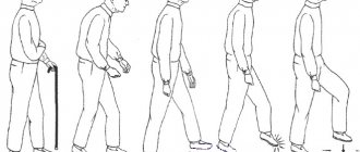
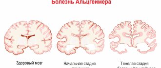
Symptoms of ischemic stroke
Almost never a cerebral infarction is asymptomatic. You may have already come across actively distributed reminders on the timely recognition of a stroke: after all, it is very important to call an ambulance and provide medical assistance to the patient as early as possible - the earlier assistance is provided, the less brain damage.
The main symptoms of ischemic stroke are:
- dizziness,
- loss of orientation in space,
- vomit,
- convulsions,
- impaired coordination, speech, vision, writing, reading, swallowing,
- inability to move individual limbs and/or perform simple manipulations such as raising two arms at the same time, brushing teeth, or turning pages of a book.
The symptoms are extremely varied. They depend primarily on which particular part of the brain was deprived of blood supply - then exactly the function for which this part is responsible will be disrupted.
At the same time, all the symptoms do not appear; you may notice one or more - and this is a good reason to immediately call an ambulance.
Diagnosis of thrombosis
Primary diagnosis is based on a detailed history and anthropometry (calf or thigh circumference).
The Wells scale is used to diagnose acute thrombosis and diagnose pulmonary embolism [8,9]. Instrumental diagnostics include compression or duplex scanning of veins, Doppler ultrasound with compression of veins, impedance plethysmography, pulmonary angiography, radiocontrast or MRI venography [6,9], CT and MRI angiography [7,9].
To diagnose arterial thrombosis, physical tests are used (6-minute walk test, treadmill test), determination of pulsation of superficial arteries (arteries of the dorsum of the foot), duplex scanning of the arteries of the extremities, angiography (X-ray of a vessel filled with a radiopaque substance) and measurement of transcutaneous oxygen tension [ 7].
Recommendations and contraindications for thrombophlebitis
The doctor advises you to follow a diet and wear low-heeled shoes with a comfortable last.
Forbidden:
- wear stilettos and heels higher than 3 cm;
- massage limbs;
- go to the gym and lift weights;
- remove leg hair with wax or epilator;
- go to the bathhouse and sunbathe;
- wear socks, stockings, and knee socks with a thick elastic band.
It is necessary to exclude the use of hormonal drugs and choose a different method of contraception.
Tests for thrombosis
Laboratory indicators play a significant role in the timely diagnosis of thrombosis.
Thus, guidelines for the management of patients with a new coronavirus infection provide for stratification of the risk of coagulopathy in patients with COVID-19 based on simple laboratory tests: D-dimer, prothrombin time, platelet count, fibrinogen level [1,9]. A clinical blood test can detect inflammation. It also determines the level of platelets, that is, the very substrate of thrombosis.
Additionally, the level of inflammation in the blood and the risk of thrombosis is indicated by an increased level of C-reactive protein.
Biochemical analysis primarily demonstrates blood glucose levels. It can be used to judge the presence of diabetes, one of the most serious risk factors for thrombosis.
Also, a biochemical analysis can determine the level of protein C, which also characterizes the severity of the risk of thrombosis.
Elevated levels of homocysteine in the blood are also a currently proven risk of thrombosis, leading to miscarriage and cardiovascular events (heart attacks and strokes).
D-dimer is a laboratory marker of fibrin formation [8]. It also indicates the presence of inflammation, just like C-reactive protein. The level of D-dimer is a control indicator of COVID-19 and its complications, including those associated with thrombosis.
You can take tests under the comprehensive Thrombosis program, which includes determining the levels of Antithrombin-III, D-dimer and genetic factors of cardiac diseases and platelet levels. This program allows you to determine the fact of thrombosis occurring somewhere in the body, as well as determine the genetic predisposition to it. This program, like other tests, is offered by the CITILAB network of clinics.
Additional determination of homocysteine and C-reactive protein levels will help determine the biochemical risk of thrombosis.
Indications for intervention
- Narrowing of the vascular lumen by more than 50-60%.
- Symptoms of microstroke and stroke.
- High risk of complications with other vascular interventions.
The operation is also performed for patients who have already undergone surgery to remove plaques, but are faced with a relapse of narrowing of the arterial lumen.
Stenting is not performed if:
- complete blockage of the carotid artery,
- allergies to the drugs used.
Also, surgery is not prescribed for cerebral hemorrhages that occurred within the last 2 months or for heart rhythm disturbances.
| Type of intervention | Price |
| Stenting of cerebral vessels | 300,000 - 450,000 rub. |
Request a call back Get a free consultation
Treatment and prevention of thrombosis
Treatment of thrombosis includes anticoagulant and antiplatelet therapy, thrombolytic therapy, installation of an inferior vena cava cava filter, and surgical removal of the thrombus [5].
Complications of anticoagulant therapy must be kept in mind: major bleeding, heparin-induced thrombocytopenia and warfarin-induced skin necrosis [5]. To reduce the risk of continued thrombus formation, NSAIDs are used [2]. For the purpose of secondary prevention, small doses of heparin are prescribed. Non-drug treatment methods are also prescribed - elastic bandaging, compression hosiery, local hypothermia and exercise therapy [2, 4].
Prevention of thrombosis includes a number of measures used in situations of increased risk of thrombosis.
Primary prevention of atherothrombosis:
- systematic physical activity in the form of walking or morning exercises;
- blood pressure control, maintaining working blood pressure below 140/90 mmHg;
- control of blood sugar levels (less than 6 Mmol/l), early detection and treatment of diabetes mellitus;
- weight loss, body mass index less than 25 kg per m2;
- a diet limited in cholesterol and high-density fat (total cholesterol less than 5 mmol/l), fruits and vegetables;
- smoking cessation [3,7].
Primary prevention of venous thrombosis:
- compression underwear;
- bandaging with elastic bandages;
- drinking plenty of fluids, especially after surgery;
- regular exercise, walking, especially when traveling;
- prohibition of taking alcohol and sleeping pills in large doses;
- prohibition of the use of compressive shoes and clothing [2,5,6].
Sometimes, during periods of particular risk, anticoagulants are prescribed several days before the flight. There is no point in taking aspirin in such cases [5].
Briefly about the treatment method
For ischemic stroke, the most effective and reliable method of removing blood clots from the arteries of the brain is thrombectomy. There are several options for the procedure: - Aspiration thrombectomy. During the operation, a micro-incision is made through which a special catheter is inserted. Through it, saline solution flows under pressure into the affected vessel. Under the influence of the solution and salt, the thrombus softens, is divided into pieces and aspirated (suctioned out) using a syringe. Thanks to this method, the number of injuries to the internal walls of blood vessels is reduced. — Percutaneous mechanical thrombectomy. A catheter with a stent is inserted through a large artery. With the help of a stent, the clot is captured and removed through an incision in the vessel. This method is most often used in the treatment of stroke caused by blockage of blood vessels in the brain.
Bibliography
- Consensus position of experts of the Eurasian Association of Therapists on some new mechanisms of the pathogenesis of COVID-19: focus on hemostasis, issues of blood transfusion and the blood gas transport system / G.P. Arutyunov, N.A. Koziolova, E.I. Tarlovskaya, A.G. Arutyunov, N.Yu Grigorieva, etc. // Cardiology. 2020;60(6). DOI: 10.18087/cardio.2020.5.n1132.
- Thrombophlebitis (thrombosis of superficial veins): modern standards of diagnosis and treatment / V.Yu. Bogachev, B.V. Boldin, O.V. Jenina, V.N. Lobanov//Hospital-substituting technologies: Outpatient surgery. 2016.- 3-4 (63-64)- P.16-23.
- Modern problems of thrombosis of arteries and veins / I.N. Bokarev, L.V. Popova // Practical Medicine, 2014. - No. 6 (82) – P13-17.
- Treatment of thrombophlebitis. Current recommendations and clinical practice / P.F. Kravtsov, K.V. Mazaishvili, S.M. Markin, H.M. Kurginyan // Thrombosis, hemostasis and rheology, 2020 No. 2 – P 68-72.
- Venous thrombosis: modern treatment / P.S. Laguta // Atherothrombosis, 2015 - No. 2 - P. 7-16.
- Deep vein thrombosis of the lower extremities / A.K. Lebedev, O.Yu. Kuznetsova // Russian family doctor, 2015.
- National recommendations for the diagnosis and treatment of diseases of the arteries of the lower extremities, Association of Cardiovascular Surgeons of Russia, Russian Society of Angiologists and Vascular Surgeons, Russian Society of Surgeons, Russian Society of Cardiology, Russian Association of Endocrinologists, M, 2021.
- Diagnosis and pharmacotherapy of acute venous thrombosis / N.V. Sturov, G.N. Kobylyanu// Difficult Patient, 2013., No. 12., T. 11. –P.19-22.
- Principles of management of patients with venous thromboembolism during the COVID-19 pandemic // V.Ya. Khryshchanovich // Surgery News, 2021.- t28 No. 3 – P329-338.
Stages
The sequence of development of the disease is as follows:
- Acute stage
. Clinical signs of thrombophlebitis are bright and appear unexpectedly. The temperature rises, the skin around the affected area turns red, and the patient shudders. If treatment is started within two weeks, the development of the disease can be avoided. The blood clot will dissolve. The duration of exacerbation is up to one month. - Subacute stage
. Lasts up to two months. Signs of the disease subside, but do not fade away. The skin remains red and the swollen veins turn blue. The swelling goes away, but the seals remain. - Chronic stage
. Diagnosed two to three months after the disease is detected. Symptoms are either absent or worsened. During remission, the limbs remain swollen, quickly become tired and itchy, but there is no pain. Discomfort occurs in the lower extremities when walking for a long time.
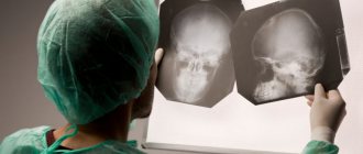
How is preparation for the operation carried out?
Diagnostics
The treatment regimen and tactics of vascular surgery are always selected individually. To make an accurate diagnosis and determine the stage of development of the pathology, a number of examinations are carried out:
- Ultrasound Dopplerography of the main arteries.
- CT (computed tomography).
- Electroencephalography of the brain.
- Angiography.
Aspirin is also prescribed before surgery. It helps reduce blood clotting.
Stenting
The operation is performed under local anesthesia. The patient is connected to tracking equipment. It controls blood pressure and heart rate. During the operation, the doctor communicates with the patient. This allows you to control all brain functions.
The site of the intervention is numbed. A special drug is administered to reduce blood clotting. A catheter is inserted through the femoral vessel (artery). At the end it has an inflatable balloon. The end of the catheter reaches the site of narrowing of the vessel. Due to this, the arterial lumen expands. In this case, the patient does not experience pain. The can is installed. Then it expands. After this, the stent itself is installed. In a compressed form, it is introduced into the lumen of the vessel using a second catheter. After the stent reaches the area expanded by the balloon, it relaxes and subsequently plays the role of an arterial wall. To ensure the stent is firmly in place, the balloon is inflated again. After this, the catheter is removed.
Stenting usually lasts about 2 hours.