Lev Manvelov, Candidate of Medical Sciences
, Albert Kadykov,
Doctor of Medical Sciences
“Science and Life” No. 2, 2007
General diagram of the blood supply to the brain.
Blood enters the brain through four large main arteries: two internal carotid and two vertebral. At the base of the brain stem, the vertebral arteries merge into one, the basilar artery. In the brain, the internal carotid artery divides into two main branches: the anterior cerebral artery, which supplies blood to the anterior parts of the frontal lobes, and the middle cerebral artery, which supplies parts of the frontal, temporal and parietal lobes. The vertebral and basilar arteries supply blood to the brain stem and cerebellum, and the posterior cerebral arteries supply the occipital lobes of the brain (image: Science and Life)
Headache, noise and dizziness, memory impairment, increased fatigue, decreased performance - such symptoms occur not only in the elderly, but also in middle-aged and even young people. Often, patients and some medical professionals do not take such complaints very seriously. Meanwhile, they may indicate chronic cerebral circulatory failure.
Blood supply to the brain
The normal functioning of the brain requires a large amount of energy. Nutrients and oxygen are delivered to the cells of the nervous tissue through the bloodstream. Nature has taken care to create a high degree of reliability of blood supply to the brain. It is provided by four powerful main arteries: two carotid and two vertebral. At the base of the brain, the branches of these vessels form a closed circle, called Willisian after the English physician and anatomist of the 17th century, Thomas Willis, who first described it. Thanks to this, the lack of blood supply in one of the main vessels is compensated by others. It also happens that even with serious disturbances in blood flow in three of the four main vessels, a person complains only of a slight deterioration in well-being - the compensatory capabilities of the brain are so great. Great, but, unfortunately, not unlimited. Man manages to “shatter” these perfect compensation mechanisms created by nature. It all starts with the most ordinary complaints of headache, dizziness, memory loss and fatigue.
After some time, the patient develops more serious neurological symptoms, indicating multiple brain damage. The reason for this is chronic cerebral circulatory failure, or “dyscirculatory encephalopathy.” This term was proposed in 1971 by well-known domestic scientists working at the Research Institute of Neurology of the Russian Academy of Medical Sciences, Academician of the Russian Academy of Medical Sciences E.V. Shmidt and Candidate of Medical Sciences G.A. Maksudov, and it means changes in the brain associated with disturbances in its blood supply.
The main causes of the occurrence and development of dyscirculatory encephalopathy are arterial hypertension and atherosclerosis.
More than 40% of the adult population of Russia suffers from hypertension. Men and women, old people and young people get sick. Only in 5% of cases the cause of hypertension is clear. These may be renal failure, endocrine disorders, atherosclerosis and some other diseases. In 95% of cases, the cause of hypertension remains unclear, which is why it is called essential (literally, hypertension itself). With hypertension, the walls of blood vessels become denser, local narrowings (stenoses) and tortuosity are formed. All this leads to circulatory disorders, including blood supply to the brain. Sometimes it comes to occlusion - complete closure of the lumen of the vessel.
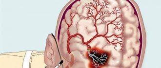
Blood clots, thrombi, develop in the area of atherosclerotic plaques that form on the inner walls of the vessel. Blood clots can completely block even large vessels, causing serious cerebrovascular accidents. (Image: Science and Life)
Unlike hypertension, the cause of atherosclerosis is known - it is a disorder of lipid metabolism. In patients with atherosclerosis, the level of fat-like substances in the blood increases - cholesterol, low-density lipoproteins, triglycerides, which are deposited on the walls of blood vessels, forming lipid stains. Then the spots grow into so-called plaques. Due to the deposition of calcium salts, the plaques become denser and ultimately narrow or even close the lumen of the blood vessels. Then they begin to disintegrate, their particles - emboli - enter the bloodstream and sometimes clog other small and large vessels.
Sometimes the development of dyscirculatory encephalopathy is facilitated by osteochondrosis, since in this disease, due to deformation of the intervertebral discs, the vertebral arteries that supply the brain with blood can be pinched.
Impaired blood supply leads to the gradual death of neurons in various parts of the brain, and the patient experiences neurological symptoms. Discirculatory encephalopathy is most characterized by emotional and personal disturbances. At the onset of the disease, asthenic conditions are noted: general weakness, irritability, poor sleep. Asthenia is often accompanied by depression. Gradually, such painful personality traits as egocentrism and periodically occurring causeless agitation begin to appear, which can be pronounced and manifest itself in inappropriate behavior. With further development of the disease, emotional reactivity decreases and gradually turns into dullness and apathy.
Once it begins, the disease steadily progresses, although during its course both sharp periodic deterioration (paroxysmal course) and periods of slow increase in symptoms of the disease can be observed.
We should not forget that dyscirculatory encephalopathy increases the risk of many severe brain diseases and, above all, stroke - an acute circulatory disorder of the brain (Manvelov A., Candidate of Medical Sciences; Kadykov A., Doctor of Medical Sciences. “Stroke is a social problem and medical” // “Science and Life” 2002, No. 5.). In Russia, strokes are registered in more than 400 thousand people per year. Of these, 35% die in the first three weeks of the disease, and only half of the patients reach the annual milestone. The possibility of epileptic seizures occurring against the background of developing discirculatory encephalopathy should not be excluded.
When is it time to go to the doctor?
Only a medical specialist can understand what is happening. If discomfort in the head, neck or upper back appears sporadically, for example, when the weather changes, and then disappears without a trace for a long time, there is usually no cause for concern. If you suspect a cold, headache from fatigue, physical or mental stress, just rest. But if you experience the following symptoms, you should definitely go to the doctor:
- previous head injuries with and without complications;
- feverish condition;
- blurred perception;
- muscle tension in the neck or back of the head;
- sudden painful shootings;
- loss of sensation in the arms or legs;
- convulsions;
- problems concentrating your eyes or thoughts;
- bulging of the fontanelle in the newborn period.
Even short-term loss of vision or hearing is also a serious reason for medical intervention.
Types of chronic insufficiency of blood supply to the brain
Brain with blood vessels ( bottom view
).
The branches of the main vessels of the brain at its base form a vicious circle called the circle of Willis.
Thanks to this, if one of the vessels is narrowed or blocked, the blood supply to the brain is fully or partially restored. (Image: Science and Life) There are three main types of cerebrovascular accidents.
In Binswanger's disease, due to thickening of the walls and narrowing of the lumen of small arteries, diffuse damage to the internal structures of the brain occurs - the so-called white matter. Multiple small lesions are areas of dead neurons. In patients, circadian (daily) pressure fluctuations are disrupted: at night it either drops too sharply, or, conversely, increases, although the pressure should decrease slightly at night. One of the main symptoms of the disease is sleep disturbance. The patient has trouble falling asleep or sleeps with frequent awakenings. Other typical signs are the slow progression of memory and intelligence impairments up to dementia (dementia); increasing gait disturbances, urination and defecation disorders. It is known that Binswanger's disease can occur even at a relatively young age - up to 35 years.
Another type of dyscirculatory encephalopathy - the so-called multi-infarct conditions - is characterized by multiple small infarctions in the brain (micro-strokes). This means that in a certain area of the brain, due to blockage of the vessel, necrosis of the nervous tissue occurs. This affects both the superficial (gray matter) and deep (white matter) structures of the brain.
The main reason for the development of multi-infarction conditions is the narrowing and hardening of intracerebral arteries during arterial hypertension. Another common cause is heart disease accompanied by atrial fibrillation. In such patients, blood clots form in the cavities of the heart - thrombi, which can clog the vessels supplying the brain with blood. Increased blood clotting also contributes to the formation of blood clots. Another cause of multi-infarction conditions is atherosclerotic damage to intracerebral arteries.
Discirculatory ecephalopathy also develops with damage to the main (carotid and vertebral) arteries, which are not located inside the brain, but provide blood flow to the brain. Lesions can have different natures and causes - thrombosis, stenosis, bends and kinks of various etiologies.
There are three stages of dyscirculatory encephalopathy. The duration of each of them may be different. Much depends on the degree of hypertension or atherosclerosis, lifestyle, habits, heredity, concomitant diseases, etc. At the initial stage of the disease, people often complain of headaches, dizziness, noise in the head, decreased memory (non-professional) and performance. Patients are absent-minded, irritable, tearful, and their mood is often depressed. They usually have difficulty switching from one activity to another.
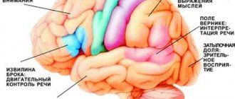
Functional areas of the brain.
When the blood supply to certain areas of the brain is disrupted, patients experience corresponding neurological symptoms (image: Science and Life)
At the next stage of the disease, memory impairment, including professional memory, progresses. The range of interests narrows, rigidity of thinking (obsession on some problem), incompatibility appear, the intellect suffers, and a change in personality occurs. Such patients are characterized by daytime sleepiness and poor night sleep. Neurological symptoms intensify, movements slow down, their coordination is impaired, mild speech disturbances appear, staggering when walking, and performance is significantly reduced.
At the last stage of the disease, gross changes in brain tissue make neurological symptoms even more pronounced, and mental disorders intensify, including dementia (dementia). Patients completely lose their ability to work, stop recognizing loved ones, perform inappropriate actions, and may get lost when going for a walk.
Common Causes
At the initial stages of diseases, it is very difficult to detect them, since most of them occur without visible symptoms, or are manifested by minor pain.
The following internal and external causes can lead to the disease:
- severe infections that entered through wounds, insect bites, respiratory tract or blood/lymph from other infected areas of the body;
- trauma to the skull;
- chemical poisoning;
- excessive exposure to radiation;
- adherence to bad habits, systematic intoxication with alcohol, tobacco, drugs or certain medications;
- poor quality of food;
- genetic predisposition to brain destruction;
- congenital disorders.
Diagnosis of encephalopathy
When examined, the vast majority of patients with discirculatory encephalopathy reveal characteristic diseases or physiological characteristics and habits. These risk factors include:
- arterial hypertension (blood pressure from 140/90 mm Hg and above);
- heart diseases (coronary disease, rheumatic lesions, heart rhythm disturbances, etc.);
- diabetes;
- excess body weight;
- sedentary lifestyle;
- hypercholesterolemia (total cholesterol above 6.2 mmol/l);
- long-term and frequent neuropsychic overstrain (stress);
- family history of cardiovascular diseases (stroke, myocardial infarction or arterial hypertension in close relatives);
- smoking;
- alcohol abuse.
Men with rapidly progressing dyscirculatory encephalopathy usually have a history of psycho-emotional stress, a sedentary lifestyle, alcohol abuse, lack of regular treatment and the presence of two or more concomitant diseases. In women, in addition to the listed factors, excess body weight often contributes to the unfavorable course of the disease.
If patients with arterial hypertension and atherosclerosis (or representatives of other risk groups) have complaints of headache, dizziness, decreased performance, memory impairment, then the initial stage of dyscirculatory encephalopathy can be suspected. Patients with such symptoms should, first of all, constantly monitor blood pressure, undergo an electrocardiographic examination, complete general blood and urine tests, and blood tests for sugar and lipids. A psychological study to assess the state of memory, intelligence, attention and speech would not hurt.
Even small nonspecific changes in the electrocardiogram can be harbingers of cardiovascular diseases, manifested in impaired blood circulation in the brain. By the way, normal electrocardiograms or echocardiograms do not exclude the presence of the disease, since changes can only be noticeable at the time of myocardial ischemia (anemia) or an attack of angina. An electrocardiogram taken during physical activity provides important information. Daily monitoring of heart function also allows you to identify abnormalities.
Information about the condition of the fundus (the back wall of the eye), the cells of which are directly connected to the neurons of the brain, is important for making a diagnosis. Changes in the blood vessels and nerve cells of the fundus make it possible to judge about disturbances in the structure of the brain tissue. In patients with discirculatory encephalopathy, hearing is often reduced, the swallowing reflex and sense of smell are impaired. Therefore, to make a diagnosis, it is necessary to conduct an otoneurological study that reveals disorders of the vestibular apparatus, auditory, olfactory and taste perceptions.
Useful information is provided by studying the rheological properties of blood - its fluidity. The main factor influencing the fluid properties of blood and the degree of its saturation with oxygen is considered to be hematocrit - the ratio of the volume of red blood cells to the volume of plasma. Its increase increases blood viscosity and worsens blood circulation. There is a direct connection between high hematocrit and cerebral infarctions.
After preliminary studies, the patient is usually referred for an X-ray examination of the cerebral vessels - angiography. Doctors consider angiography to be the “gold standard” with which the results of other research methods are compared. After the administration of a special contrast agent, X-ray images of the brain vessels are obtained. Angiography provides information about the duration and sequence of filling of blood vessels, about the formed “bypass” circulatory pathways in case of blockage or narrowing of cerebral vessels. The results of the study are important when deciding on the feasibility of surgery.
Electroencephalography is an old and very common method of studying the brain, based on recording its electrical potentials. Changes in the encephalogram indicate organic changes in the brain tissue, therefore, at the initial stage of the disease with dyscirculatory encephalopathy, encephalography may not reveal any abnormalities.
A real revolution in brain research was made by the advent of computed tomography, which combines the achievements of radiography and computer data processing methods. With its help, you can obtain not indirect, but direct data about brain structures and their changes. The method allows you to determine the location and size of brain lesions and their nature.
Magnetic resonance imaging of the brain.
Image of a healthy brain (A); changes in the brain matter in Binswanger's disease - rarefaction of the white matter of the brain (B); hydrocephalus - accumulation of fluid in the brain tissue - manifests itself in the form of expansion of the grooves and ventricles of the brain (shown by arrows) (B); multi-infarct condition - dead nerve tissue appears as small dark spots (shown by arrows) (D). Image: Science and Life
Recently, magnetic resonance methods have been used to diagnose cerebral circulatory disorders: nuclear magnetic resonance, magnetic resonance imaging and magnetic resonance angiography. Nuclear magnetic resonance provides information about the physicochemical properties of brain structures, making it possible to distinguish healthy tissues from altered ones. Magnetic resonance imaging allows you to obtain images of the brain, determine the location, size, shape and number of lesions, and study cerebral blood flow. Magnetic resonance angiography is a modification of magnetic resonance imaging. With its help, you can study the passage and “caliber” of extracranial and intracranial arteries and veins.
Currently, highly informative methods for obtaining three-dimensional images of brain structures have been created and are successfully used: single-photon emission computed tomography and positron emission tomography.
Duplex scanning of the internal carotid artery.
A formed small atherosclerotic plaque is visible, the lumen of the vessel is slightly narrowed (A); later stage of atherosclerosis - the lumen of the vessel of the internal carotid artery is partially blocked by a large plaque (B); occlusion - complete closure of the lumen of the vessel with a plaque (B); artery tortuosity (D) (Image: Science and Life)
Ultrasound methods are widely used to examine patients not only in hospitals, but also in outpatient settings: Dopplerography and echotomography, duplex scanning and transcranial Dopplerography. Doppler ultrasound is used to identify lesions of the carotid and vertebral arteries. It makes it possible to obtain information about the profile of blood flow in the vessels. With duplex scanning, color contrast of flows allows you to more clearly distinguish between moving (blood) and stationary (vascular walls) objects. The main vascular lesions detected by transcranial Doppler ultrasound are blockages, stenoses, spasms and aneurysms. The most complete information about the state of the vascular system of the brain can be obtained by comparing data from various ultrasound methods. Recently, a new method of ultrasound diagnostics has appeared - transcranial sonography with color Doppler coding. With its help, you can “see” the structures of the brain through the bones of the skull.
Treatment for inflammation of the facial nerve
Drug treatment
Treatment of trigeminal neuritis is complex. The disease is first treated with medication - the patient is prescribed drugs that will alleviate the situation. These include painkillers, decongestants, vasodilators and B vitamins. Most often, the recommended medications are tablets, but you can speed up the recovery process by using ointments and gels. Sometimes doctors prescribe intramuscular injections.
In special cases, the recovery process of the facial nerve may be slowed down. Then the patient is prescribed glucocorticosteroids, which improve the metabolic processes of nervous tissue. Various biostimulants and hyaluronidases also contribute to a speedy recovery.
You cannot prescribe medications for yourself. Be sure to see a neurologist or neuropathologist at the first symptoms to determine the diagnosis and treatment strategy. Recovery medications are recommended to patients on a case-by-case basis, paying attention to the presence of chronic diseases, symptoms, and so on.
Surgery
Another way to treat the facial nerve is surgery. However, doctors turn to this option quite rarely - only when the trigeminal nerve is ruptured. Surgery is also required if there is no effect from the conservative method after six months or a year. Surgical intervention is only relevant during the first year of the presence of the disease; later, the muscles on the face irreversibly atrophy.
The surgical process involves suturing the damaged area of the facial nerve to restore its motor function.
Massage
The next treatment method is massage for the treatment of the facial nerve. The purpose of this method is to remove swelling, improve blood circulation, restore sensitivity and conduction of nerve impulses. Massage is contraindicated for tuberculosis, oncology, atherosclerosis and elevated temperature.
Initially, the massage therapist works only with the healthy side of the face, collar area, neck and area above the shoulders. Basically, the master uses rubbing, stroking, kneading and vibration.
For noticeable desired changes, it is necessary to conduct ten to twenty massage sessions from five to fifteen minutes. The duration is determined based on the degree of inflammation of the trigeminal nerve, the goals of therapy and the dynamics of recovery.
Physiotherapy
The next treatment method is physical therapy. It alleviates the severity of symptoms, helps to activate metabolic processes in tissues and restore the functions of the facial nerve.
Doctors prescribe this course of treatment from the first days of the onset of neuritis. The list of physical procedures includes:
- Ultrasound
- Laser irradiation of blood
- Electrophoresis of drugs
- Microwave therapy
- Exposure to ultra-high frequency electricity
- Ozocerite treatment
- Myoelectrostimulation
- Darsonvalization
This complex is indicated for the first week of treatment. Doctors prescribe it together with medication. This tandem helps speed up the process of restoration of the facial nerve. And its most important advantages are the absence of side effects and painlessness.
Alternative Methods
There are also alternative treatment methods. These are procedures aimed at restoring facial muscles and eliminating the symptoms of facial neuritis. Such procedures include:
- Clay or paraffin masks
- Acupuncture
- Reflexology
- Injections to eliminate muscle disorders
- Therapeutic baths
- Taping – stretching the face using adhesive plasters
- Immunosorption – purification of blood from antigens and antibodies
- Biofeedback – facial muscle training
Gymnastics for the face
Also, in conjunction with complex treatment, you can do facial exercises. Before this, you need to consult with a specialist; the doctor will draw up an individual list of exercises based on the severity of the process, location of the lesion and symptoms. Typically, such gymnastics takes about ten minutes a day.
A standard set of exercises includes relaxing and tensing individual facial muscles. For example, to restore articulation, it is recommended to pronounce the sounds “u”, “o”, “and”. Afterwards, you need to bring your lower lip under your upper teeth and reproduce the sounds “v” and “a”.
Gymnastics for inflammation of the trigeminal nerve:
- Close eyes
- Raise your eyebrows
- Frown
- Squint
- Smile with your mouth closed
- Smile with your mouth open
- Puff up your cheeks
- Pull them back
- Whistle
- Widen your nostrils
- Curl your lips
- Raise your upper lip and return to the starting position
- Lower your lower lip and return to the starting position
- Take water into your mouth
- Rinse your mouth
- Close your mouth
- Run the tip of your tongue along your gums
- Move your tongue right and left
Disease prevention
Doctors recommend eliminating effects on the body that cause inflammation of the trigeminal nerve. Here are some recommendations to help avoid illness:
- Avoid drafts and hypothermia
- Keep your head warm during the cold season
- Monitor your blood pressure
- Timely treatment of infectious and bacterial diseases
- Have a routine check-up with an oncologist
- Avoid skull and head injuries
You can sign up for an individual consultation, take tests or undergo treatment at the Medunion private clinic. You can easily make an appointment with us by calling 202-95-54 or online, directly on the website, by clicking on the “Online booking” button.
We have been working in Krasnoyarsk since 2006 and provide high-quality medical services to the population. The staff consists of highly qualified doctors of broad and narrow specialization.