HEPATIC ENCEPHALOPATHY in the practice of an emergency physician
What clinical symptoms allow one to diagnose PE? What are the manifestations of neurological disorders at different stages of development of PE? What are the features of the development of PE in alcoholism? What mistakes should a doctor avoid when treating PE?
Etiology and pathogenesis
Hepatic encephalopathy (HE) is a potentially reversible disorder of nervous and mental activity that occurs in any liver disease that occurs with insufficiency of hepatic cellular function. In most cases, PE complicates the course of the terminal stage of chronic diffuse liver diseases or acute necrotizing (fulminant) hepatitis. It has been established that with liver damage of any etiology, PE can lead to the development of coma and become the direct cause of death of the patient (see Table 1).
| Table 1. Causes of development of PE. | ||
| Encephalopathy variant | Survival | Etiology |
| Acute encephalopathy without liver fibrosis/cirrhosis | 20–45% | Viral hepatitis. Alcoholic hepatitis. Acute poisoning (chemical and biological poisons, drugs). Withdrawal syndrome |
| Acute encephalopathy due to fibrosis/cirrhosis of the liver | 70-80% | Forced diuresis Vomiting, diarrhea Bleeding Gastrointestinal tract Infections Alcohol intake Withdrawal syndrome Constipation Excess protein in the diet Surgical interventions Taking sedatives |
| Chronic portosystemic encephalopathy in end-stage liver disease | 100% | Portosystemic shunting Intestinal contamination Excess protein in the diet |
| Figure 1. Mechanism of development of PE. |
The pathogenesis of PE is not fully established. It is believed that the development of HE is the result of a combined effect on the central nervous system of several mechanisms, the activity of which is initiated and maintained by a pronounced impairment of the hepatic clearance of toxins and metabolites (see Figure 1). The most important factors for the development of PE are considered to be:
- a rapidly increasing increase in plasma ammonia concentration;
- an increase in plasma concentration and an imbalance between the synthesis and catabolism of neurotransmitters and their precursors in the central nervous system.
Clinical symptoms and diagnosis of PE. The type and amount of excess, “toxic” metabolites circulating in the plasma and central nervous system, to varying degrees, correlate with various symptoms of PE, which include:
- disturbance of consciousness;
- personality change;
- intellectual disorder;
- actual neurological disorders (see Table 2).
| Table 2. Glasgow Consciousness Scale (GCS). | ||
| Symptom | Symptom severity | Point |
| Consciousness | oriented | 5 |
| confusion/lethargy | 4 | |
| Verbal reaction | the answer is out of place | 3 |
| slurred sounds | 2 | |
| No answer | 1 | |
| executes commands | 6 | |
| purposeful response to pain | 5 | |
| undirected response to pain | 4 | |
| Motor reaction | flexion response to pain | 3 |
| extensor response to pain | 2 | |
| no reaction | 1 | |
| spontaneous | 4 | |
| Eye reaction | per voice | 3 |
| for pain | 2 | |
| No | 1 | |
| Total indicator (10–15 points): stupor, 5–10 points: precoma, 0–5 points: coma. | ||
Mechanism of development of PE
Early signs of disturbances of consciousness (US) in PE include a decrease in spontaneous movements, a fixed gaze, lethargy, and apathy. Impaired consciousness in PE is, in principle, characterized by drowsiness and inversion of the normal rhythm of sleep and wakefulness. In this case, deterioration of the condition and transition from drowsiness to coma can occur within a very short time. To determine the degree of NS, the Glasgow scale is often used (see Table 2). However, in clinical practice, to assess the depth of NS specifically in PE, a simpler qualitative scale is used, according to which:
- Stage I of NS is characterized by weakened concentration, euphoria and anxiety;
- stage II is characterized by the appearance of drowsiness, disorientation, personality changes and inappropriate behavior;
- at stage III of NS, stupor, hypersomnia and confusion develop, but the patient can follow simple commands and pronounce words clearly;
- with IV degree NS, the patient falls into a coma and contact with him is impossible.
Neurological disorders in PE are generally not specific and can also develop with uremia, severe respiratory and heart failure. However, one of the most pathognomonic neurological symptoms of PE is considered to be the development of “flapping” tremor (asterixis), the distinctive feature of which is the patient’s inability to maintain a fixed position. The greatest severity of hyperkinesis of the muscles of the limbs when maintaining a constant posture and its decrease during movement make it possible to differentiate asterixis from tremor in alcoholic delirium and neuroencephalopathy.
The most reliable way to determine the degree of intellectual impairment in PE is to conduct a number-binding test (Reitan test). Changes in personality and intelligence in PE are the most difficult to diagnose, since they are always superimposed on constitutional, previously acquired disorders and always require dynamic assessment. The greatest difficulties in clinical practice are encountered in the differential diagnosis of PE and personality disorders that develop with chronic alcohol abuse (toxic or alcoholic encephalopathy, the presence of which does not directly affect the outcome of acute PE, but determines the risk of developing delirium against the background of alcohol withdrawal syndrome). It is important that in alcoholism, acute PE can develop with any of the clinical and morphological forms of alcoholic liver disease (see Figure 2).
| Figure 2. Natural history of alcoholic liver disease in chronic alcohol abuse. |
PE in patients with alcoholism generally has the same characteristic signs as in other cases, however, they often have muscle rigidity, hyperreflexia, and foot clonus. Alcoholic delirium differs from “pure” PE by prolonged motor agitation, increased activity of the autonomic nervous system, insomnia, frightening hallucinations and rapid small tremor (see Table 3). Severe anorexia is often observed, accompanied by nausea and vomiting.
| Table 3. Characteristic features of HE and delirium in alcohol withdrawal syndrome. | |
| Hepatic encephalopathy | Alcohol delirium |
|
|
A characteristic feature of PE is the variability of the clinical picture. It is easy to diagnose PE, for example, in a patient with liver cirrhosis,
with massive gastrointestinal bleeding or sepsis, the examination of which reveals confusion and flapping tremor. In cases where obvious causes of deterioration cannot be identified and there are no signs of liver cirrhosis, it is impossible to recognize the onset of PE unless due importance is given to the subtle signs of the syndrome. Anamnesis data obtained from family members who have noticed changes in the patient’s condition or behavior can be of great importance.
In patients with acute PE without signs of chronic portal hypertension (varicose veins of the anterior abdominal wall, edematous-ascitic syndrome, splenomegaly, liver disease or a history of alcoholism), prehospital diagnosis is especially difficult. In these cases, it should be based, firstly, on a thorough examination of the anamnesis, if possible; secondly, to analyze the effectiveness of standard nonspecific therapy for coma. Differential diagnosis in patients without cirrhosis of the liver must be carried out with a number of diseases leading to a sudden and severe impairment of consciousness (see Table 4).
| Table 4. Causes of acute disturbances of consciousness in patients without signs of portosystemic shunting. | ||
| Disease | Characteristic symptoms and anamnestic data | Emergency treatment |
| Hypoglycemia | History of hypotension, bradycardia, diabetes mellitus or hypoglycemic therapy | Administration of concentrated dextrose |
| Acute alcohol poisoning | Smell of alcohol, hypertension, shortness of breath, facial flushing, tachycardia, indications of alcohol intake | Massive infusion and maintenance therapy |
| Alcohol withdrawal syndrome with delirium | Chronic alcohol abuse, last alcohol intake no more than three to five days ago, acute psychosis with agitation and hallucinations | Administration of mannitol, concentrated glucose and diazepam, maintenance therapy |
| Acute diazepine poisoning | Profound disturbance of consciousness, indication of psycho-emotional problems, suicide attempts | Administration of flumazenil, maintenance therapy |
| Acute paracetamol poisoning | Indication of recent inflammatory disease, psycho-emotional problems, suicide attempts | Administration of acetylcysteine, maintenance therapy |
| Acute opiate poisoning | Indication of drug addiction, symptoms of opiate intoxication | Administration of naloxone, maintenance therapy |
| Wernicke's encephalopathy | Nystagmus, bilateral paresis of the abductor muscles, indications of fasting, history of alcoholism | Administration of thiamine |
| Intracranial pathological processes (trauma, heart attack, aneurysm, meningitis, encephalitis) | Anamnestic indications, characteristic symptoms, lack of effect from emergency treatment of coma | Maintenance therapy, emergency hospitalization |
| Endogenous intoxications and hypoxic conditions (uremia, ARDS, acute infectious diseases) | Anamnestic indications, characteristic symptoms, lack of effect from emergency treatment of coma | Maintenance therapy, emergency hospitalization |
In patients with liver cirrhosis and active portosystemic shunting, the most important diagnostic issue is also to determine the causes that led to the development of PE (see Table 5).
| Table 5. Factors contributing to the development of acute HE in liver cirrhosis. |
Metabolic:
|
Bleeding and blood loss:
|
Influence of chemical and pharmacological factors:
|
Infectious diseases:
|
| Constipation |
| High Protein Diet |
In most cases, any of the above factors either directly suppresses the functions of the central nervous system or disrupts them indirectly - inhibiting liver function, increasing the concentration of nitrogen-containing products in the intestine and the amount of blood flowing bypassing the liver through portocaval anastomoses.
Laboratory data occupy a rather modest place in the diagnosis of PE. It is known that neither liver function tests nor elevated plasma ammonia concentrations directly correlate with the severity of disturbances of consciousness.
Treatment of PE
Treatment of PE has three basic goals:
- identification and immediate elimination of factors provoking PE;
- reducing the amount of ammonia and other toxins formed in the intestines during digestion and the activity of microbial flora;
- normalization of neurotransmitter metabolism.
The treatment of PE (stages III and IV) is schematically presented in Table 6.
| Table 6. Treatment of hepatic encephalopathy. |
|
The scope of therapeutic measures for PE is one of the standard emergency care algorithms verified by clinical practice, since the effectiveness of treatment directly depends on the earliest possible start of therapy. In this sense, pharmacotherapy for PE directly relates to the presumption of emergency medical care. However, in practice it turns out that patients with PE, both at the prehospital stage and in the hospital, often do not receive adequate treatment. Moreover, in many cases, patients are prescribed pharmacotherapy, which can provoke a deterioration in the state of consciousness and disruption of autonomic functions. The most common treatment errors include:
- insufficient infusion of concentrated glucose solutions, which, in addition to energy-carrying activity, also have a strong osmotic effect, which is important for threatening cerebral edema;
- unreasonable and uncontrolled administration of isotonic and other non-concentrated polyionic solutions, which entails a direct threat of the development of cerebral and pulmonary edema;
- underestimation of the importance of the planned administration of osmotic laxatives and emergency bowel cleansing with high osmotic enemas (effectively reduce the absorption of ammonia in the intestine and its bacterial contamination, help reduce portal pressure);
- underestimation of the danger of diuretic therapy, massive laparocentesis and the prescription of sedatives in relation to the deepening of the stage of PE.
One of the most significant reasons for inadequate treatment is often the often unsatisfactory diagnosis of PE. Paradoxically, today in all levels of healthcare, against the backdrop of actually existing documents regulating medical activities, the identification and precise qualification of PE as a life-threatening complication of the underlying liver disease has largely lost its practical meaning. For example, in alcoholic liver disease, both in emergency medical services and in the hospital, PE is often not diagnosed at all, since it is included in the rarely used categories of “acute or subacute liver failure”, “acute hepatitis”, “acute alcohol poisoning” or “withdrawal condition”. In the overwhelming majority of cases, when alcoholic liver disease with impaired consciousness is detected, in practice, diagnoses are made that correspond to the headings K.70.0 - K.70.2, which in the case of PE does not at all reflect the real state of affairs (see Table 7). In addition, the term “PE” during diagnosis is often replaced by the term “toxic (alcoholic) encephalopathy,” which entails an underestimation of the severity of the patient’s condition and the risk of death and a late start of specific treatment.
| Table 7. Definition of alcoholic liver and brain disease according to ICD-10. |
Alcoholic liver disease
Alcohol diseases NS
Mental disorders
|
If PE is detected at the prehospital stage, patients with stages III and IV of impaired consciousness should be immediately hospitalized in the intensive care unit.
PE prognosis
The prognosis of PE depends entirely on the severity of hepatic cellular failure and the time of initiation of treatment. In liver cirrhosis, patients with relatively preserved parenchymal function and intense collateral circulation have a better prognosis, while patients with acute hepatitis have a worse prognosis. The prognosis improves significantly if it is possible to quickly eliminate the factors leading to worsening HE: infection, diuretic overdose or blood loss. However, the most effective way to improve the prognosis is to diagnose PE as accurately as possible and start treatment early.
Literature
- Mayer K.-P. Hepatitis and consequences of hepatitis. - M.: GEOTAR - MED., 2001.
- Sherlock S., Dooley J.. Liver diseases. - M.: GEOTAR - MED., 1999.
- Bailey B., Amre D. K, Gaudreault P. Crit Care Med 2003 Jan; 31(1):299-305.
- Carthy MM, Wilkinson ML BMJ 1999 May; 318:1256-9.
- Fedosiewicz-Wasiluk M. Pol Merkuriusz Lek 2002 Aug; 13(74):151-3.
- DW World J Gastroenterol 2002 Dec;8(6):961-5.
- Helewski KJ, Kowalczyk-Ziomek GI, Konecki JJ Wiad Lek 2002;55(5-6):301-9.
- McGuire BM Semin Gastrointest Dis 2003 Jan;14(1):39-42.
- Kelly DA Postgrad Med J 2002 Nov;78(925):660-7.
- Samuel DJ Gastroenterol Hepatol 2002 Dec;17 Suppl 3:S274-S279.
E. I. Vovk MGMSU, NNPOSMP, Moscow
Total information
Liver pathology is extremely rarely complicated by hepatic encephalopathy. However, if the disease is nevertheless detected, its outcome is almost always tragic: for 2/3 of those affected, encephalopathy ends in death. Chronic liver diseases are more often complicated by hepatic encephalopathy than acute ones. They are potentially reversible, but significantly affect the patient’s ability to work and lifestyle.
Modern medical science does not yet have a clear idea of the causes of the development of the disease, but recent research allows us to hope for a solution to this problem in the near future. Discovering the mechanisms of the disease will contribute to the development of an effective treatment plan that will not only reduce the number of deaths, but will significantly improve the quality of life of those affected.
Therapy
Therapy for this disease is a complex process that requires as quickly as possible clarification of the causes of its occurrence. The course of treatment includes: diet therapy, bowel cleansing, lowering nitrogen levels and symptomatic therapy.
It is necessary to reduce the amount of protein coming from food. It is necessary to adhere to this diet for a long time, since an increase in the amount of protein in the diet of a cured patient can provoke a relapse of the disease.
To speed up the excretion of ammonia in feces, it is necessary to achieve at least two bowel movements per day. For this purpose, cleansing enemas are used.
Antibacterial therapy involves taking antimicrobial drugs, the action of which is especially effective in the intestinal lumen (vancomycin, metronidazole, neomycin).
To achieve a sedative effect, benzodiazepine drugs are used, with preference given to haloperidol.
Sources
- I. L. Klyaritskaya Hepatic encephalopathy. 2005. Crimean therapeutic journal;
- Polukhina A.V., Khaimenova T.Yu., Vinnitskaya E.V. Hepatic encephalopathy: the problem of pharmacotherapy. 2021. Medical Council;
- Ivanikov I. O., Syutkin V. E. Hepatic (porto-systemic) encephalopathy. 2003. Practical medicine;
- Podymova Svetlana Dmitrievna New approaches to the pathogenesis, clinical picture, treatment of hepatic encephalopathy. 2021. Therapeutic Archive;
- Tsapyak T. A., Klopotiy E. V., Viltsanyuk I. A. Hepatic encephalopathy and the possibilities of its drug correction. 2007. Crimean therapeutic journal;
- Polunina T. E., Maev I. V. Hepatic (porto-systemic) encephalopathy. 2011. Medical advice.
Video from YouTube on the topic of the article:
Forecast and prevention of the disease
The prognosis for the development of the disease, although it depends on a whole range of factors, is almost always negative. The survival rate of patients is higher if the disease develops against the background of chronic liver pathologies. In cirrhosis, the prognosis is seriously worsened in the presence of jaundice and ascites.
In acute liver failure, the prognosis for the development of the disease worsens in children under 10 years of age and adults after 40 years of age due to viral infection. The mortality rate in the first two stages reaches 30%, and in the last stages it exceeds 80%.
Prevention comes down to normalizing lifestyle, avoiding excessive use of medications and timely treatment of diseases that provoke the development of encephalopathy.
Due to the fact that the lethality of the disease directly depends on the stage of its development, timely detection of the disease is of great importance. To do this, it is important, at the slightest suspicion of the presence of the disease, to seek advice from a specialist, as well as to do a CT and MRI. You can undergo diagnostics at one of the clinics located in the service database. The site has collected information about hundreds of diagnostic clinics. A consultant is ready to help you choose a clinic over the phone and make an appointment for a convenient date.
Causes of hepatoencephalopathy in alcohol dependence
The basis of brain disorders in this disease is liver failure against the background of hepatitis caused by alcohol. Ethanol has a direct damaging effect on liver cells. Hepatocytes are destroyed and cease to perform a number of their basic functions. The acute process is especially dangerous when drinking large quantities of alcoholic beverages.
Malfunction of the liver leads to:
- accumulation of toxins selectively acting on neurons;
- changes in the acid-base properties of blood, hemostasis;
- deviations in the values of oncotic (created by plasma proteins) and hydrostatic pressure;
- disturbance of the electrolyte and water balance of the body.
These pathological processes cause inhibition of the function of astrocytes - cells that act as a natural defense between the tissues of the nervous system and blood.
As a result of the destruction of hepatocytes and astrocytes in the central and peripheral nervous system:
- Unneutralized poisons accumulate;
- electrolyte imbalance occurs;
- a malfunction of neurotransmitters—impulse transmitters—is formed;
- the production of cerebrospinal fluid (CSF) increases sharply, leading to a pronounced increase in intracranial pressure;
- There are increasing signs of cerebral edema.
The following has a particularly destructive effect on nervous tissue:
- ammonia;
- the appearance of false neurotransmitters - substances that replace the main mediators of the nervous system, which leads to insufficient functions of the latter;
- some types of fatty acids and amino acids.
Symptoms of the disease
In the clinical picture of the disease, two types of disorders are distinguished: neurological and mental.
These include:
- disorders of consciousness;
- changes in the sleep cycle (severe drowsiness occurs during the daytime, and insomnia occurs at night);
- behavioral deviations (sharp changes in mood, causeless apathy, indifference and detachment);
- intellectual disorders (forgetfulness, confusion of thoughts, impaired fine motor skills, writing and memory);
- lack of emotional coloring of speech.
In addition, due to disturbances in the liver, the smell of acetone from the mouth may appear. Its appearance is due to the fact that the body, trying in every way to get rid of decay products, removes them using the respiratory system.
In some cases, the presence of liver failure can be judged by the appearance of asterixis - asymmetrical sweeping arrhythmic muscle twitching that occurs in the neck and limbs after their tonic tension. In some cases, encephalopathy affects the part of the brain responsible for regulating body temperature, which can cause a sudden change in temperature.
Based on the speed of onset of symptoms, the disease is divided into acute and chronic forms. The acute form develops lightning fast and quickly, and can lead to a coma. The chronic form develops gradually over time.
In its development, the disease sequentially goes through several stages:
- The first stage is subcompensation. This stage is characterized by the development of the disease, mental changes occur (apathy, sleep disturbances and sudden changes in mood), accompanied by lesions of the skin and mucous membranes.
- The second stage is decompensation. At this stage, mental disorders worsen, the patient becomes irritable and aggressive. Fainting and inappropriate behavior may occur.
- The third stage is terminal. It is accompanied by depression of consciousness up to a state of complete detachment, but the reaction to the stimulus (especially painful ones) may persist.
- The fourth stage is coma, characterized by a complete lack of response to stimuli, convulsions appear. At this stage, one patient out of 100 survives.
Diagnosis of the disease
The main goal of diagnosis is to identify characteristic symptoms, determine the severity of the lesion and the stage of development of the disease. A detailed medical history can play a huge role (it is important to find out whether the patient has previously suffered from hepatitis, abused alcohol or taken uncontrolled medications).
A consultation with a gastroenterologist should be carried out in the early stages of the disease. It is very important that the gastroenterologist pays attention to symptoms of a neurological and psychological nature. Identification of symptoms of brain tissue damage in a patient in a comatose state indicates the possibility of an imminent death.
To confirm the diagnosis, the following laboratory diagnostic methods are used:
- blood test (diagnoses anemia, inhibition of platelet production, leukocytosis);
- coagulogram (detects coagulopathy, after development, which is DIC syndrome);
- liver tests (an increase in all markers characterizing the condition of the liver is noted, without exception).
If necessary, diagnostic methods are used to identify lesions in other organs and systems of the body.
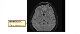
To determine the extent of damage to the liver and other internal organs, hardware diagnostic methods such as ultrasound, MRI and CT are used. If encephalopathy is suspected, a biopsy of liver tissue is required (allows us to find out the cause of the disease). Electroencephalography is used to assess the condition of brain tissue.