Depression is a persistent negative emotional state characterized by depressed mood, decreased interest in others, and an inability to experience pleasure. Its characteristic manifestations are feelings of melancholy, self-deprecation, indifference, tearfulness, decreased activity and fatigue, psychomotor retardation or, on the contrary, agitation. Sleep disturbances in the form of insomnia or, conversely, hypersomnia are typical for depression; changes in appetite with accompanying changes in body weight.
Depression is an important medical problem, since numerous studies [10, 57] have demonstrated a significant relationship between its severity, morbidity and mortality due to cardiovascular and other somatic diseases. Depression is an independent unfavorable prognostic factor during the postoperative period in surgical patients, associated with a high risk of infectious and neurological complications [28, 42]. The emotional state is one of the most important criteria for the quality of life, and therefore the concept of “health,” as defined by the World Health Organization (WHO), includes the psychological well-being of a person, including the absence of depression [1]. Considering the potentially curable nature of depression, understanding the mechanisms of its development, as well as the development of objective approaches to diagnosing this disorder are of great scientific and practical importance.
The functioning of the central nervous system in depression has significant differences from the norm. A number of experimental studies [41, 57] have shown the tendency of depressed patients to negatively perceive neutral or even positive stimuli and/or situations. In particular, patients with depression are significantly more likely to perceive a neutral facial expression in portraits as a manifestation of sadness or anger [14, 31, 41]. In addition, numerous neurophysiological studies have revealed that α-rhythm power asymmetry in the frontal leads is typical for patients with depression [17, 27]. Finally, studies of heart rate indicate a significant connection between depression and power deficits in the high- and low-frequency components of the spectrum of its variability [2, 7].
This review examines the results of modern neuroimaging studies in depression. It is important to note that in the vast majority of cited works, the severity of depressive disorders met the criteria for “major” depression according to DSM-IV. In addition to data obtained using magnetic resonance imaging, we also cite electroencephalographic studies of depression, the results of which became the basis for the formation of modern concepts of its pathogenesis.
To conclude the review, we provide a description of modern concepts of emotion regulation (the concept of the “negative brain”, cognitive regulation of emotions and deficits in activation of the left frontal cortex, which consider depression as a special mode of brain functioning.
Neuroimaging and pathological studies of depression
Structural and anatomical correlates of depression
Most neuroimaging and pathological studies in patients with major depression have identified gray matter deficits in the prefrontal cortex and limbic system structures.
The most typical finding in such cases is a deficiency of gray matter in the anterior cingulate cortex, localized ventral to the genu of the corpus callosum (popliteal cingulate cortex) [49]. Thus, in a study by M. Ballmaier et al. [4] gray matter volume in the left and right cingulate cortex in elderly patients with depression was reduced by 18 and 20%, respectively. In addition, the researchers found a decrease in the volume of white matter in the same brain structure on the left by 14%, on the right by 21%. Similar white matter deficits in the left anterior cingulate cortex in elderly patients with depression were noted in a study by S. Bell-McGinty et al. [5]. K. Cullen et al. [16] identified deficits in the myelinated tracts connecting the popliteal cingulate cortex to the amygdala in the right hemisphere of the brain in adolescents with depression. At the same time, M. Ries et al. [52], who found gray matter deficits in the posterior cingulate cortex and adjacent areas of the cortex in elderly people with moderate depression, did not find significant differences in the volume of the anterior cingulate cortex compared to the control group. According to some researchers [9, 35, 49], pathomorphological changes in the popliteal cingulate cortex are more of a predisposing factor than a consequence of depression, since this anomaly was detected at the earliest stages of the disease, as well as in healthy individuals with a high family history of affective disorders . Thus, P. Keedwell et al. [35] revealed differences in the microstructure (decrease in fractional anisotropy) of the cingulate fasciculus in healthy students with a family history of major depression compared to students who did not have relatives with depression. Intergroup differences were most pronounced in the popliteal area of the left cingulate band. Interestingly, the ability to experience pleasure (hedonia) was positively correlated with fractional anisotropy of the left and, to a lesser extent, the right cingulate fasciculus. It is important that a number of longitudinal studies [49] have demonstrated a progressive decrease in the volume of the popliteal cingulate cortex in patients with psychotic depression, as well as significant correlations between the severity of depressive disorders and the volume deficit of the popliteal cingulate cortex.
The volume of the orbitofrontal cortex is also reduced in patients with severe depression compared to controls [4, 49]. Thus, in a study by M. Ballmaier et al. [4] patients with depression showed a smaller volume of the medial sections of the orbitofrontal cortex by 19% on the left and 24% on the right and lateral sections by 12% compared to the control group. At the same time, some studies have noted an excess of normative values for gray matter volume in a number of areas of the orbitofrontal cortex in patients with depression. Thus, according to J. Phillips et al. [48], in patients in remission during treatment with antidepressants, there was a general increase in brain volume, most pronounced in the orbitofrontal cortex and inferotemporal gyrus of the right hemisphere, although in patients who did not achieve remission during the same period, a decrease in tissue volume was recorded in the left hemisphere .
In a number of studies [5, 49], the volume of gray and/or white matter in patients with depression was reduced in the ventrolateral prefrontal cortex, frontal pole cortex, and dorsolateral prefrontal cortex. H. Soennesyn et al. [60] revealed in elderly patients a significant positive correlation between the volume of hyperintense foci in the white matter of the deep parts of the frontal lobes of the brain and the severity of depression, as well as a tendency towards the persistence of depressive disorders. Similar data were obtained in a longitudinal 3-year study by M. Firbank et al. [22], who noted an association between the progression of hyperintense changes in the white matter of the brain and the occurrence of depression in older people.
Deficits in the volume of prefrontal cortex structures have also been regularly identified in populations at high risk of developing depression. Thus, A. Carballedo et al. [9] found a deficit in the volume of prefrontal cortex structures in a group of healthy people with a family history of depression. J. Lagopoulos et al. [40] revealed a significant connection between a decrease in the size of the frontal cerebral cortex and the severity of depressive disorders in young patients already at the initial stage of the disease.
Many studies have also identified gray matter deficits in the hippocampus, parahippocampal gyri, and temporal cortex [49, 63]. Thus, in a study by S. Bell-McGinty et al. [5] reported smaller gray matter volume in the right hippocampus and medial frontal lobes in both hemispheres in elderly patients with depression compared to controls.
The same study revealed a significant relationship between the duration of depression and a decrease in the volume of the hippocampal-entorhinal cortex. Similar data were published by K. Sawyer et al. [58], who found a decrease in the volume of the right hippocampus during a 4-year follow-up of elderly patients with depression. At the same time, in a study by I. Rosso et al. [55], the volume of gray matter in the hippocampus of adolescents with depression did not differ from the control group.
According to a number of researchers, the size of the amygdala (subcortical nucleus located in the deep parts of the temporal lobes) in patients with depression is also changed. Thus, smaller amygdala sizes compared to the control group were found in children with depression [41, 55]. For example, I. Rosso et al. [55] noted corresponding changes in adolescent girls with major depression: the amygdala in both hemispheres was 12% smaller in volume compared to the control group. At the same time, in adult patients, the size of the tonsils is sometimes slightly larger compared to the age norm [41]. A possible explanation for such discrepancies is the tendency for gray matter volumes to decrease as the brain matures, and this is most pronounced in the subcortical nuclei. Accordingly, large volumes of gray matter in populations with mental disorders may be a correlate of delayed brain development.
It should be noted that in many neuroimaging and pathological studies in cases of severe depression, gray matter deficits were more pronounced in the left hemisphere compared to the right [49, 63]. Thus, in a study by P. Tu et al. [63] patients with depression had significantly smaller volumes of the left temporal, entorhinal, and lingual cortices compared to the control group. We also note the higher incidence of depression with damage to the left frontotemporal region compared to damage to the right hemisphere in patients after traumatic brain injuries [8], strokes [65] and other brain diseases. It is important to emphasize that the connection between left hemisphere brain damage and depression is recorded mainly in cases of small damage [18].
Functional and anatomical correlates of depression
Currently, dozens of studies have been published that have examined the patterns of brain activity in patients with depression using functional magnetic resonance, single-photon, or positron emission tomography. In most of these studies, brain activity is assessed during functional tests with an impact on the emotional sphere. The most common test is to present the patient with portraits of faces with expressions of sadness, fear, anger or joy, as well as photographs of people in a neutral emotional state.
Most of these studies have shown that depressed patients, when presented with sad or fearful faces, tend to respond with hyperactivation of limbic system structures such as the amygdala, anterior cingulate cortex, and ventromedial prefrontal cortex [41, 49]. At the same time, presenting happy faces to patients with depression does not cause them to activate the listed brain structures, which is characteristic of healthy people. In addition, patients with depression are characterized by deficits in the synchronization of activity between the amygdala and the medial prefrontal cortex. The listed functional disorders correlate with the intensity of depressive experiences, and the improvement in the psychological state of such patients is associated with the normalization of the functions of these brain structures. Relapses of depression are accompanied by increased metabolism in the popliteal region of the anterior cingulate cortex, the ventromedial part of the frontal cortex and the amygdala [49].
Many studies have noted that abnormalities in the activation of limbic system structures in patients with depression tend to lateralize. Thus, J. Almeida et al. [3] observed in patients with depression a lack of interaction between the left hemisphere ventromedial, popliteal cingulate cortex and the amygdala in the process of assessing happy faces. At the same time, the presentation of photographs with frightened faces was accompanied by an excessive expression of the conjugation of activity in the left hemisphere popliteal cortex and amygdala. Z. Mingtian et al. [43] also studied the activation features of brain structures when presented with photographs of fearful faces in young patients with early onset depressive disorders. Researchers have observed similar overactivation of the left hemisphere structures - amygdala, thalamus, prefrontal and temporal cortices, combined with underactivation of the right prefrontal cortex in patients with depression. At the same time, in a study by G. Perlman et al. [47] found that the presentation of negative emotional images to adolescents with depression was accompanied by hyperactivation of the right hemisphere amygdala with less interaction of this subcortical structure with the insular cortex and medial prefrontal cortex compared to the control group. J. Fournier et al. [24] recorded hyperactivation of the right hemisphere amygdala in adult patients with depression when presented with photographs of angry faces. In a study by R. Tao et al. [61] in patients with depression, bilateral hyperactivation of the amygdala was recorded in combination with hyperactivation of the popliteal cingulate and orbitofrontal cortices when presented with photographs of fearful faces.
E. Rose et al. [54] observed in patients with depression who performed a test to memorize visuospatial information, significantly less deactivation of the popliteal cingulate cortex and the adjacent medial orbitofrontal cortex. The authors suggested that excessive activation of the popliteal cingulate cortex in depression is directly related to negative experiences in such patients.
Insufficient activation of the ventral striatum during reward is also characteristic of patients with depression [49]. Thus, in a study by O. Robinson et al. [53] in patients with depression, a reduced ability to learn when rewarded was noted against the background of normal indicators of learning when punished. In the same study, behavioral deficits in patients with depression were accompanied by deficits in activation of the ventral striatum when receiving reward.
Resting brain activity in depressed patients is also altered. Thus, M. Gaffrey et al. [25] observed a lack of coordinated activity between the posterior cingulate cortex and areas of the middle temporal gyrus, inferior parietal lobule and cerebellum in children with depression compared to age-related norms.
At the same time, coordination of activity in the posterior cingulate and anterior popliteal cortex was increased in children with depression. A similar lack of conjugation of activity in the posterior cingulate cortex and adjacent cortical centers in young patients with depression was observed by X. Zhu et al. [68]. In addition, this same patient population showed excessive coordination of activity in the anterior cingulate and medial prefrontal cortex. M. Wu et al. [66] observed excessive synchronization of the activity of the dorsomedial prefrontal and orbitofrontal cortices in elderly patients with depression with a deficit in synchronization of the popliteal cingulate cortex. The listed abnormalities in brain activity at rest correlated with a number of psychopathological phenomena characteristic of depression. Specifically, excessive synchronization of activity in the anterior cingulate and medial prefrontal cortices was correlated with obsessional susceptibility in a study by X. Zhu et al. [68]. In a study by S. Green et al. [29] a lack of coordinated activity between the popliteal cingulate and temporal cortices correlated with ideas of self-blame and feelings of guilt.
A number of studies have identified changes in the activity of the amygdala and prefrontal-cingulate cortex in patients in remission. P. Kanske et al. [34] observed excessive activity of the amygdala in patients in remission when viewing stories with negative content. In addition, R. Elliott et al. [20] revealed in such patients a lack of activation of the frontal poles and lateral orbitofrontal cortex when they were presented with images with social stories of both negative and positive content. Hypoactivation of the left hemisphere orbitofrontal and dorsolateral prefrontal cortex when viewing photographs with fearful faces in patients in remission was also demonstrated in a study by R. Kerestes et al. [36].
In the same study, activation of the right hemispheric dorsolateral prefrontal cortex was positively correlated with the duration of euthymia.
H. Cremers et al. [13] studied the correlation between neuroticism (in this case, it was considered a risk factor for the development of depression) in healthy middle-aged people and the activity of brain structures when assessing the expression of anger, fear and sadness in portrait photographs. The authors found a significant direct correlation between neuroticism and the activity of the right hemisphere dorsomedial prefrontal cortex, while in individuals with elevated levels of neuroticism, activity in this region of the prefrontal cortex correlated with activity in the right amygdala; in the group with a high level of neuroticism, the activity of the left amygdala was less correlated with the right hemispheric cingulate gyrus compared to subjects with a low level of neuroticism. Another research group [21] found a positive relationship between neuroticism and excessive activation of the insular cortex during decision-making under conditions of information deficiency.
Neurophysiological correlates of depression
R. Davidson [17] was the first to point out a connection between the severity of interhemispheric asymmetry of activation of the frontal cortex and susceptibility to severe depression. He proposed a method for measuring interhemispheric asymmetry as the difference between the power of α-activity (8-13 Hz) in the right (F4) and left (F3) frontal leads. Based on his own research, the author identified a relative pattern of hypoactivation of the left frontal cortex in patients with severe depression, demonstrating a connection between activation of the left frontal cortex and positive affective reactions, on the one hand, and activation of the right frontal cortex and negative affective reactions, on the other.
Subsequently, a number of research groups confirmed the connection between hypoactivation of the left frontal cortex and the severity of depression. Thus, I. Gotlib et al. [27] noted hypoactivation of the left frontal cortex not only in individuals with severe depression, but also in cases where there were episodes of depression in the anamnesis (during the examination period, the patients were in remission). The authors did not reveal significant correlations between the asymmetry of activation of the frontal cortex and the severity of unrealistic ideas about oneself or the opinions of others, i.e. correlations with cognitive factors that reduce stress resistance. There was also no connection between interhemispheric asymmetry and performance of the well-known Stroop test. The researchers concluded that hypoactivation of the left frontal region is an important predisposing factor in the pathogenesis of depression. However, left frontal hypoactivation was not associated with current depression or stress-reducing cognitive factors.
Although the connection between depression and interhemispheric asymmetry of α-activity in the frontal leads has been established in many studies, in some studies it was not found. Thus, S. Reid et al. [51] did not reveal intergroup differences in the distribution of α-activity power in the frontal leads. However, in this study, “classical” intergroup differences in indicators of interhemispheric asymmetry of α-activity in the frontal leads were identified during a separate analysis of data relating to the first minutes of EEG recording. It is also important that, unlike other researchers, the authors used too “soft” criteria for inclusion in the study of both patients and control group participants, which could lead to “erosion” of intergroup differences.
Interestingly, in a study by D. Smit et al. [59], the relationship between hemispheric asymmetry and the risk of developing depressive and anxiety disorders was significant only in the subgroup of young women. The same study revealed the influence of heredity on the severity of interhemispheric asymmetry in a group of both young women and young men.
At the same time, in middle-aged subjects the severity of interhemispheric asymmetry was significantly higher compared to young ones, but the influence of heredity on interhemispheric asymmetry was not detected in this group.
Thus, the results of neuroimaging studies indicate three leading features of brain functioning in patients with depression: firstly, depression is characterized by excessive activity of such structures of the limbic system as the amygdala, popliteal cingulate and ventromedial prefrontal cortex, with the maximum severity of such changes observed when exposed to negative incentives. Secondly, patients with depression are characterized by a deficiency of gray matter and abnormal functioning of the frontotemporal structures of the cerebral cortex. Thirdly, with depression there is a tendency towards more pronounced abnormalities in the functioning of the structures of the left hemisphere of the brain, including a typical finding of electroencephalographic studies is a deficit in activation of the left prefrontal cortex in patients with depression. The listed features correspond to modern concepts of emotion regulation, which we discuss below.
Modern concepts of emotion regulation
The concept of "negative brain"
The term "negative brain" was proposed by L. Carretié et al. [11] based on data from experimental and neuroimaging studies indicating activation of a number of brain structures in response to negative stimuli. The fundamental function of the structures that make up the “negative brain” is to provide rapid and intense responses to dangerous, harmful and potentially destructive events. Obviously, an effective response to such stimuli has a more important adaptive value compared to a response to neutral or positive (attractive) stimuli.
In dangerous situations, the body is required to urgently mobilize appropriate resources, and the evolutionary process supported the formation of neurological mechanisms capable of increasing adaptive capabilities in difficult conditions. The speed of information processing in brain structures can only be achieved by reducing the number of connections between neuronal centers, i.e. the relative specialization of such centers in processing similar information patterns. Thus, in the structure of the brain, a number of neuronal centers specialize primarily in processing information about threatening and destructive events. Data from modern research indicate the relative autonomy of the “negative brain”, provided by its own sensory subcortical centers, cortical analyzer and executive structures [11].
The amygdala is the most studied structure of the “negative brain.” It receives direct projections from the thalamus and sensory cortex, which allows it to form rapid and independent responses to negative stimuli from higher cortical centers [11, 38]. The speed of the amygdala’s responses to negative stimuli is also ensured by direct connections of these structures with a number of executive autonomic and motor centers of the brain.
Numerous experimental studies have demonstrated the leading role of the amygdala in the formation of conditioned reflex manifestations of fear and anxiety [19]. In healthy people, activation of the amygdala is recorded predominantly when viewing faces with an expression of fear or sadness, while this tendency is most pronounced after induction of a sad mood in patients [26, 64]. The studies presented in the first part of the review indicate the maximum severity of the listed trends in depression.
In addition to the amygdala, to brain structures predominantly involved in the processing of negative information, L. Carretié et al. [11] include the ventromedial prefrontal cortex and the adjacent anterior cingulate cortex, the anterior parts of the insula and some other brain structures, the functions of which are less studied. Some researchers [62] suggest that the medial regions of the prefrontal cortex, including the anterior cingulate, form a subject's cognitive representation of current affective experiences. Other authors [33] point to the direct involvement of the ventromedial cortex in the formation of associations between threatening events and the context in which they occur. Finally, there is an opinion that activation of the popliteal cingulate cortex during the processing of negative information is aimed at finding a decision about further actions [54]. Probably, a deficiency of gray matter in the popliteal cingulate cortex and the adjacent ventromedial prefrontal cortex reduces the computational capabilities of these brain structures in depressed patients and leads to stereotyping of the “decisions” being formed.
The hyperactivation of “negative brain” structures found in patients with depression explains such clinical characteristics of depression as the persistent tendency of depressed patients to perceive neutral stimuli as negative [13, 31, 41] and a deficit in heart rate variability [2, 7]. The latter is explained by the direct participation of the amygdala and anterior cingulate cortex in the regulation of heart rate [15, 67].
Concept of cognitive regulation of emotions
Cognitive deficits are a fairly common finding in neuropsychological examinations of patients with depression. A number of studies have found that patients with depression have a lower level of education compared to corresponding control groups [5, 52]. The most pronounced cognitive deficit is recorded in patients with psychotic depression (with delusional ideas) [23]. In the work of K. Blankstein et al. [6], who examined students with moderate depression, found that they had great difficulties in resolving interpersonal conflicts, as well as in organizing their time, everyday life and the educational process. Thus, cognitive deficits can be considered a common component of depressive disorders and possibly an unfavorable prognostic factor.
J. Gross and R. Thompson [30] formulated the main postulates of the concept of cognitive regulation of emotions. In their opinion, the emotional system of the brain is capable of self-regulation, but at the same time, the “meaningfulness” of emotional processes has a significant impact on the emotional state of the subject. The ability to make the right choice, taking into account the long-term outcomes of a particular form of behavior, requires significant cognitive effort and is based on previous experience. Active transformation of the current situation, the ability to shift attention from unpleasant but inevitable moments, to see the positive in an ambiguous context are important cognitive mechanisms that allow an individual to adapt to emotionally difficult conditions. Accordingly, cognitive deficit deprives an individual of the ability to adapt to difficult conditions, which in many cases leads to depression.
A number of experimental studies [37, 39] have established a direct connection between being in an information-enriched environment and neuroplastic changes in the “cognitive” cortex. Neuroplastic changes in brain structures include thickening of the gray matter, lengthening and increased branching of dendrites, and an increase in the density of dendritic spines and the density of synapses on them. At the same time, stressful situations inhibit neuroplastic processes and cause reverse changes in brain structures [50]. This can be demonstrated by the results of an experimental study by J. Radley et al. [50]: 3-week stress led to a decrease in the length and “branching” of dendrites of neurons in the medial prefrontal cortex by 20% and a decrease in the number of synapses on pyramidal neurons by 33%. The authors suggest that hypotrophic changes due to stress are a consequence of hypercortisolemia, which is a typical characteristic of depressive disorders. These kinds of results from experimental studies of stress are consistent with the results of neuroimaging studies of depression, which have shown a decrease in the volume of gray matter in a number of brain structures as the duration of the disease increases [5, 58].
Thus, with depressive disorders, a vicious circle is formed. Cognitive deficit reduces adaptive capabilities and contributes to the subject getting into stressful situations. At the same time, chronic stress and hypercortisolemia aggravate hypotrophic changes in brain structures, which leads to worsening cognitive deficits and depression.
Concept of left frontal cortex activation deficit
The concept of deficits in left frontal cortex activation in depression is a special case of the concept of cognitive emotion regulation. However, modern research indicates the importance of this factor in the pathogenesis of affective disorders. Thus, K. Ochsner et al. [46] recorded pronounced activation of the left convexital prefrontal cortex during the process of “rethinking” the meaning of an emotionally negative story picture in healthy subjects. The main finding of this study was the inverse correlation between the degree of activation of the left hemisphere dorsolateral prefrontal cortex and the degree of activation of the right hemisphere amygdala and left orbitofrontal cortex. In other words, the researchers observed an “inhibitory” effect of activation of the left dorsolateral prefrontal cortex on the structures of the “negative” brain. Currently, the inhibitory (braking) effect of the prefrontal cortex on the activity of the amygdala is a generally accepted fact, as it has been demonstrated in numerous studies, including neuroimaging studies [18, 32].
A number of neuroimaging and neurophysiological studies have established a tendency towards greater activation of the left frontal cortex during the experience of positive emotions in healthy people and the right frontal cortex - negative ones [18, 44]. In a study by J. Coan and J. Allen [12], the predominance of activation of the left frontal cortex compared to the right hemisphere significantly correlated with the tendency to active behavior in healthy people. Thus, deficits in activation of the left prefrontal cortex, documented in numerous studies of depressed patients, explain the deficits in positive emotions and apathy in a significant proportion of such patients.
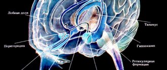
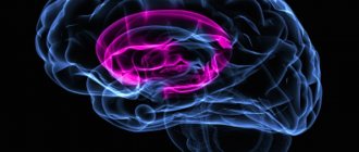
Cingulate gyrus
The cingulate gyrus is a relatively large brain structure, but in the clinic of psychoneurological disorders its anterior part plays the most significant role. The latter includes cortical areas 23, 24 and 25 according to Brodmann. In clinical practice, damage to these zones affects motivation and movements because it has developed connections with the dorsal and ventral striatum (striatum). Previously, researchers have found significant differences in cytoarchitecture between the anterior granular cortex (area 32) and the posterior agranular cortex (area 24). Currently, four functional zones of the cingulate gyrus are distinguished: viscero-motor (including area 32), cognitive-effector and motor (area 24) and sensory, if we consider the cingulate gyrus gradually moving from the front to the back. In animals, the cingulate cortex is a limbic region that is associated with vocalizations in primates, occurring in response to appropriate stimulation. The cingulate gyrus is involved in processes of autonomic activity, including changes in the frequency and depth of breathing, pulse rate, sexual activity and automated oral reactions. “Wing epilepsy” is characterized by short episodes of loss (change) of consciousness, with manifestations of vocalisms, rapid motor activity (axial flexion and tension of the limbs), as well as automatisms of gestures. In humans, the cingulate cortex is involved in nociceptive processes, possibly related to thalamic afferentation, and extending to affective responses, including fear, euphoria, depression and aggression. , manifested in response to stimulation and externally expressed in certain behavior: disinhibition, hypersexuality, tick-like movements, and obsessive-compulsive activity. Damage to the anterior cingulate cortex underlies emotional flattening and decreased motivation; a combination of such symptoms is sometimes referred to as “frontal lobe syndrome.” Activation of area 25 (popliteal cingulate zone) is noted in severe depression. Work on deep stimulation of these structures or its surgical excision is being carried out in the treatment of severe depression. This area has reciprocal connections with other frontal areas, with the hypothalamus, ventral striatum, amygdala and autonomic outputs to the brainstem.
The posterior part of the cingulate cortex has been studied to a lesser extent than the anterior one. It is likely that the posterior portion of the cingulate gyrus is less involved in motor functioning and more involved in visual-spatial functions, learning and memory. Recently, projections of the cingulate gyrus to the base of the brain (subiculum) have been shown. The posterior cingulate area passes into the area of the parietal cortex, as a result of which it is possible that it is related to self-awareness and the structures responsible for this phenomenon (the precuneus of the cerebral hemispheres).