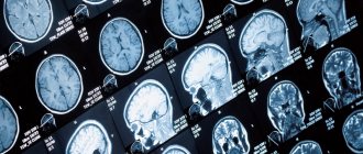
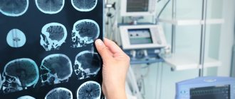
Stereotactic biopsy of a brain tumor is a fairly common procedure in neurosurgery, allowing the doctor to diagnose the nature of changes in the brain substance. Situations arise when, based on magnetic resonance imaging (MRI), positron emission tomography (PET CT) and other non-invasive research methods, it is not possible to determine the nature of a brain tumor. If the doctor suspects a brain tumor and for one reason or another open surgical intervention is not indicated at this stage (the tumor is located in hard-to-reach vital structures of the brain, is multiple in nature, the nature of the changes is assumed to be non-tumor, etc.), the patient is offered a high-precision neurosurgical intervention - stereotactic biopsy of a brain tumor. After receiving the sample, the pathologist examines the obtained tumor tissue and makes a histological diagnosis. After establishing the nature of the neoplasm, further treatment tactics become clearer.
To carry out these interventions, specialized stereotactic equipment is used, which is capable of localizing in three-dimensional space the place of greatest interest for performing a biopsy. The equipment allows you to obtain projections of brain structures (neoplasms, convolutions, vessels, etc.) in a three-dimensional coordinate system. This allows the neurosurgeon to select appropriate target coordinates for guiding the biopsy needle.
The decision to perform a tumor biopsy rather than attempt open surgery is made taking into account many factors, often in collaboration with other colleagues - neurosurgeons, radiologists, oncologists, neuroimaging specialists.
What is a biopsy?
A biopsy is the removal of a small area of tissue to be examined for signs of a disease.
The tissue sample can be taken from any organ. Biopsies are performed in a variety of ways. In some cases, a hollow needle is used to remove a small piece of tissue, while in other situations, surgical removal of the entire suspicious lesion is required.
Often, to remove tissue, a needle is inserted into the area of pathological cellular changes through the skin. To safely perform a biopsy, imaging guidance is often used: ultrasound, fluoroscopy, computed tomography (CT) or magnetic resonance imaging (MRI). Such studies make it possible to verify the exact position of the needle inside the pathological formation.
Up
WHAT IS THE SURGICAL INTERVENTION?
It is possible to perform an open biopsy using craniotomy, as when removing a tumor. In this case, a window is opened in the skull bone by removing a fragment, which is subsequently replaced in its original place. However, this approach is not always practiced and is limited to superficial lesions in prominent areas of the brain or when samples need to be taken from the cranial bone or meninges.
If possible, partial resection of the lesion is performed and a sample of this tissue is taken for analysis. Minimally invasive biopsy methods are chosen: for diffuse damage to brain tissue without clear boundaries for small and deep lesions for significant lesions in a very visible area of the brain with a high risk of serious neurological consequences To do this, a trepanation or a single small hole is made as an entry point into the skull, through which a cannula is inserted and directed to specific, pre-planned coordinates.
The classic method, which is still used in many centers and for other types of operations, is stereotaxy. Stereotaxy involves obtaining target coordinates by performing a CT scan of the skull immediately before entering the operating room. A metal frame is attached to the patient's skull to serve as a reference for obtaining coordinates, which will later be attached to another component on the operating table.
Because this procedure is not particularly pleasant for the patient, has higher logistical requirements, and takes longer to prepare, an alternative technique has been developed. The cannula is inserted into specific coordinates in the brain using neuronavigation technology (frameless stereotactic brain tumor biopsy), which is used at the Instituto Clavel. This method provides maximum accuracy and safety.
During neuronavigation biopsy of brain tumors, the patient's head must be in a fixed position relative to the landmark of the navigation device. An accurate, high-quality MRI of the patient's skull is loaded onto the console of this device and a target is determined to hit.
The neuronavigator combines the downloaded image with the marked target and shows on the screen the position of the biopsy cannula in relation to the target, allowing neurosurgeons to reach it safely. The path is selected in the image in such a way as to avoid risk areas inside the brain and reach the desired depth.
Each time biopsy samples are taken from brain tumors, a fresh sample is sent to the laboratory to be analyzed and assessed for viability and the presence of abnormal tissue before sending additional samples for final examination and wound closure.
In what areas is biopsy used?
When a suspicious area of tissue is detected, an instrumental examination is carried out, which in most cases allows us to determine its nature: benign or malignant. If such a diagnosis is unsuccessful, a biopsy may be required.
Typically, a biopsy is used to detect certain diseases. In addition, it is used to examine tissues before organ transplantation. Most often, a biopsy helps diagnose cancer, but it is also used to detect other conditions, such as infections or autoimmune diseases.
Tissue samples can be taken from all organs for a variety of reasons. The following are the main types of biopsies and the reasons for performing them:
- Bone biopsy: Used to diagnose cancer or bone infection. This biopsy can be done using a needle that is inserted through the skin or surgically.
- Bone marrow biopsy: Used to diagnose blood cancers such as leukemia. In this process, the doctor uses a needle to remove a small sample of bone tissue and red bone marrow. In some cases, only bone marrow testing is required.
- Breast biopsy: used to determine the nature (benign or malignant) of lumps in the breast. There are several types of similar procedures: Stereotactic biopsy (mammography-guided)
- Ultrasound-guided biopsy
- MRI-guided biopsy
If necessary, it is possible to perform a biopsy of almost any organ, including the bladder, heart, prostate gland, tissues and organs of the neck, parathyroid glands, etc.
Up
1.What is a stereotactic biopsy?
Stereotactic biopsy
is essentially a minimally invasive neurosurgical operation performed for the purpose of selecting tissue samples of a neoplasm (tumor) for laboratory research.
The samples obtained using this procedure allow the doctor to draw conclusions about the nature of the tumor, its level of malignancy, and the prospects for its “behavior.” It is on the basis of laboratory tests that the doctor makes the final decision on the choice of tactics and treatment strategy - the surgical technique, the need for radiation therapy, chemotherapy, etc.
A must read! Help with treatment and hospitalization!
How should you prepare for research?
In most cases, the biopsy is performed on an outpatient basis and does not require any special preparation for the patient.
A surgical biopsy may require you to temporarily stop taking certain medications, which you should ask your doctor about.
It is usually recommended that you stop eating and drinking 8 hours before the biopsy. However, oral use of drugs is allowed with a small amount of water. Patients receiving insulin injections for diabetes should consult their doctor about changing the dose of the drug.
Before performing a biopsy, it is very important to inform the doctor about all medications that the patient is taking, including herbal ones, as well as any allergies, especially to local anesthetics. Three days before the procedure, you should stop taking aspirin or other blood thinners.
You should also tell your doctor if you have any recent illnesses or other conditions.
During the examination, you must wear a special hospital gown. Women should always inform their doctor about any possibility of pregnancy. Some instrumental studies using imaging techniques are not performed during pregnancy to avoid negative effects on the fetus.
It is advisable to come to the hospital with a relative or friend who will help the patient get home. When using sedatives, this condition is necessary.
Preparing a child for a biopsy is carried out in a similar way. Consult your doctor for more detailed instructions.
Up
RECOVERY AND REHABILITATION AFTER BIOPSY OF BRAIN TUMORS
Generally, a one-night hospital stay is required for a minimally invasive biopsy unless complications occur and the patient's condition is worse than when admitted to the clinic.
The patient's recovery is usually influenced not so much by the operation itself as by the course of the disease, age and previous condition of the patient. It is a painless and short procedure (about an hour), allowing the patient to almost immediately regain his autonomy for basic daily activities.
If surgery with craniotomy is required, the patient usually spends the first night in the intensive care unit for observation. Since the aggression on brain tissue is minimal, post-operative symptoms are not noticeable and the risks are limited, so the patient can leave the hospital after two nights.
What does the diagnostic equipment look like?
There are quite a large number of biopsy techniques. Moreover, the equipment used is very different.
In a needle biopsy, a tissue sample is removed using a needle.
The biopsy needle is usually several centimeters long, and the syringe barrel is the diameter of a wide paper clip. The needle is hollow inside, allowing the tissue sample to be grasped and removed.
Several types of needles are used when performing a biopsy:
- Thin needle attached to a syringe: Thinner in diameter than needles used to draw blood from a vein.
- Cutting automatic needle with spring mechanism: consists of an internal "thick" needle, which is inserted into a locking cell, covered with a protective sheath and attached to a spring mechanism.
- Vacuum aspiration biopsy device: allows you to obtain a fairly large tissue sample.
A puncture biopsy is often performed under the guidance of CT, fluoroscopy, ultrasound or MRI.
Mammograph
It is a rectangular apparatus equipped with an X-ray tube. The device is used exclusively for x-ray examination of the mammary glands and has special additional devices that ensure that radiation reaches only the gland. A device is attached to the X-ray machine to position and compress (squeeze) the breast, which helps obtain images from different angles.
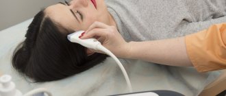
Ultrasound scanner
consists of a console that includes a computer and electronic equipment, as well as a video display and an ultrasound probe used for scanning. The sensor is a small, portable device that resembles a microphone and is attached to the scanner using an electrical cord. The ultrasound sensor sends high-frequency sound signals inaudible to the human ear and picks up echoes reflected from the internal structures of the body. The principle of operation of the device is similar to sonars that are used on submarines. In this case, an image instantly appears on a monitor resembling a television or computer screen. Its appearance depends on the amplitude (strength), frequency and time it takes for the sound signal to return from the patient's body to the transducer, as well as the type and condition of the tissue through which the sound travels.
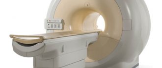
Computer tomograph
usually a massive rectangular apparatus with a hole, or short tunnel, in the middle. During the procedure, the patient is placed on a narrow table that slides inside the tunnel. The X-ray tube and electronic X-ray detectors are located opposite each other inside a ring-shaped structure called a gantry. In a separate office there is a computer workstation where the resulting image is processed. There is also a doctor or technologist here who monitors the operation of the tomograph and the progress of the examination.
Vacuum device for aspiration biopsy
is an instrument in which a tissue sample is taken into a needle under pressure.
When performing a surgical biopsy
the pathological area is often marked with special thin wires or contrast material, which is introduced into the patient’s body under the control of ultrasound, CT or X-ray.
Other sterile instruments are also used during the procedure: syringes, forceps, scalpels, sponges and microscopic glasses or tissue containers.
Up
What is the basis for the research?
The features of the biopsy procedure itself depend on the location of the tissue and organ that needs to be examined.
In most cases, a biopsy can be performed using a needle, which is the most minimally invasive procedure and allows the patient to go home the same day. In this case, visual control allows you to select the best point for inserting the needle and bring the instrument to the site of pathological changes: fluoroscopy, ultrasound, CT or MRI.
If the focus of pathological changes is located in hard-to-reach places, then a surgical biopsy in the operating room may be required. In this case, the removal of a tissue sample requires the presence of a surgeon who performs a minor operation. To determine the location of the lesion and collect tissue, the doctor uses an instrument equipped with a small camera.
Image guidance allows the doctor to insert a needle through the skin and guide it to the suspicious area of tissue.
A sample of cells and tissues is collected using one of the following methods:
- A fine-needle aspiration biopsy uses a small-gauge needle and syringe to remove fluid or a small sample of cells.
- During a core needle biopsy, an automatic spring mechanism is activated, which “pushes” the needle deep into the tissue. After returning the needle, the hollow part of the instrument is filled with a tissue sample in the form of a column. The cutting element of the instrument immediately cuts off the tissue and holds it inside the syringe. The process is repeated several times.
- In a biopsy using a vacuum device, the needle is first placed into the area of pathological changes. After this, a vacuum device is activated, which “sucks” the tissue sample under pressure into the hollow part of the needle. The tissue is cut using a cutting element and passes through a needle into a special reservoir. The process is repeated several times.
Up
How is the research conducted?
As a rule, a needle biopsy is performed on an outpatient basis.
Typically, minimally invasive image-guided procedures, such as core biopsies, are performed with an experienced radiologist, interventional radiologist, or neuroradiologist (as appropriate).
To administer sedatives during the procedure, the nurse inserts an intravenous catheter. In addition, a sedative may be prescribed orally.
The area where the biopsy needle is inserted is numbed with a local anesthetic. In some cases, such as breast or thyroid biopsies, sedation is not required.
In the case of a biopsy, children, especially young children, may be given general anesthesia, which makes the procedure more comfortable for both the child and the doctor.
If the study is carried out under fluoroscopy control, then during the procedure the patient lies or stands in front of the screen.
During a CT or MRI-guided biopsy, the patient is positioned horizontally.
The use of tomography allows one to determine the exact location of the pathological formation and the safest route for needle insertion. Once the lesion is located, the needle insertion point is marked with a marker. Hair is carefully removed from the skin, it is disinfected, and a sterile surgical drape is applied.
A tiny puncture is made in the skin where the biopsy needle is inserted. Using imaging guidance, the doctor inserts a needle through the skin, advances it to the suspicious area, and removes a tissue sample. A complete analysis may require several samples.
After the procedure is completed, the needle is removed, bleeding is stopped, and a pressure bandage is applied to the wound. No stitches are required.
For several hours after the biopsy, the patient is under the supervision of medical personnel. X-ray(s) or other testing is done to monitor for possible complications.
The patient can return home immediately after the examination, which, however, depends on the type of biopsy performed.
The procedure usually takes about an hour. Depending on the characteristics of the study, the doctor may leave the patient under observation for several hours.
For children, the biopsy is performed in a similar way.
During a stereotactic breast biopsy, the patient lies face down or sits on a movable table while the gland to be examined is placed in a special hole in the table.
A fine-needle breast biopsy uses an extremely small needle and syringe to remove fluid or a small sample of cells.
During a core needle biopsy, an automatic spring mechanism is activated, which “pushes” the needle deep into the tissue. After returning the needle, the hollow part of the instrument is filled with a tissue sample in the form of a column. The cutting element of the instrument immediately cuts off the tissue and holds it inside the syringe. The process is repeated several times.
In some cases, a device for vacuum aspiration of tissue is used during breast biopsy. In this case, the tissue sample is “sucked” into the needle under pressure and ends up in a special reservoir. The needle then automatically changes position in the breast tissue without the need to remove it and reinsert it, and an additional sample is collected. Typically, vacuum aspiration allows you to obtain 8-10 tissue samples around the suspicious area. After completion of the study, the needle is removed.
During a surgical biopsy, a special guide wire is inserted into the pathological area to facilitate the surgeon's work. You can also insert a small tag into the suspicious area, allowing it to be detected if necessary in the future.
After the biopsy is completed, bleeding is stopped and a pressure bandage is applied to the wound.
Up
RISKS OF BIOPSY OF BRAIN TUMORS
The main risk with biopsies of brain tumors is bleeding, although most are not large enough to be of major concern. The risk of moderate or severe bleeding is less than 1%, and the lesions with the highest risk of bleeding are considered to be high-grade gliomas.
Potential neurological deficits depend on the point of entry into the brain, but especially on the location of the lesion. Depending on where the target is located, one of the safe entry points into the cerebral cortex will be determined. For this purpose, not the most noticeable areas are selected, that is, those whose functions are less significant, and damage to their neurons will be invisible to the patient (unlike other, more noticeable areas, for example, those that control movement).
The main risks of neurological complications are associated with voluntary movements that are controlled in certain areas of the brain, such injuries usually require a minimally invasive procedure, and are less likely to affect the sensory organs or swallowing.
Some possible post-operative symptoms that may not be related to complications are headache, dizziness and nausea, provided they are mild and respond well to medications. In some cases, the patient may be somewhat disoriented and unstable in the first hours. The latter usually occurs in older people.
What should you expect during and after the procedure?
During a puncture biopsy, insertion of the needle is accompanied by short-term acute pain. During a surgical biopsy, local anesthesia and general anesthesia help relieve pain.
The introduction of a local anesthetic to numb the skin is accompanied by a light injection, after which the sensitivity of the skin disappears for a while. There is a feeling of slight pressure as the biopsy needle is inserted.
Before the procedure, the doctor may prescribe the patient a mild sedative in tablets. In addition, sedatives are administered intravenously, if necessary, throughout the procedure.
There may be some soreness where the needle was inserted for a few days after the biopsy. For severe pain, analgesics can be used.
How to care for your skin after a biopsy may vary, but you can usually remove the bandage one day after the procedure and then shower or bathe.
Up
Who reviews the biopsy results and where can they be obtained?
After the sample is collected, the tissue is sent to the laboratory for further analysis.
The sample taken is examined by a histologist or pathologist under a microscope. After a few days, the specialist sends a report to the attending physician.
You should ask the doctor who performed the biopsy about where you can pick up the report. With a breast biopsy, the results of the procedure can in some cases be discussed with a radiologist.
After completion of the procedure or other treatment, the specialist may recommend that the patient undergo a dynamic follow-up examination, during which an objective examination, blood tests or other tests, and instrumental examination are performed. During this examination, the patient can discuss with the doctor any changes or side effects that have appeared after the treatment.
Up
Bone marrow biopsy
A bone marrow biopsy is a medical test that helps doctors check the health of your blood cells. Health care professionals will be able to perform the procedure even if there are problems with blood cell production.
A healthcare professional inserts a thin needle into a large bone. The needle takes a sample of bone marrow. The tissue sample is then sent to a laboratory for analysis.
Below are answers to frequently asked questions about biopsies.
What is bone marrow?
Bone marrow is the soft tissue found inside the largest bones. Bone marrow produces many of the body's blood cells, including red blood cells, white blood cells, and platelets.
Stem cells in the bone marrow produce various blood cells. There are two main types of stem cells in the bone marrow: myeloid cells and lymphoid cells.
Myeloid cells are involved in the creation of red blood cells, white blood cells and platelets. Lymphoid stem cells produce a specific type of white blood cell responsible for immunity.
Blood is made up of various components and plays an important role in maintaining health. The bone marrow creates these components. Red blood cells play a critical role in transporting oxygen throughout the body. White blood cells, of which there are several types, play an important role in helping the body fight infection. Platelets help stop bleeding by clotting the blood.
What is the purpose of a bone marrow biopsy?
Doctors order a bone marrow biopsy if there are signs or symptoms indicating abnormal blood cell production.
Bone marrow biopsy is also used during treatment of patients with blood cancer to monitor the progress of treatment and chemotherapy.
There are many symptoms and diseases that can be diagnosed and evaluated using a bone marrow biopsy. Some of these diseases and conditions include the following:
- Anemia is a lack of red blood cells.
- Abnormal bleeding or blood clotting.
- Cancers of the blood and bone marrow, such as leukemia, lymphoma, or multiple myeloma.
- Cancer that has spread to the bone marrow.
What to Expect During a Bone Marrow Biopsy
The procedure is performed in a doctor's office, hospital, or clinic. The biopsy itself lasts about 10 minutes, and the total time is between 30 and 45 minutes.
Before the procedure
Before the bone marrow biopsy, your doctor will ask questions to ensure a safe procedure. Preparing a list of questions and a history can help speed up the process.
The specialist will ask questions about medications that may increase bleeding, since a bone marrow biopsy carries this risk.
Common pain relievers such as aspirin, ibuprofen, and naproxen may also increase bleeding. Anticoagulants or blood thinners such as heparin and warfarin are known to increase these risks.
The patient will be instructed whether to continue taking medications or stop them before the procedure.
The presence of allergies in the patient can cause serious concerns in connection with the biopsy. The doctor will ask about allergies, especially to anesthetics and latex, which is found in surgical gloves.
Anesthetics may be used during the procedure, so patients should ask a friend or family member to drive them home.
During the procedure
The procedure depends on the doctor. Typically, the process is carried out in two stages:
- Aspiration: The doctor removes fluid from the bone marrow.
- Biopsy: The doctor removes a piece of bone and bone marrow tissue.
A bone marrow biopsy is usually performed in an outpatient setting, but some patients may have the procedure done in a hospital. The procedure is usually done from the pelvic bone, but other bones may be used.
The stages of a bone marrow biopsy are usually as follows:
Before the procedure, the patient is changed into hospital clothes. The healthcare provider will ask the patient to lie on their side or stomach. Positions may vary depending on location selected. The healthcare provider then cleans the biopsy site with an antiseptic.
The doctor applies an anesthetic with a needle to numb the biopsy area. Some pain may occur when the needle is used and the anesthetic gets into such an area.
After the biopsy site is numb, the doctor makes a small incision. Bone marrow aspiration is often performed first. The doctor will use a syringe to take a sample of fluid from the bone marrow cells.
After aspiration, the doctor performs a bone marrow puncture. This process involves the use of larger needles than suction. The doctor guides the needle into the bone, rotates it, and then takes a sample of the bone tissue.
Is a bone marrow biopsy harmful?
Pain is usually experienced during and after the procedure.
Research has identified ways to make bone marrow taps more comfortable. The experience of the doctor plays an important role in reducing pain. Pain relievers such as lidocaine may also help relieve pain during the procedure.
Anxiety and worry often make the procedure more painful. People who are nervous about a biopsy should discuss it with their doctor. Doctors are familiar with various options to relieve pain or anxiety during the procedure.
What happens after a bone marrow biopsy
Results may be available within a few days after the biopsy, although it may take longer. A hematologist who specializes in blood will analyze the samples and explain the results. Additional tests may be required.
The biopsy areas may be sore for several days. Patients should follow their doctor's instructions regarding acceptable pain medications to use. Some pain relievers, including aspirin, increase the risk of bleeding.
The doctor will give hygiene instructions and schedule a time when the protective bandage can be removed. The dressing usually remains for 1-2 days.
Some symptoms that occur after the procedure indicate an infection or complication. For the following conditions, patients should talk to their doctor:
- Fever.
- Bleeding or other discharge.
- Increased pain.
- Any symptom that suggests infection.
Risks of a Bone Marrow Biopsy
A bone marrow biopsy is generally safe, but there is a risk of complications that include the following:
- Bruising and pain at the procedure site.
- Prolonged bleeding.
- Infection.
Depending on the patient's clinical condition, other risks may occur.
Is there an alternative to a bone marrow biopsy?
Direct examination of the bone marrow is important in the evaluation of certain blood diseases, including cancer. The doctor receives information that is useful for choosing the right treatment. No other tests can provide such accurate information.
New tools such as automatic drills are under development. Clinical trials have shown that this method reduces the time of the biopsy procedure and, according to patients, is less painful.
Benefits and risks of having a biopsy
- A needle biopsy is a reliable method of obtaining tissue samples, which allows you to confirm the benign or malignant nature of the tumor.
- A needle biopsy is a minimally invasive procedure that does not have the disadvantages of a surgical biopsy, including the need for large skin incisions and the use of general anesthesia.
- In general, the procedure is not accompanied by severe pain, and the accuracy of its results is comparable to a surgical biopsy.
- Recovery after a needle biopsy is fairly quick, and patients can soon return to their normal lives.
Up
2.Indications and objectives for performing a brain biopsy
The essence of stereotactic biopsy is extremely precise puncturing of the required brain segment
under the control of a computer or magnetic resonance imaging scanner.
In addition to the collection of histological samples discussed above, stereotactic procedures also provide the opportunity to solve the following problems:
- determination of the exact location and structure of the neoplasm;
- drainage of brain cysts and abscesses;
- drainage of the cerebral ventricles against the background of hydrocephalus and after a hemorrhagic stroke;
- introduction of the Omaya reservoir for chemotherapy;
- determination of the degree of infectious damage to the brain substance.
Visit our Neurosurgery page