In medicine, the term sinus durae matris - sinuses of the dura mater, implies vascular collectors located between the plates of the dura mater. These are peculiar triangular ducts with endothelium on the surface, formed in the splits of the hard layer of the brain. They are supplied with blood from the internal and superficial vessels of the brain, and participate in the reabsorption of cerebrospinal fluid from the cavity between the arachnoid and non-solid medulla.
Doctors' recommendations for pathologies
The main recommendation of doctors is a timely visit to a specialist to clarify the picture and nature of symptomatic manifestations.
As well as preventing mechanical head injuries and protection from external factors, such as weather conditions. Prevention of brain diseases is possible only if you visit a doctor and get rid of chronic diseases, in particular those that are associated with an increase in the viscosity of hemostasis or stratification of the walls of blood vessels. In addition, it is necessary to treat infectious pathologies in a timely manner; they are the ones that mostly cause deviations.
- Violations of structures
- Treatment of cavernous sinus thrombosis:
- Violations of structures
- Nerves
- Channel localization
- Prevention, complications, prognosis
- Treatment
- Venous sinuses
- Pathology of intracranial sinuses
- Causes
- Vascular (soft) MO
- Sinus transversus
- Connection with elements of the cerebellum
- General information
- Indications and contraindications
Functions of sines
There are specific tasks for the venous sinuses.
They perform the function of uninterrupted supply of blood and oxygen to the vessels of the brain. It is through them that blood directly flows from the head organ to several double veins located in the neck, which carry blood away from the upper part of the body. The sinuses of the dura mater perform the functions of blood vessels, and in addition take part in the metabolism of cerebrospinal fluid. The structure is very different from the cerebral vessels.
Successful drainage of blood from the cerebral vessels often saves from the occurrence of fatal pathologies. In cases where difficulties arise in the field of vascular blood circulation, it becomes possible to quickly eliminate it, thanks to the recanalization of blood vessels and the formation of collaterals.
Violations of structures
Inflammation of the structures of the main human organ can appear when infectious agents penetrate into the bloodstream (all kinds of unconnected vascular substrate, solid, liquid or vapor, circulating through the bloodstream, uncharacteristic in a normal state, capable of provoking blockage of an artery at a fairly large distance from the site of occurrence).
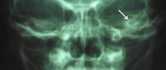
In some cases, neurosurgeons can determine damage to the base of the skull by seeing signs of intense exophthalmos. When a fracture occurs, the integrity of the internal carotid artery, which is in contact with the cavernous duct, is disrupted. The flow of venous blood, penetrating into the ophthalmic veins related to this reservoir, provokes pulsation, obvious hyperemia and protrusion of the apple of the optic organ.
Treatment of cavernous sinus thrombosis:
Children with rhinogenic intracranial complications undergo complex intensive therapy. Urgent surgical intervention is carried out with the aim of wide opening of all lesions. Indications for surgery are absolute. During surgery on the frontal sinus, a fairly extensive exposure of the dura mater of the anterior cranial fossa to the border with healthy tissue is shown. For an epidural abscess, all pathologically altered tissues are consistently and carefully opened and removed tissues, the abscess is widely opened, washed with an antibiotic solution and favorable conditions are created for drainage of the wound. With a subdural abscess of rhinogenic origin, characteristic local changes in the dura mater are revealed: thinning, yellowish color and bulging, absence of pulsation, granulation, fibrinous plaque, swishes, in the direction of which open, and then wash or drain the abscess cavity. In the absence of a fistula, a subdural abscess is diagnosed by puncture through the dura mater in 3-4 directions to a depth of no more than 4 cm. Exposure of the meninges and drainage of rhinogenic abscesses is carried out after external opening of the frontal sinuses in older children age. With a well-formed thick capsule, small depth and size of the abscess, it is removed entirely along with the capsule. In children, removal of the abscess is difficult due to the very thin capsule, its adhesion to the dura mater and bone structures. If the abscess capsule is thin, the brain abscess is large and located deep in the brain, a puncture method of treatment is used. An approach to the abscess is possible through the outer covering of the head and directly from the primary purulent focus in the paranasal sinuses. In case of thrombosis of the superior sagittal sinus, it is exposed, opened and the thrombus is removed. To stop bleeding, a tampon is inserted between the sinus and the bone. In case of thrombosis of the cavernous sinus, a surgical approach to it is impossible due to topographical features and surgical intervention is limited to a wide opening of the affected paranasal sinuses, elimination and reliable drainage of the primary pathological focus. Massive antibacterial therapy is carried out with the introduction of optimal doses of broad-spectrum antibiotics spectrum of action, in severe cases of rhinogenic meningitis, eidolumbar (sometimes suboccipital) administration of antibiotics or metronidazole solution is additionally prescribed. Detoxification therapy is carried out (intravenous administration of plasma, hemodez, rheopolyglucin) using the latest methods of extracorporeal detoxification (plasmapheresis, ultraviolet irradiation of autologous blood, hemosorption of blood, hyperbaric oxygenation).Dehydration is carried out by intramuscular administration of magnesium sulfate, intravenous infusions of 40% glucose solution, prescription of diuretics. Hyposensitizing and restorative treatment, immunotherapy (anti-staphylococcal plasma, gamma globulin, serum polyglobulin, staphylococcal toxoid) are carried out. In the complex therapy of cavernous and thrombosis superior sagittal sinuses must include direct (heparin) and indirect (dicoumarin, nitrofarsin, syncumar, phenylin) anticoagulants in order to disrupt the biosynthesis of prothrombin and other blood coagulation factors under constant thromboelastogram monitoring. Treatment with anticoagulants is stopped gradually, increasing the interval between doses, since rapid cancellation may cause a sharp compensatory increase in the concentration of prothrombin with the risk of thrombosis. In severe cases, endovascular (intracarotid) regional infusion therapy is used using activators of endogenous fibrinolysis in combination with antibiotics, which accelerates recanalization of the sinuses. At the same time, blockers of proteolytic enzymes secreted by staphylococcus (contrical, trazylol) are used ). Epsilon-aminocaproic acid has a lesser effect. The creation of a constant high concentration of thrombolytic and antibacterial drugs in the vascular basin of the pathological focus promotes rapid thrombolysis, restoration of blood flow in the cavernous sinus, and relief of the inflammatory process.
Violations of structures
Pathologies of these choroid plexuses arise due to their blockage, which in turn is provoked by thrombosis, thrombophlebitis or compressive neoplasm of intracranial veins and arteries.
Inflammation of the structures of the main human organ can appear when infectious agents penetrate into the bloodstream (all kinds of unconnected vascular substrate, solid, liquid or vapor, circulating through the bloodstream, uncharacteristic in a normal state, capable of provoking blockage of an artery at a fairly large distance from the site of occurrence). The pathological agent can enter the meninges and the vascular beds of the head bone tissue on its surface. In this case, symptoms of peak manifestations of meningitis and other pathologies are likely to appear. In preschool children, a picture of neuropoisoning appears.
In some cases, neurosurgeons can determine damage to the base of the skull by seeing signs of intense exophthalmos. When a fracture occurs, the integrity of the internal carotid artery, which is in contact with the cavernous duct, is disrupted. The flow of venous blood, penetrating into the ophthalmic veins related to this reservoir, provokes pulsation, obvious hyperemia and protrusion of the apple of the optic organ. This deviation is otherwise called the carotid-cavernous anastomosis, and this is one of the extremely rare pathologies when listening to the skull with a phonendoscope makes it possible to hear the noise of blood flow in the area where the vessels join.
Formation of cerebrospinal fluid. Fluid flow inside GM and SM
Cerebrospinal fluid is formed in the choroid plexuses of the lateral ventricles (due to the good permeability of the capillary walls).
Fluid flow: Lateral ventricles => lateral foramina => III ventricle => aqueduct => IV ventricle => subarachnoid space (pontine-cerebellar cistern) => pachyonic granulations => venous sinuses.
Cerebrospinal fluid swells through the inguinal granulations => venous sinuses => Dura Mater veins => through the perineural spaces of the sense of smell and in the mucous membranes of the nasal cavity => lymphatic bed of the nasal mucosa => lymphatic vessels.
Nerves
The dura mater is innervated by various branches. In particular, branches of the vagus and trigeminal nerves approach it. In addition, innervation is provided by sympathetic fibers. They enter the hard shell in the thickness of the outer wall of blood vessels. In the area of the cranial anterior fossa, the dura mater receives processes from the optic nerve.
Its branch, the tentorial, provides innervation to the cerebellar tentorium and the falx cerebri. The cranial middle fossa is supplied by the meningeal process of the maxillary and part of the mandibular nerves. Most of the branches lie along the vessels of the membrane. In the tentorium cerebellum, however, the situation is somewhat different. There are few vessels there, and the branches of the nerves are located in it independently of them.
Important Newborn sleeps poorly: causes of sleep disturbances day and night
What processes does the dura mater of the brain have? List them
Dura Mater GM produces shoots located between the parts of GM :
1) Falx cerebrum (falx cerebri) - between the hemispheres of the cerebrum (sagittal plane). It goes in especially deeply with its front part.
Starts in front of the Crista gallae of the ethmoid bone => attaches (with its convex edge) to the lateral ribs of the Sulcus sinus sagittalis superioris => reaches the Protuberantia occipitalis interna => passes into the upper surface of the tentorium of the cerebellum.
2) Falx cerebellum (falx cerebelli) - between the hemispheres of the cerebellum in the area of its posterior notch.
Starts from Protuberantia occipitalis interna => Crista occipitalis interna => posterior edge of Foramen magnum (there is a transition into two folds limiting the opening at the back).
3) Tentorium cerebelli - a horizontal plate between the occipital lobes of the cerebrum and the cerebellum (the middle part is pulled upward).
The anterior free edge is concave—notch of the tentorium. Limits the opening of the tentorium. This is where the brainstem passes through. It begins above the posterior cranial fossa - between the upper edges of the pars petrosa of the temporal bones and the sulcus sinus occipitalis superioris.
4) Saddle diaphragm (diaphragma sellae) - goes above the sella turcica, forming a roof. Below it is the pituitary gland. There is a hole in the middle of the seat diaphragm. A funnel passes through it. The pituitary gland hangs on it.
In the region of Impressio trigemini (top of the pyramid) Dura Mater GM splits into two leaves, forming the trigeminal cavity (cavum trigeminale). It contains the trigeminal nerve ganglion.
Channel localization
The sinuses of the dura mater of the brain are classified according to intracranial localization and the presence of intersinus connections. The words “sinus” and “sinus”, as well as “reservoir” are synonymous and mean the same thing.
Superior sagittal sinus
The superior sagittal sinus is characterized by considerable length and complex structure. The falx cerebri is involved in its formation. This is what is called the crescent-shaped plate. It is formed by the dura mater. The process begins from the crest of the ethmoid bone and goes backward along the midline, filling the interhemispheric gap that separates the hemispheres from each other. The groove of the superior sagittal sinus is the base of the falx.
This canal forms numerous lateral lacunae. This is the name given to small cavities that communicate with the venous network of hard leaves.
The superior sagittal sinus is equipped with the following vascular connections:
- The anterior sections of the sinus are connected to the veins of the nasal cavity.
- The middle sections have a connection with the venous vessels of the parietal lobes of the brain.
This vascular reservoir gradually increases in volume and expands. Its posterior section enters the common sinus drainage.
Inferior sagittal reservoir
The inferior sagittal sinus is referred to in the medical literature as sinus sagittalis inferior. It is so called because it is located in the lower segment of the falx. Compared to the upper sinus, it is much smaller in size. Due to numerous venous anastomoses, it is connected to the straight sinus.
Direct sinus
The straight sinus is located at the junction of the falx and tentorium, which covers the cerebellum. Has a sagittal direction. The great cerebral vein flows into it. The blood flow from it is directed towards the transverse venous sinus.
Transverse sinus
The transverse sinus occupies a wide groove of the same name on the surface of the occipital bone. It is located in the area where the cerebellar tentorium extends from the dura mater. It is the largest of all venous reservoirs and continues into the sigmoid venous sinuses.
Sigmoid venous reservoir
The sigmoid sinus occupies sigmoid grooves on both sides, shaped like the letter S. The external cerebral veins are connected to it. At the level of the jugular foramen, the blood flow from the sigmoid canals is directed into the bed of the internal jugular vein.
Cavernous sinus
The cavernous sinus is localized on the sides of the sella turcica, in appearance it resembles a triangle, in the upper part of which the oculomotor nerve is located, in the lateral part there is a branch of the trigeminal nerve. Its anatomy is distinguished by a large number of internal partitions. This explains its other name - cavernous sinus.
The internal part of the structure is occupied by the abducens nerve. Inside the sinus is a section of the internal carotid artery, surrounded by the sympathetic nerve plexus. Paired ophthalmic venous vessels flow into this canal. The sphenoparietal sinuses of the dura mater are associated with it.
The cavernous sinuses are connected by venous branches running along the contours of the sella turcica. Such complex vascular relationships allow the vessels to form a fairly large sinus surrounding the pituitary gland lying in the center of the sella turcica.
A continuation of this sinus are two venous reservoirs surrounding the temporal pyramids above and below. These are called the superior and inferior petrosal sinuses. Connected by numerous venous vessels, the stony sinuses participate in the formation of the main plexus of venous vessels located in the occipital lobe of the brain.
Occipital venous canal
The occipital sinus is located at the base of the falx and the internal crest of the occipital bones. At the top it is connected to a transverse channel. In the lower section, this sinus is divided into two branches that surround the foramen magnum. They are connected to the right and left sigmoid sinuses. The superficial veins of the brain and the vertebral plexus of veins are connected to the occipital sinus.
The sinuses of the brain create a venous confluence, or drain. In Latin, this reservoir of venous blood is called “confluens sinuum”. It is located in the area of the cruciate eminence inside the occipital bone. The flow of venous blood from all intracranial vessels and reservoirs is directed to the jugular vein.
Thus, the structure of the human cerebral venous system is very complex. All venous channels are in one way or another interconnected not only with each other, but also with other cerebral structures.
Types of dura sinuses
Nature very thoughtfully created humans, providing the dura mater with indentations to provide the main organ with oxygen and nutritional compounds.
Superior sagittal sinus
This cranial sinus is characterized by a large space with a complex structure. The sickle of the main human organ takes a significant part in its development. This is a crescent leaf. It is made of dura mater. The process originates from the top of the ethmoid bone, passes in the middle back, penetrating into the interhemispheric foramen, separating the brain regions from each other. The groove-like growth of the superior sagittal sinus is essentially the base of the falx bone.
This duct provides numerous lacunae on the sides. These are small cavities that are connected to a venous network of strong plates.
The superior sagittal reservoir has the following venous connections:
- the anterior parts belong to the vessels of the labial cavity (near the nose);
- the middle parts belong to the venous beds of the parietal fragments of the brain.
As a person grows, this collector of arteries and veins becomes larger and wider in terms of mass capacity. Its posterior fragment protrudes into the combined sinus drain.
Inferior sagittal sinus
This cistern of the structure of the cranium is presented in medical annals as sinus sagittalis inferior. It was so named because it is located in the lower location of the cerebral arch. Compared to the upper reservoir, it has a significantly small volume. Thanks to the large number of venous anastomoses, it is attached to the rectum.
Direct sine
This fragment of the skull is, in fact, the so-called continuation of the lower cistern from the rear side. It connects the rear sections of the superior tanks and the lower manifold. Along with the superior one, a large vessel is included in the anterior part of the nondual sinus. The posterior section of the cavity flows into the median fragment of the double descending duct, which developed due to the divergence of the dura mater of the skull, which is located in the groove of the hard tissue of the back of the head, extended laterally and towards the bottom, attached to the sinus. This fragment is called the sine drain.
Sigmoid venous sinus
This reservoir is the most significant and extensive. On the surface inside the scales of the occipital bone tissue, it is presented in a large groove. The venous reservoir then flows into the sigmoid sinus. Then it goes deeper into the mouth of the largest vessel, which carries out venous drainage from the head. Thus, the transverse sinus and sigmoid sinus are characterized as the main venous reservoirs. In addition, all other pockets go into the first one. Some vein sinuses are included in it directly, some through a smooth transition. On the temporal sides, the transverse recess continues with the sigmoid recess of the proper side. The place where the venous expansions of the sagittal, rectal and occipital sinuses are included in it is called the common drain.
Cavernous reservoir
It acquired this name because it has a large number of partitions. They provide the reservoir with an appropriate structure. The abducens, ophthalmic, trochlear nerve fibers that move the eyes, and in addition the carotid artery (which is inside) along with the sympathetic intertwining (autonomic nerves in the thoracolumbar region) are stretched through the cavernous sinus. Between the right and left localizations of space there are communicative connections. They are provided in the posterior and anterior intercavernous. Accordingly, a venous ring develops in the location of the sella turcica. The cavernous sinus (its flanking fragments) passes into the space of the sphenoid-parietal sinus, which lies on the border of the small branch of the bone in the form of a wedge.
Occipital venous sinus
The occipital cistern is located at the base of the arch and the upper part of the occipital region located inside. From above it refers to the transverse duct. At the bottom, this pocket is divided into two branches that encircle the joint at the back of the head. They are interconnected by sigmoid sinuses on both sides. The superficial veins of the main human organ and the veins and vessels of the spine are related to the occipital space.
Prevention, complications, prognosis
It is very important, if a person notices any changes or pain in his condition, to consult a doctor in time, because the sooner this disease is identified, the sooner and faster doctors will be able to help the sick patient. Therefore, there is no need to wait until the last moment, because waiting can often lead to a stroke or cerebral edema, or death. Other complications include blurred vision, deterioration in hormone production, possible formation of cysts in the lining of the brain and other cardiovascular diseases in humans
Other complications include blurred vision, deterioration in hormone production, possible formation of cysts in the lining of the brain and other cardiovascular diseases in humans.
Of course, the prognosis for recovery is influenced by the stage of the disease at which a person is; it all depends on how quickly the person turned to specialists. In any case, the mortality rate usually does not exceed twenty percent in advanced stages.
So, in order not to undergo such a difficult test, you need to adhere to preventive measures and check regularly with a doctor.
Diagnostic methods
Diagnostic measures include the following:
- A number of immunological studies.
- Laboratory tests - blood, urine.
- Blood chemistry.
- Clinical studies of patient biomaterial.
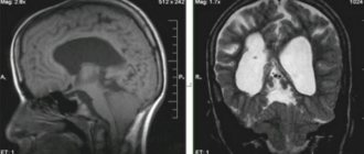
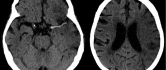
- MRI.
- CT scan.
- Differential diagnosis.
To ensure the correct diagnosis, a histological examination is performed. A fragment taken from the site of the rash for study is separated by biopsy.
It is not possible to carry out a diagnosis at home or without consulting a competent doctor, so there is no need to hesitate and go to the hospital at the first sign.
Treatment
The development of cerebral and focal neurological symptoms in thrombosis of the cerebral veins and venous sinuses is based on changes in brain tissue and the development of intracranial hypertension. This combination is potentially dangerous and may be associated with an unfavorable outcome of the disease.
Therefore, it is necessary to carry out complex treatment, including pathogenetic (recanalization of venous sinuses) and symptomatic therapy (correction of intracranial hypertension, anti-inflammatory treatment).
The main goal of treatment for thrombosis of the cerebral veins and venous sinuses is to restore their patency. There are indications of the successful use of thrombolysis, but the number of hemorrhagic complications increases significantly. Currently, the drugs of choice are anticoagulants, in particular low molecular weight heparins.
According to various studies, the use of direct anticoagulants in the acute period of thrombosis of the cerebral veins and venous sinuses improves the outcome and reduces the risk of death and disability. According to ISCVT, 80 out of 624 patients with thrombosis of the cerebral veins and venous sinuses received low molecular weight heparins. 79% of these patients recovered, 8% had mild symptoms, 5% had significant neurological impairment, and 8% of patients died. These data indicate the effectiveness and safety of low molecular weight heparins in the acute period of thrombosis of the cerebral veins and venous sinuses.
Important Why does insomnia occur and how can you fight it?
In cases of infectious thrombosis of the venous sinuses, antibacterial therapy should be carried out using high doses of broad-spectrum antibiotics, such as cephalosporins (ceftriaxone 2 g/day IV), meropenem, ceftazidine (6 g/day IV), vancomycin (2 g /day i.v.).
The primary source of infection should be revised, and surgical methods should be used if necessary. It is not advisable to perform surgical treatment before prescribing drug therapy. First of all, you need to carry out antibiotic therapy. The decision to undergo surgical intervention on the ear or sinus is possible when control of the infection is achieved. There is no consensus on the feasibility and safety of anticoagulant therapy, although most authors adhere to the management of such patients using low molecular weight heparins.
At the end of the acute period of thrombosis of the cerebral veins and venous sinuses, it is recommended to prescribe indirect oral anticoagulants (acenocoumarol, warfarin) with maintaining the international normalized ratio (INR) within 2-3. In this case, direct anticoagulants are used until the INR reaches target values.
In case of thrombosis of the cerebral veins and venous sinuses during pregnancy, the administration of indirect anticoagulants should be avoided due to their teratogenic properties and the possibility of penetration through the placental barrier. In such cases, treatment with direct anticoagulants is recommended.
Currently, there are no studies clearly regulating the duration of use of oral anticoagulants. According to the EFNS recommendations (2006), indirect anticoagulants should be used for 3 months. with secondary thrombosis of the cerebral veins and venous sinuses, which developed in the presence of a so-called transient risk factor, within 6-12 months. in patients with idiopathic thrombosis and in “minor” thrombophilic conditions, such as deficiency of proteins C and S, heterozygous mutation of Leiden factor or mutations in the prothrombin gene (G20210A). Lifelong anticoagulant therapy is recommended for patients with recurrent venous sinus thrombosis, as well as for congenital thrombophilic conditions (homozygous Leiden factor mutation, antithrombin III deficiency).
In addition to basic therapy, measures should be taken to prevent complications such as seizures and intracranial hypertension. These conditions require the prescription of anticonvulsants, mechanical ventilation in hyperventilation mode with positive expiratory pressure, and the administration of osmotic diuretics. However, it should be remembered that excessive dehydration, in turn, worsens the rheological properties of the blood, thereby promoting further thrombus formation.
The effectiveness of glucocorticoids for cerebral edema resulting from thrombosis of the cerebral veins and venous sinuses has not been proven. In some cases, in severe forms of CVST, complicated by dislocation of brain structures, the issue of decompressive hemicraniotomy, which is a life-saving operation, may be considered.
Venous sinuses
- The superior sagittal sinus
(lat. sinus sagittalis superior) is located along the upper edge of the falciform process of the dura mater, ending at the back at the level of the internal occipital protrusion, where it most often opens into the right transverse sinus. - The inferior sagittal sinus
(lat. sinus sagittalis inferior) - spreads along the lower edge of the falx, flows into the straight sinus. - The straight sinus
(lat. sinus rectus) is located along the junction of the falciform process with the tentorium of the cerebellum. It has a tetrahedral shape, goes from the posterior edge of the inferior sagittal sinus to the internal occipital protrusion, opening into the transverse sinus. - The transverse sinus
(lat. sinus transversus) is a pair, located in the transverse groove of the skull bones, located along the posterior edge of the tentorium of the cerebellum.
At the level of the internal occipital protrusion, the transverse sinuses communicate with each other. In the region of the mastoid angles of the parietal bones, the transverse sinuses become sigmoid sinuses
, each of which opens through the jugular foramen into the jugular bulb. - The occipital sinus
(lat. sinus occipitalis) is located in the thickness of the edge of the falx of the cerebellum, extending to the foramen magnum, then splits, and in the form of marginal sinuses opens into the sigmoid sinus or directly into the superior bulb of the jugular vein. - The cavernous (cavernous) sinus
(lat. sinus cavernosus) is a paired sinus, located on the sides of the sella turcica. The cavity of the cavernous sinus contains the internal carotid artery with the surrounding sympathetic plexus and the abducens nerve. The oculomotor, trochlear and ophthalmic nerves pass through the walls of the sinus. The cavernous sinuses are connected to each other by intercavernous sinuses. Through the superior and inferior petrosal sinuses they connect, respectively, to the transverse and sigmoid sinuses. - Intercavernous sinuses
(lat. sinus intercavernosi) - are located around the sella turcica, forming a closed venous ring with the cavernous sinuses. - The sphenoparietal sinus
(lat. sinus sphenoparietalis) is a pair, directed along the small wings of the sphenoid bone, opening into the cavernous sinus. - The superior stony sinus
(lat. sinus petrosus superior) is paired, runs from the cavernous sinus along the superior stony groove of the temporal bone and opens into the transverse sinus. - The lower stony sinus
(lat. sinus petrosus inferior) is paired, lies in the lower stony groove of the occipital and temporal bones, connects the cavernous sinus with the sigmoid sinus.
The structure of the sinuses of the solid MO
The development of TMO collectors occurs due to their division into two sheets, which are similar to channels. These ducts are designed to distribute the venous flow of blood from the main human organ, which is subsequently sent to several double vessels that are located in the neck and transfer blood from the brain.
The dura mater plates that make up the sinus look like tightly stretched ropes that do not lose tension. This structure allows blood to flow freely from the head and neck, without in any way affecting the state of intracranial pressure.
The following types of dura mater reservoirs have been established in humans:
- Superior or inferior sagittal. The first is located longitudinally along the upper border of the falx bone and ends on the occipital fragment, and the next is longitudinally along the border of the falx below and flows into the straight sinus;
- Straight. It is located longitudinally of the fragment, where the falciform process passes into the cerebellar tentorium;
- Transverse (double). Formed on the transverse growth of the skull, located longitudinally along the posterior border of the cerebellar groove;
- Occipital. It is located in the cavity of the cerebellar arch, and then extends to the occipital junction;
- Sigmoid. Located in the division in the ventral fragment of the head bone tissue;
- Cavernous (double). It is located on the sides of the formation of a wedge-shaped bone in the body (sella turcica);
- Sphenoparietal sinus (double). Refers to a small wedge-shaped border of bone and ends in a cavernous reservoir.
Stony (double). Located close to both borders of the pyramidal bone of the temples.
The collectors of the medulla begin to collect anastomoses with venous vessels on the surface of the brain, through venous branches that connect the vascular sinuses of the dura mater with the external circulatory vessels of the head. These depressions begin to communicate with the diploic processes, which are typically located in the cranial vault and then pass into the vessels of the head. Then the blood tends to pass through the venous plexuses and then flows into the dura mater reservoirs.
Pathology of intracranial sinuses
Diseases of these vascular formations are most often caused by their occlusion, which can be caused by thrombosis, thrombophlebitis, or tumor compression of intracranial vessels.
Inflammatory diseases of brain structures can occur when infectious agents enter the venous blood stream (purulent emboli). The infection can be brought to the membranes of the brain from the superficial venous vessels of the skull. In this case, the development of a clinical picture of acute meningitis and encephalitis is possible. In young children, a picture of neurotoxicosis develops.
Sometimes neurosurgeons may suspect a fracture of the base of the skull when they see a picture of pulsating exophthalmos. When injured, the internal carotid artery associated with the cavernous canal is damaged. A stream of arterial blood, entering the ophthalmic veins associated with this sinus, causes pulsation, severe redness and protrusion of the eyeball. This pathology is otherwise called carotid-cavernous anastomosis, and this is one of the rarest conditions when listening to the head with a phonendoscope allows you to hear the sounds of blood in the area of the anastomosis.
When the walls of the sinus are damaged, a number of neurological symptoms appear due to damage to nearby branches and nuclei of the cranial nerves. With pathology of the cavernous sinus, oculomotor disorders and the development of trigeminal neuralgia may occur.
If the patient suffers from frequent attacks of headaches or intracranial hypertension, the development of reverse (retrograde) blood flow is possible - from the brain cavity to the superficial veins of the skull. Therefore, in children with intracranial hypertension, the pattern of veins on the scalp is clearly visible. Due to the flow of blood, the pressure inside the skull decreases. This is a compensatory mechanism for reducing intracranial pressure.
Important Erythromelalgia (Mitchell's disease)
The cerebral sinuses are an important component of the cerebral venous network. Knowing their functions, structural features and localization, experts can assume the development of pathology in a certain area of the brain. To clarify the diagnosis, magnetic resonance imaging with intravascular injection of a contrast agent is necessary.
Rate this article:
- 4.21
Total votes: 96
Possible pathologies
Most diseases have not yet been fully studied, although they are detected in the early stages thanks to modern diagnostic methods.
| Name of disease / Which collector is affected | Reasons for development | Symptoms or how it manifests itself | Complications |
| Thrombosis/cerebral | Past infectious disorders. Intrauterine fetal hypoxia. Late toxicosis. Systemic diseases of an inflammatory nature. | Seemingly causeless convulsions. Constant nervousness. Headache attacks | Without proper treatment, the prognosis is poor: disability or death |
| Occlusion/sagitral | The main causes are infections that cause severe inflammation. | Weakness in the legs. Severe headaches. Bleeding from the nose | Decreased vision percentage. Ptosis of the eyelids. Decreased production of pituitary hormones |
| Embolism / all GM vessels | Heart diseases. Diabetes. Oncological diseases | Pain syndrome in the chest area. Constant shortness of breath | Pulmonary infarction. Paradoxical embolism. Increased pressure in the human lungs |
| Cerebral atherosclerosis / cerebral | Liver disorders. Excessive smoking and drinking alcohol. Genetic predisposition. Hormonal disorders | Temporary amnesia. Mental disorders. Chronic fatigue | Without treatment, there is a possibility of developing complete amnesia, death, or severe mental impairment of the person. |
| Stenosis / all collectors | Diabetes mellitus increased body weight Smoking genetic predisposition | Emotional instability. Memory loss | Loss of coordination. Uncontrolled urination. Death. It all depends on the stage of the disease. |
GM pathologies are varied. The table above covers only common vascular diseases.
Causes
The most common cause is infections that cause inflammation of the ear, tonsils, teeth, maxillary sinuses, facial tissues, eyes, as well as septic conditions due to viral, bacterial (including tuberculosis), and fungal infections.
In the absence of infectious diseases, sinus thrombosis occurs under the influence of the following factors:
- traumatic brain injury,
- operations on bones, skull tissues, internal organs,
- intracranial or other tumors,
- puncture of the spinal canal for diagnosis or pain relief,
- toxicosis of the second half of pregnancy with hypertension,
- caesarean section or natural birth,
- taking estrogen drugs (including contraceptives),
- extreme degree of dehydration,
- autoimmune diseases,
- increased blood clotting activity,
- circulatory disorders,
- diabetes.
Vascular (soft) MO
Pia mater encephali directly covers the brain surface. It is presented in the form of a transparent two-layer plate, which extends into the cracks and grooves. The vascular MO contains chromatophores—pigment cells. Especially many of them have been identified at the base of the brain. In addition, there are lymphoid, mast cells, fibroblasts, numerous nerve fibers and their receptors. Parts of soft MO accompany arterial vessels (medium and large), reaching arterioles. Between their walls and the shell there are Virchow-Robin spaces. They are filled with cerebrospinal fluid and communicate with the subarachnoid space. Elastic and collagen fibrils are thrown through them. Vessels are suspended from them, through which conditions are created for their displacement during pulsation without affecting the brain matter.
Subarachnoid space: location, tanks. Subdural space GM
Subarachnoid space of various sizes: above the gyri - narrow, above the grooves - expansions (cisterns):
- The cistern magna (cerebellomedullary) is the largest cistern, bounded by the cerebellum, medulla oblongata and occipital bone. Cerebrospinal fluid is collected from this area. The puncture is made between the upper edge of C1 and the foramen magnum.
- The pontine cistern (prepontine) is located anterior to the pons and contains the basilar artery. It communicates posteriorly with the subarachnoid space of the spinal cord + cerebellocerebral cistern, and anteriorly with the interpeduncular cistern.
- The basal cistern (supracellar) is pentagonal in shape. Includes the interpeduncular and chiasm cistern (optic nerves).
- The quadrigeminal cistern (veins of Galen) (quadrigeminalis) is located between the corpus callosum and the cerebellum. Arachnoid cysts may be located in its area.
- Bypass (encompassing, bypassing) tank (ambient) is an irregularly shaped channel. It runs along the sides of the cerebral peduncles and the roof of the midbrain. It communicates with the pontine and interpeduncular cistern ( anteriorly ) and the quadrigeminal cistern ( posteriorly ).
- The cistern of the lateral fossa of the cerebrum (fossae lateralis cerebri) is located in the lateral groove of the cerebrum.
All tanks are located on the side of the base of the GM so that the GM does not hit the bones of the base.
The subdural space of the SM is between Dura Mater and Arachnoidea. Contains : liquid. It resembles lymph or tissue fluid, but it is not cerebrospinal fluid.
Sinus transversus
This sinus is the largest and widest. On the inside of the scales of the occipital bone, it corresponds to a wide groove. Next, the sinus transversus becomes the sigmoid sinus. Then it goes into the mouth of the internal jugular vessel. Sinus transversus and Sinus sigmoideus thus act as the main venous collectors. At the same time, all other sinuses flow into the first. Some venous sinuses enter it directly, some indirectly. On the right and left, the transverse sinus continues into the sinus sigmoideus of the corresponding side. The area where the venous sinuses sagittalis, rectus and occipitalis flow into it is called the drain.
Connection with elements of the cerebellum
In the anterior part, the sickle is fused with the cockscomb on the ethmoid bone. The posterior region of the process at the level of the occipital internal protrusion connects with the tentorium of the cerebellum. He, in turn, hangs over the cranial fossa like a gable tent. It contains the cerebellum. Its tentorium penetrates the transverse fissure in the cerebrum. Here it separates the cerebellar hemispheres from the occipital lobes. There are irregularities on the leading edge of the tentorium. Here a notch is formed, to which the brain stem adjoins in front. The lateral portions of the tentorium fuse with the edges of the groove in the posterior sections on the transverse sinus of the occipital bone and with the upper edges of the pyramids on the temporal bones. The connection extends to the posterior processes of the wedge-shaped element in the anterior parts on each side. The cerebellar falx is located in the sagittal plane. Its leading edge is free. It separates the cerebellar hemispheres. The posterior part of the falx is located along the occipital internal crest. It extends to the edge of the large hole and covers it with two legs on both sides. At the base of the falx there is the occipital sinus.
What is the structure of the dura mater of the brain?
Dura Mater GM:
- It consists of two welded layers (SM has separate sheets). The Dura Mater GM layer is thicker than Dura Mater SM = 0.5 mm.
- Outer layer - fuses with the bones of the skull from the outside (periosteum)
- The inner layer is shiny, adjacent to the brain.
Dura Mater GM is loosely fused with the vaults, but tightly fused with the base (in case of epidural hemorrhage, the blood clot is in the vault area).
Beginning - from the lower edge of the foramen magnum => to S2 => downwards continues around the spinal cord => merges with other membranes => formation of the external terminal filament.
General information
The brain contains 25 billion neurons that make up the gray matter. The weight of the organ varies depending on gender. For example, in men its weight is about 1375 g, in women - 1245 g. On average, its share in the total body weight is 2%. At the same time, scientists have found that the level of intellectual development is not related to brain mass. Mental abilities are affected by the number of connections created by the organ. Brain cells are neurons and glia. The former generate and transmit impulses, the latter perform additional functions. There are cavities inside the brain. They are called ventricles. The cranial nerves extend from the organ we are considering to different parts of the human body. They are paired. In total, 12 pairs of nerves depart from the brain. The brain is covered by three membranes: soft, hard and arachnoid. There are spaces between them. They circulate cerebrospinal fluid. It acts as an external hydrostatic environment for the central nervous system and also ensures the removal of metabolic products. The membranes of the brain differ in their structure and the number of vessels passing through them. However, they all provide protection for the contents of the upper part of the skull from mechanical damage.
Features of shells and intershell spaces of SM
Dura Mater SM - consists of 2 separate sheets (for GM - welded layers). The layer of Dura Mater SM is thinner than Dura Mater GM.
Intershell spaces SM:
1) Epidural space of the SM - between the outer and inner layers of Dura Mater SM (foramen magnum => S2)
- The outer leaf is the periosteum of the vertebrae.
- The inner leaf is closer to the SM.
- Contains : fatty tissue + internal vertebral venous plexus. Performs an elastic function : fatty tissue is a soft lining,
- venous plexus – hydraulic seal.
2) Subdural space of the SM - between Dura Mater and Arachnoidea.
- Contains : liquid. It resembles lymph or tissue fluid, but it is not cerebrospinal fluid.
3) Subarachnoid space SM - between Arachnoidea and Dura Mater.
- Contains : cerebrospinal fluid.