History of the study
In Germany in 1909, the physician-scientist Korbinian Brodmann first constructed and described maps of Brodmann's cytoarchitectonic fields.
Ten years later, O. Vogt and C. Vogt studied and described more than a hundred myelo-architectonic areas in the cerebral hemispheres. As a result, at the State Scientific Center for Neurology I.N. Filimonov and S.A. Sarkisov released a map of the brain hemispheres according to Brodmann, which describes 47 fields.
Brodmann fields are most often used in studying the neural organization of the brain and its functioning. The division of one or another area of the brain into a specific section of the fields was carried out on the basis of histological analysis - Nissl color staining.
The concept of structural and functional blocks of the brain A. R. Luria
A. R. Luria proposed a structural-functional model of the brain as a substrate of mental activity. This model characterizes the most general patterns of the functioning of the brain as a whole and allows us to explain its integrative function (E. D. Chomskaya).
According to this model, the entire brain can be divided into three structural and functional blocks:
a) Energy block,
b) Block for receiving, processing and storing exteroceptive information,
c) Block of programming, regulation and control of complex forms of activity.
Any VPF is carried out with the mandatory participation of all three blocks. Each block is characterized by structural features, the physiological principles underlying its work, and the role it plays in ensuring mental functions.
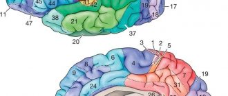
Groups of brain fields according to Brodmann
Description of Brodmann fields in the cerebral cortex by zone:
- The first zone is motor , responsible for reactions of active movements. This zone includes 4, 6, 8, 9 Brodmann fields. 4 is responsible for motor skills and is located in the precentral gyrus. 6 is allocated in the anterior regions of the precentral gyrus and in the area of the middle frontal gyrus. 8 coordinates voluntary eye movement and is located in the posterior parts of the superior and middle frontal gyri. 9 is located in the prefrontal region.
- The second zone is affective . Includes areas of the cerebral cortex posterior to the Rolandic fissure. Here are fields 1, 2, 3, 5 and 7. The upper part of the zone is responsible for tactile sensations of the legs and genitals. The underlying areas are for sensations in the hands, skull and mouth. The second zone directly interacts with the first. The first zone contains afferent nerve cells that receive stimuli from proprioceptors - these are motor sensory areas. The second zone contains motor components - these are sensorimotor areas that regulate the formation and intensity of pain sensations.
- The third zone is visual , located in the occipital region of the cerebral cortex. It includes 17, 18, 19 Brodmann fields. The 17th belongs to the primary visual area, and the 18th and 19th to the secondary visual area. From the secondary areas, visual stimuli come to the primary areas and are already processed there. With pathologies of the 17th Brodmann area, cortical blindness occurs - loss of visual perception. In case of violation of 18, the function of identification and perception of written speech is affected. In pathologies 19, hallucinations and disturbances in figurative memory are noted.
- The fourth zone is auditory , located in the temporal zone of the cerebral cortex. It includes 22, 41, 42, 52 fields. With damage to 22, auditory hallucinations are noted, orientation to sound is affected, and musical deafness occurs. With pathologies 42, sound recognition suffers, and lesion 41 causes cortical deafness, that is, a complete loss of auditory perception. 52 is a zone that is responsible for the spatial perception of voices, sounds and speech.
- The fifth, olfactory zone, includes fields 11 and 29, which are located in the pyriform gyrus. Responsible for recognizing different odors.
- The sixth zone is the taste zone , which includes the 43rd field.
- The seventh zone is speech , in right-handed people it is located in the left hemisphere. This includes field 22 - Wernicke's speech center, field 37 - controls voluntary speech and its understanding, field 47 - singing areas, 44 and 45 - Brocca's speech centers.
- fields 24, 25 and 26 perform the task of recognizing mismatches and errors.
Brodmann field diagram:
The first functional block of the brain
The first block is the block for regulating energy tone and wakefulness.
It has been proven (I.P. Pavlov, A.R. Luria, M.N. Livanov) that for normal mental activity the body must be in a state of wakefulness (in other words, the cerebral cortex must be in a state of tone, i.e. have a certain level of arousal).
Only under conditions of optimal wakefulness can a person best receive and process information, recall the necessary systems of connections in memory, program activities, and exercise control over them.
It was found that the devices that provide and regulate the tone of the cortex are not located in the cortex itself, but in the underlying stem and cortical parts of the brain .
This apparatus consists of nonspecific structures of different levels:
- reticular formation of the brain stem,
- nonspecific structures of the midbrain,
- limbic system,
- hippocampal region.
The reticular formation is a nerve network in which the bodies of neurons with short axons are interspersed.
The reticular formation has a number of structural and functional features that ensure its main functions:
- firstly, it consists of an ascending and descending part. Along the fibers of the ascending reticular formation, excitation is directed upward, ending in the higher formations (hypothalamus, ancient cortex and new cortex). The fibers of the descending reticular formation have the opposite direction: starting from the neocortex, they transmit excitation to the structures of the midbrain and brain stem.
- In addition, the neurons of the reticular formation work on the principle of “gradual accumulation of excitation,” i.e. excitation does not spread in individual impulses, but gradually, gradually changing its level and thus modulating the state of the entire nervous system.
- And finally, fibers (collaterals) from all analytical systems, as well as fibers from the cerebral cortex and cerebellum, converge to the reticular formation.
The presence of numerous connections in the reticular formation itself, the convergence of all nerve pathways on most of its neurons create additional opportunities for the wide and simultaneous spread of excitation waves to the primary, secondary and tertiary zones of the cortex, as well as other brain structures.
As you know, the nervous system is always in a state of certain activity and its presence is mandatory for any manifestation of vital activity.
It is customary to distinguish several sources of activity: first of all, the metabolic processes of the body that underlie homeostasis (protein, carbohydrate, etc.). Then there is a direct influx of information entering the body from the outside world (from exteroceptors).
It is known that in a state of sensory deprivation a person falls into a sleep, from which he can only be brought out by the arrival of new information.
The listed sources of activity are characteristic of both humans and animals. But in addition to this, a significant part of a person’s activity is determined by his plans, intentions, and programs. Formed in the process of conscious life, they are social in nature and are realized with the close participation of first external and then internal speech.
The functional significance of the first block in ensuring mental functions is
- Firstly, in the regulation of activation processes, in maintaining the general tone of the central nervous system necessary for any mental activity (activating function).
- Secondly, in transmitting the regulatory influence of the cerebral cortex on the underlying brain stem formations (modulating function): Due to the descending fibers of the reticular formation, the higher parts of the cortex control the work of the underlying apparatus, modulating their work and providing complex forms of conscious activity.
When the reticular formation is damaged, the productivity of all HMFs decreases (primarily involuntary attention and memory), activity and sleep are disrupted.
In the case of massive lesions of the reticular formation, the line between sleep and wakefulness is blurred, the person is in a half-asleep state, and his orientation in time and place suffers.
Distinctive diagnostic signs of damage to the reticular formation are a simultaneous decrease in the productivity of absolutely all mental processes, as well as the possibility of partial compensation of the defect by complicating the task.
The involvement of voluntary processes and special motivation make it possible to briefly increase the efficiency of mental processes.
Thus, the first block of the brain is involved in ensuring mental activity, primarily in the organization of attention, memory, emotional state and consciousness in general.
In addition, the first block of the brain is involved in the regulation of emotional (fear, pain, pleasure, anger) and motivational states.
The limbic structures of the brain included in this block occupy a central place in the organization of emotional and motivational states.
In this regard, the first block of the brain perceives and processes a variety of interoceptive information about the state of internal organs and regulates these states.
Centers of the first signal system
The first signaling system is found both in Homo sapiens and in other living beings. It performs the functions of understanding and analyzing irritations emanating from the surrounding world and manifesting itself in the form of sensations and ideas.
The centers of the first signaling system are located in the nuclei of sensory sensitivity analyzers, motor analyzers, auditory, skin, visual and olfactory analyzers.
They are indicated in the superior and inferior parietal regions of the brain, in the precentral and supramarginal sulcus, as well as in the thickness of the lateral sulcus (auditory nuclei).
The purpose of this system is presented in the table of Brodmann fields:
| Brodmann fields | Analyzer cores | Functions |
| 1, 2, 3, 5, 7 | Cortical | Responsible for the perception of temperature and pain, as well as tactile sensations. Affective pathways extending to the cerebral cortex intersect in the region of the spinal cord and medulla oblongata. Because of this, the functions of either hemisphere are controlled by the diametrically inverted part of the body. |
| 6 and 4 | Motor | Neurons are released here, the reactions from which control the muscles of the lower body and facial muscles. |
| 8 | Skulls and eyes | Nuclei that control head and eye movements. |
| 40 | Motor | They control the voluntary, purposeful movements of an animal or person. |
| 18, 17 and 19 | Visual | Responsible for visual memory, perception of images and orientation in unfamiliar places. |
| 7 | Skin | Performs tactile functions of recognizing objects and surfaces by touch and other types of skin sensitivity. |
| 41, 42 and 52 | Auditory | Perceiving and remembering sounds from the outside. |
| 43 | Olfactory | Phylogenetically the oldest region of the cerebral cortex. Provides the function of perception and memory of odors. Closely related to taste sensations. |
The picture shows the fields of the cerebral cortex according to Brodmann and their functions:
Second functional block of the brain
The second block - the block for receiving, processing and storing information - is located in the outer sections of the new cortex (neocortex) and occupies its posterior sections, including the apparatus of the occipital, temporal and parietal cortices.
The structural and anatomical feature of this brain block is the six-layer structure of the cortex .
It includes:
- primary zones (providing reception and analysis of information coming from outside),
- secondary zones (performing the functions of synthesizing information from one analyzer) and
- tertiary zones (the main task of which is a comprehensive synthesis of information).
A distinctive feature of the devices of the second block is modal specificity .
Experiments recording the activity of individual neurons showed that nerve cells of the primary zones are distinguished by high modal specificity and narrow specialization.
The first means that they respond to stimulation of only one modality (one type), for example, only visual or only auditory.
The second assumes that these neurons respond only to a single feature of a stimulus of one type (for example, only to the width of a line or the angle of inclination, etc.).
Thanks to this, the devices of the second functional block of the brain perform the functions of receiving and analyzing information coming from external receptors and synthesizing this information.
Basic laws of the construction of the cortex, which is part of the second block of the brain
A. R. Luria identifies the basic laws of the structure of the cortex, which is part of the second block of the brain.
1. The law of the hierarchical structure of cortical zones.
According to this law, the ratio of primary, secondary and tertiary zones of the cortex carries out an increasingly complex synthesis of information.
More complexly organized cortical areas provide more complex functions.
A. R. Luria emphasizes that the ratio of primary, secondary and tertiary zones in an adult and a child is different.
For the normal development of secondary zones in a child, it is necessary that primary zones be formed, and for the development of tertiary zones, secondary zones.
Therefore, damage to the primary zones in early childhood can lead to gross disturbances in the development of secondary and, especially, tertiary zones.
In an adult, when the cortical zones are formed, the tertiary, the most organized, control the function of the underlying secondary and primary zones. Therefore, in an adult, the interaction of cortical zones occurs from top to bottom.
In this case, damage to the primary zones does not lead to noticeable impairments in mental functions and can be compensated by the work of nearby structures.
2. The law of decreasing modal specificity of hierarchically constructed cortical zones.
This law suggests that as we move from primary to tertiary zones, the manifestation of their modal specificity decreases.
The primary zones of each of the brain lobes included in the second brain block have maximum modal specificity (due to the huge number of neurons with a highly differentiated, modality-specific function).
Secondary zones, in which the upper layers with associative neurons predominate, have modal specificity to a much lesser extent.
Modal specificity is even less characteristic of the tertiary zones of the described block (“overlap zones” of the cortical ends of various analyzers).
Thus, this law describes the transition from a fractional reflection of private, modality-specific features to a synthetic reflection of more general schemes of the perceived world.
3. The law of progressive lateralization of functions.
The law of progressive lateralization of functions explains the connection of functions with a particular hemisphere (as we move from primary zones to tertiary zones).
The primary zones of both hemispheres of the brain are equivalent. At the level of secondary zones, some of the functions performed by the left and right hemispheres remain the same, but some of the functions of the left hemisphere are already different from the functions performed by the right hemisphere of the brain.
The functions of the tertiary zones of the left hemisphere are already fundamentally different from the functions of similar zones of the right hemisphere of the brain.
When the devices of the second block of the brain are damaged, the dysfunction depends on which areas are affected.
When the primary zones are damaged, a disturbance in the perception of individual signs of a perceived stimulus of one modality occurs (blind spot, hemeanopsia, tone scale disturbance, anesthesia, etc.).
When the secondary zones of the cortex are damaged, there is a violation of the synthesis of individual signs of the perceived stimulus into a holistic image of one modality (agnosia, aphasia).
Damage to the tertiary zones leads to disruption of the complex synthesis of stimuli coming from different analyzers, which manifests itself in a violation of orientation in space.
Moreover, according to the law of progressive lateralization, when the tertiary zones of the right hemisphere are damaged, object orientation in space is disrupted, and when similar zones of the left hemisphere are damaged, symbolic orientation in space suffers.